|
Axon Terminal
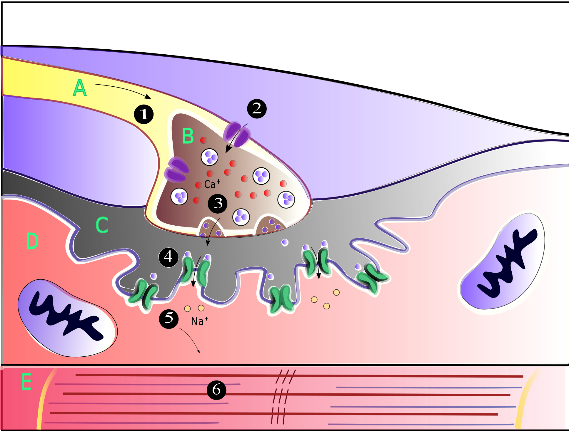
Axon terminals (also called terminal boutons, synaptic boutons, end-feet, or presynaptic terminals) are distal terminations of the branches of an axon. An axon, also called a nerve fiber, is a long, slender projection of a Neuron, nerve cell that conducts electrical impulses called action potentials away from the neuron's cell body to transmit those impulses to other neurons, muscle cells, or glands. Most presynaptic terminals in the central nervous system are formed along the axons (en passant boutons), not at their ends (terminal boutons). Functionally, the axon terminal converts an electrical signal into a chemical signal. When an action potential arrives at an axon terminal (A), neurotransmitter, the neurotransmitter is released and diffuses across the synaptic cleft. If the postsynaptic cell (B) is also a neuron, Neurotransmitter receptor, neurotransmitter receptors generate a small electrical current that changes the postsynaptic potential. If the postsynaptic cell (B) is ... [...More Info...] [...Related Items...] OR: [Wikipedia] [Google] [Baidu] |
Synapse Diag1
In the nervous system, a synapse is a structure that allows a neuron (or nerve cell) to pass an electrical or chemical Cell signaling, signal to another neuron or a target effector cell. Synapses can be classified as either chemical or electrical, depending on the mechanism of signal transmission between neurons. In the case of electrical synapses, neurons are coupled bidirectionally with each other through gap junctions and have a connected cytoplasmic milieu. These types of synapses are known to produce synchronous network activity in the brain, but can also result in complicated, chaotic network level dynamics. Therefore, signal directionality cannot always be defined across electrical synapses. Chemical synapses, on the other hand, communicate through neurotransmitters released from the presynaptic neuron into the synaptic cleft. Upon release, these neurotransmitters bind to specific receptors on the postsynaptic membrane, inducing an electrical or chemical response in the ... [...More Info...] [...Related Items...] OR: [Wikipedia] [Google] [Baidu] |
Voltage-gated Calcium Channel
Voltage-gated calcium channels (VGCCs), also known as voltage-dependent calcium channels (VDCCs), are a group of voltage-gated ion channels found in the membrane of excitable cells (''e.g.'' muscle, glial cells, neurons) with a permeability to the calcium ion Ca2+. These channels are slightly permeable to sodium ions, so they are also called Ca2+–Na+ channels, but their permeability to calcium is about 1000-fold greater than to sodium under normal physiological conditions. At physiologic or resting membrane potential, VGCCs are normally closed. They are activated (''i.e.'': opened) at depolarized membrane potentials and this is the source of the "voltage-gated" epithet. The concentration of calcium (Ca2+ ions) is normally several thousand times higher outside the cell than inside. Activation of particular VGCCs allows a Ca2+ influx into the cell, which, depending on the cell type, results in activation of calcium-sensitive potassium channels, muscular contraction, excitati ... [...More Info...] [...Related Items...] OR: [Wikipedia] [Google] [Baidu] |
Endocytosis
Endocytosis is a cellular process in which Chemical substance, substances are brought into the cell. The material to be internalized is surrounded by an area of cell membrane, which then buds off inside the cell to form a Vesicle (biology and chemistry), vesicle containing the ingested materials. Endocytosis includes pinocytosis (cell drinking) and phagocytosis (cell eating). It is a form of active transport. History The term was proposed by Christian de Duve, De Duve in 1963. Phagocytosis was discovered by Élie Metchnikoff in 1882. Pathways Endocytosis pathways can be subdivided into four categories: namely, receptor-mediated endocytosis (also known as clathrin-mediated endocytosis), caveolae, pinocytosis, and phagocytosis. * Clathrin-mediated endocytosis is mediated by the production of small (approx. 100 nm in diameter) vesicles that have a morphologically characteristic coat made up of the cytosolic protein clathrin. Clathrin-coated vesicles (CCVs) are found in vir ... [...More Info...] [...Related Items...] OR: [Wikipedia] [Google] [Baidu] |
Neuromuscular Junction
A neuromuscular junction (or myoneural junction) is a chemical synapse between a motor neuron and a muscle fiber. It allows the motor neuron to transmit a signal to the muscle fiber, causing muscle contraction. Muscles require innervation to function—and even just to maintain muscle tone, avoiding atrophy. In the neuromuscular system, nerves from the central nervous system and the peripheral nervous system are linked and work together with muscles. Synaptic transmission at the neuromuscular junction begins when an action potential reaches the presynaptic terminal of a motor neuron, which activates voltage-gated calcium channels to allow calcium ions to enter the neuron. Calcium ions bind to sensor proteins (synaptotagmins) on synaptic vesicles, triggering vesicle fusion with the cell membrane and subsequent neurotransmitter release from the motor neuron into the synaptic cleft. In vertebrates, motor neurons release acetylcholine (ACh), a small molecule neurotransmitter, which ... [...More Info...] [...Related Items...] OR: [Wikipedia] [Google] [Baidu] |
Auditory System
The auditory system is the sensory system for the sense of hearing. It includes both the ear, sensory organs (the ears) and the auditory parts of the sensory system. System overview The outer ear funnels sound vibrations to the eardrum, increasing the sound pressure in the middle frequency range. The Middle ear, middle-ear ossicles further amplify the vibration pressure roughly 20 times. The base of the stapes couples vibrations into the cochlea via the oval window, which vibrates the perilymph liquid (present throughout the inner ear) and causes the round window to bulb out as the oval window bulges in. Vestibular duct, Vestibular and tympanic ducts are filled with perilymph, and the smaller cochlear duct between them is filled with endolymph, a fluid with a very different ion concentration and voltage. Vestibular duct perilymph vibrations bend organ of Corti outer cells (4 lines) causing prestin to be released in cell tips. This causes the cells to be chemically elongated an ... [...More Info...] [...Related Items...] OR: [Wikipedia] [Google] [Baidu] |
Calyx Of Held
The calyx of Held is a particularly large excitatory synapse in the mammalian auditory nervous system, so named after Hans Held who first described it in his 1893 article ''Die centrale Gehörleitung''Held, H. "Die centrale Gehörleitung" Arch. Anat. Physiol. Anat. Abt, 1893 because of its resemblance to the calyx of a flower. Globular bushy cells in the anteroventral cochlear nucleus (AVCN) send axons to the contralateral medial nucleus of the trapezoid body (MNTB), where they synapse via these calyces on MNTB principal cells. These principal cells then project to the ipsilateral lateral superior olive (LSO), where they inhibit postsynaptic neurons and provide a basis for interaural level detection (ILD), required for high frequency sound localization. This synapse has been described as the largest in the brain. The related endbulb of Held is also a large axon terminal synapse (15–30 μm in diameter) found in another auditory brainstem structure, namely the anterov ... [...More Info...] [...Related Items...] OR: [Wikipedia] [Google] [Baidu] |
Glutamic Acid
Glutamic acid (symbol Glu or E; known as glutamate in its anionic form) is an α- amino acid that is used by almost all living beings in the biosynthesis of proteins. It is a non-essential nutrient for humans, meaning that the human body can synthesize enough for its use. It is also the most abundant excitatory neurotransmitter in the vertebrate nervous system. It serves as the precursor for the synthesis of the inhibitory gamma-aminobutyric acid (GABA) in GABAergic neurons. Its molecular formula is . Glutamic acid exists in two optically isomeric forms; the dextrorotatory -form is usually obtained by hydrolysis of gluten or from the waste waters of beet-sugar manufacture or by fermentation.Webster's Third New International Dictionary of the English Language Unabridged, Third Edition, 1971. Its molecular structure could be idealized as HOOC−CH()−()2−COOH, with two carboxyl groups −COOH and one amino group −. However, in the solid state and mildly acidic water s ... [...More Info...] [...Related Items...] OR: [Wikipedia] [Google] [Baidu] |
Optogenetic Methods To Record Cellular Activity
Optogenetics began with methods to ''alter'' neuronal activity with light, using e.g. channelrhodopsins. In a broader sense, optogenetic approaches also include the use of genetically encoded biosensors to ''monitor'' the activity of neurons or other cell types by measuring fluorescence or bioluminescence. Genetically encoded calcium indicators (GECIs) are used frequently to monitor neuronal activity, but other cellular parameters such as membrane voltage or second messenger activity can also be recorded optically. The use of optogenetic sensors is not restricted to neuroscience, but plays increasingly important roles in immunology, cardiology and cancer research. History The first experiments to measure intracellular calcium levels via protein expression were based on aequorin, a bioluminescent protein from the jellyfish ''Aequorea''. To produce light, however, this enzyme needs the 'fuel' compound coelenteracine, which has to be added to the preparation. This is not practica ... [...More Info...] [...Related Items...] OR: [Wikipedia] [Google] [Baidu] |
Synapto-pHluorin
Synapto-pHluorin is a genetically encoded optical indicator of vesicle release and recycling. It is used in neuroscience to study transmitter release. It consists of a pH-sensitive form of green fluorescent protein (GFP) fused to the luminal side of a vesicle-associated membrane protein (VAMP). At the acidic pH inside transmitter vesicles, synapto-pHluorin is non-fluorescent ( quenched). When vesicles get released, synapto-pHluorin is exposed to the neutral extracellular space and the presynaptic terminal becomes brightly fluorescent. Following endocytosis, vesicles become re-acidified and the cycle can start again. Chemical alkalinization of all vesicles is often used for normalization of the synapto-pHluorin signals. Synapto-pHluorin sometimes consists of yellow fluorescent protein (YFP) to monitor the cytoplasm because its pKa is higher than GFP (7.1 versus 6.0). History Synapto-pHluorin was invented by Gero Miesenböck in 1998. In 2006, an improved version was published, u ... [...More Info...] [...Related Items...] OR: [Wikipedia] [Google] [Baidu] |
Synaptic Plasticity
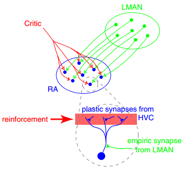
In neuroscience, synaptic plasticity is the ability of synapses to Chemical synapse#Synaptic strength, strengthen or weaken over time, in response to increases or decreases in their activity. Since memory, memories are postulated to be represented by vastly interconnected neural circuits in the brain, synaptic plasticity is one of the important neurochemical foundations of learning and memory (''see Hebbian theory''). Plastic change often results from the alteration of the number of neurotransmitter receptors located on a synapse. There are several underlying mechanisms that cooperate to achieve synaptic plasticity, including changes in the quantity of neurotransmitters released into a synapse and changes in how effectively cells respond to those neurotransmitters. Synaptic plasticity in both Excitatory synapse, excitatory and Inhibitory synapse, inhibitory synapses has been found to be dependent upon postsynaptic calcium release. Historical discoveries In 1973, Terje Lømo and ... [...More Info...] [...Related Items...] OR: [Wikipedia] [Google] [Baidu] |
Calcium Imaging
Calcium imaging is a microscopy technique to optically measure the calcium (Ca2+) status of an isolated cell, tissue or medium. Calcium imaging takes advantage of calcium indicators, fluorescent molecules that respond to the binding of Ca2+ ions by fluorescence properties. Two main classes of calcium indicators exist: chemical indicators and genetically encoded calcium indicators (GECI). This technique has allowed studies of calcium signalling in a wide variety of cell types, and can be used to measure electrical activity in hundreds of neurons in cell culture, or in living animals during ongoing behavior. Chemical indicators Chemical indicators are small molecules that can chelate calcium ions. All these molecules are based on an EGTA homologue called BAPTA, with high selectivity for calcium (Ca2+) ions versus magnesium (Mg2+) ions. This group of indicators includes fura-2, indo-1, fluo-3, fluo-4, Calcium Green-1. These dyes are often used with the chelator carboxyl grou ... [...More Info...] [...Related Items...] OR: [Wikipedia] [Google] [Baidu] |
Excitatory Synapse
An excitatory synapse is a synapse in which an action potential in a presynaptic neuron increases the probability of an action potential occurring in a postsynaptic cell. Neurons form networks through which nerve impulses travels, each neuron often making numerous connections with other cells of neurons. These electrical signals may be excitatory or inhibitory, and, if the total of excitatory influences exceeds that of the inhibitory influences, the neuron will generate a new action potential at its axon hillock, thus transmitting the information to yet another cell. This phenomenon is known as an excitatory postsynaptic potential (EPSP). It may occur via direct contact between cells (i.e., via gap junctions), as in an electrical synapse, but most commonly occurs via the vesicular release of neurotransmitters from the presynaptic axon terminal into the synaptic cleft, as in a chemical synapse. The excitatory neurotransmitters, the most common of which is glutamate, then migra ... [...More Info...] [...Related Items...] OR: [Wikipedia] [Google] [Baidu] |


