|
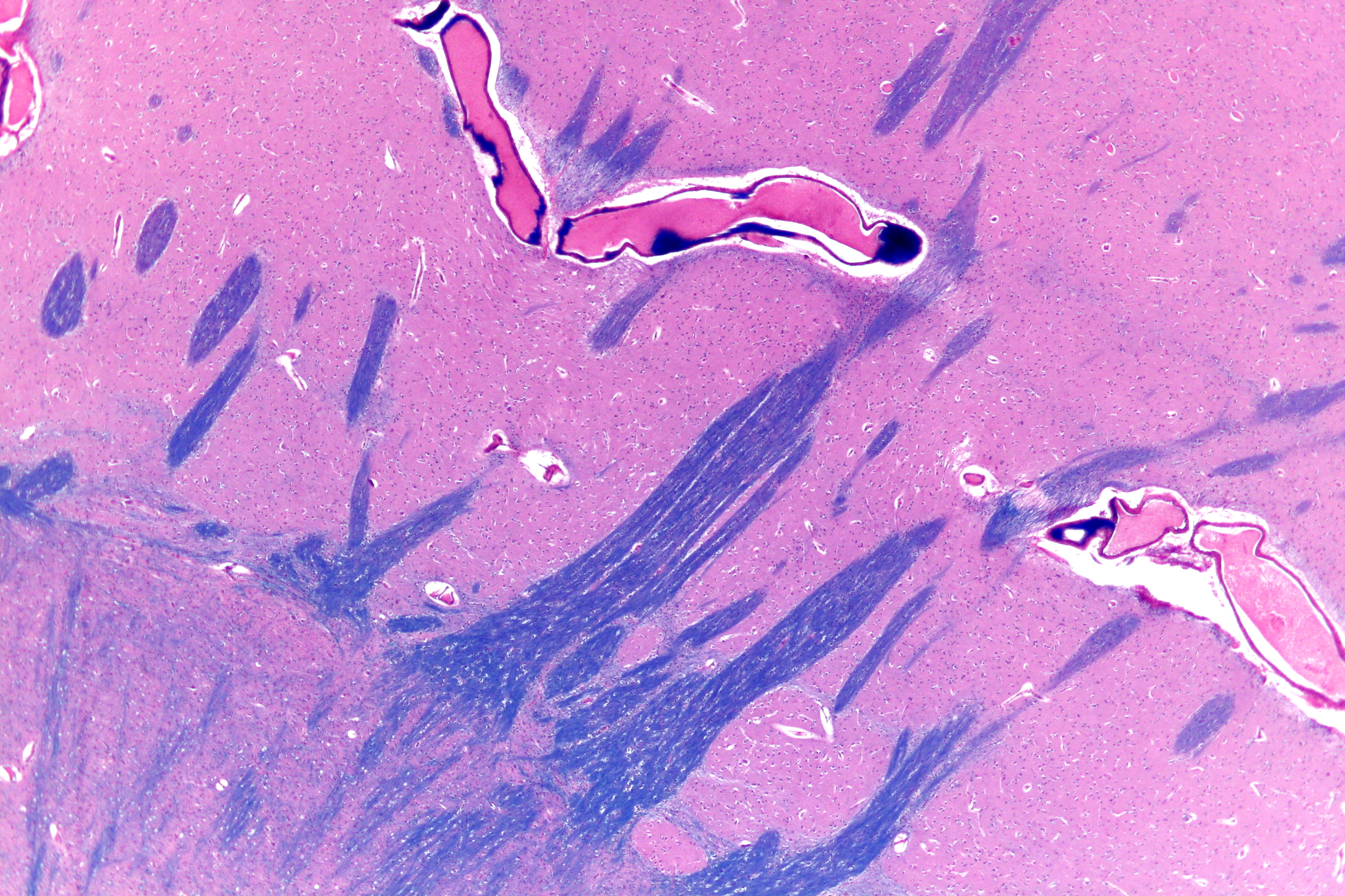
Anteromedial Central Arteries
Central arteries (or perforating or ganglionic arteries) of the brain are numerous small arteries branching from the Circle of Willis, and adjacent arteries that often enter the substance of the brain through the anterior and posterior perforated substances. They supply structures of the base of the brain and internal structures of the cerebral hemispheres. They are separated into four principal groups: anteromedial central arteries; anterolateral central arteries (lenticulostriate arteries); posteromedial central arteries (paramedian arteries); and posterolateral central arteries. Anteromedial central arteries Anteromedial central arteries (also anteromedial perforating arteries, or anteromedial ganglionic arteries) are arteries that arise from the anterior cerebral artery and anterior communicating artery, and pass into the substance of the cerebral hemispheres through the (medial portion of) the anterior perforated substance to supply the optic chiasm, ( anterior nucleus, preop ... [...More Info...] [...Related Items...] OR: [Wikipedia] [Google] [Baidu] |
Circle Of Willis
The circle of Willis (also called Willis' circle, loop of Willis, cerebral arterial circle, and Willis polygon) is a circulatory anastomosis that supplies blood to the brain and surrounding structures in reptiles, birds and mammals, including humans. It is named after Thomas Willis (1621–1675), an English physician. Structure The circle of Willis is a part of the cerebral circulation and is composed of the following arteries: * Anterior cerebral artery (left and right) at their A1 segments * Anterior communicating artery * Internal carotid artery (left and right) at its distal tip (carotid terminus) * Posterior cerebral artery (left and right) at their P1 segments * Posterior communicating artery (left and right) The middle cerebral arteries, supplying the brain, are also considered part of the Circle of Willis Origin of arteries The left and right internal carotid arteries arise from the left and right common carotid arteries. The posterior communicating artery is given ... [...More Info...] [...Related Items...] OR: [Wikipedia] [Google] [Baidu] |
Putamen
The putamen (; from Latin, meaning "nutshell") is a subcortical nucleus (neuroanatomy), nucleus with a rounded structure, in the basal ganglia nuclear group. It is located at the base of the forebrain and above the midbrain. The putamen and caudate nucleus together form the dorsal striatum. Through various pathways, the putamen is connected to the substantia nigra, the globus pallidus, the claustrum, and the thalamus, in addition to many regions of the cerebral cortex. A primary function of the putamen is to regulate movements at various stages such as in preparation and execution; and to influence various types of learning. It employs GABA, acetylcholine, and enkephalin to perform its functions. The putamen also plays a role in neurodegenerative diseases, such as Parkinson's disease. History The word "putamen" is from Latin, referring to that which "falls off in pruning", from "putare", meaning "to prune, to think, or to consider". Most MRI research was focused broadly on th ... [...More Info...] [...Related Items...] OR: [Wikipedia] [Google] [Baidu] |
Posterior Limb
The internal capsule is a paired white matter structure, as a two-way tract, carrying ascending and descending fibers, to and from the cerebral cortex. The internal capsule is situated in the inferomedial part of each cerebral hemisphere of the brain. It carries information past the subcortical basal ganglia. As it courses it separates the caudate nucleus and the thalamus from the putamen and the globus pallidus. It also separates the caudate nucleus and the putamen in the dorsal striatum, a brain region involved in motor and reward pathways. The internal capsule is V-shaped in transection forming an anterior and posterior limb, with the angle between them called the genu. The corticospinal tract constitutes a large part of the internal capsule, carrying motor information from the primary motor cortex to the lower motor neurons in the spinal cord. Above the basal ganglia the corticospinal tract is a part of the corona radiata. Below the basal ganglia the tract is called cerebr ... [...More Info...] [...Related Items...] OR: [Wikipedia] [Google] [Baidu] |
Internal Capsule
The internal capsule is a paired white matter structure, as a two-way nerve tract, tract, carrying afferent nerve fiber, ascending and efferent nerve fiber, descending axon, fibers, to and from the cerebral cortex. The internal capsule is situated in the Anatomical terms of location#Medial and lateral, inferomedial part of each cerebral hemisphere of the brain. It carries information past the subcortical basal ganglia. As it courses it separates the caudate nucleus and the thalamus from the putamen and the globus pallidus. It also separates the caudate nucleus and the putamen in the dorsal striatum, a brain region involved in motor and reward pathways. The internal capsule is V-shaped in transection forming an anterior and posterior limb, with the angle between them called the genu. The corticospinal tract constitutes a large part of the internal capsule, carrying motor information from the primary motor cortex to the lower motor neurons in the spinal cord. Above the basal gangli ... [...More Info...] [...Related Items...] OR: [Wikipedia] [Google] [Baidu] |
Globus Pallidus
The globus pallidus (GP), also known as paleostriatum or dorsal pallidum, is a major component of the Cerebral cortex, subcortical basal ganglia in the brain. It consists of two adjacent segments, one external (or lateral), known in rodents simply as the globus pallidus, and one internal (or medial). It is part of the telencephalon, but retains close functional ties with the subthalamus in the diencephalon – both of which are part of the extrapyramidal motor system. The globus pallidus receives principal inputs from the striatum, and principal direct outputs to the thalamus and the substantia nigra. The latter is made up of similar neuronal elements, has similar afferents from the striatum, similar projections to the thalamus, and has a similar synapse, synaptology. Neither receives direct cortical afferents, and both receive substantial additional inputs from the intralaminar thalamic nuclei. Globus pallidus is Latin for "pale globe". Structure Pallidal nuclei are made up of ... [...More Info...] [...Related Items...] OR: [Wikipedia] [Google] [Baidu] |
Striatum
The striatum (: striata) or corpus striatum is a cluster of interconnected nuclei that make up the largest structure of the subcortical basal ganglia. The striatum is a critical component of the motor and reward systems; receives glutamatergic and dopaminergic inputs from different sources; and serves as the primary input to the rest of the basal ganglia. Functionally, the striatum coordinates multiple aspects of cognition, including both motor and action planning, decision-making, motivation, reinforcement, and reward perception. The striatum is made up of the caudate nucleus and the lentiform nucleus. However, some authors believe it is made up of caudate nucleus, putamen, and ventral striatum. The lentiform nucleus is made up of the larger putamen, and the smaller globus pallidus. Strictly speaking the globus pallidus is part of the striatum. It is common practice, however, to implicitly exclude the globus pallidus when referring to striatal structures. In pr ... [...More Info...] [...Related Items...] OR: [Wikipedia] [Google] [Baidu] |
Lentiform Nucleus
The lentiform nucleus (or lentiform complex, lenticular nucleus, or lenticular complex) are the putamen (laterally) and the globus pallidus (medially), collectively. Due to their proximity, these two structures were formerly considered one, however, the two are separated by a thin layer of white matter—the external medullary lamina—and are functionally and connectionally distinct. The lentiform nucleus is a large, lens-shaped mass of gray matter just lateral to the internal capsule. It forms part of the basal ganglia. With the caudate nucleus, it forms the dorsal striatum. Structure When divided horizontally, it exhibits, to some extent, the appearance of a biconvex lens, while a coronal section of its central part presents a somewhat triangular outline. It is shorter than the caudate nucleus and does not extend as far forward. Relations It is deep/medial to the insular cortex, with which it is coextensive; the two are separated by intervening structures. It is lateral to ... [...More Info...] [...Related Items...] OR: [Wikipedia] [Google] [Baidu] |
End Artery
An end artery or terminal artery is an artery that is the only supply of oxygenated blood to a portion of tissue. Arteries which do not anastomose with their neighbors are called end arteries. There is no collateral circulation present besides the end arteries. Examples of an end artery include the splenic artery that supplies the spleen and the renal artery that supplies the kidneys. End arteries are of particular interest to medicine where they supply the heart or brain because if the arteries are occluded, the tissue is completely cut off, leading to a myocardial infarction or an ischaemic stroke. Other end arteries supply all or parts of the liver, intestines, fingers, toes, ears, nose, retina, penis, and other organs. Because vital tissues such as the brain or heart muscle are vulnerable to ischaemia, arteries often form anastomoses to provide alternative supplies of fresh blood. End arteries can exist when no anastomosis exists or when an anastomosis exists but is inca ... [...More Info...] [...Related Items...] OR: [Wikipedia] [Google] [Baidu] |
Basal Ganglia
The basal ganglia (BG) or basal nuclei are a group of subcortical Nucleus (neuroanatomy), nuclei found in the brains of vertebrates. In humans and other primates, differences exist, primarily in the division of the globus pallidus into external and internal regions, and in the division of the striatum. Positioned at the base of the forebrain and the top of the midbrain, they have strong connections with the cerebral cortex, thalamus, brainstem and other brain areas. The basal ganglia are associated with a variety of functions, including regulating voluntary motor control, motor movements, procedural memory, procedural learning, habituation, habit formation, conditional learning, eye movements, cognition, and emotion. The main functional components of the basal ganglia include the striatum, consisting of both the dorsal striatum (caudate nucleus and putamen) and the ventral striatum (nucleus accumbens and olfactory tubercle), the globus pallidus, the ventral pallidum, the substa ... [...More Info...] [...Related Items...] OR: [Wikipedia] [Google] [Baidu] |
Middle Cerebral Artery
The middle cerebral artery (MCA) is one of the three major paired cerebral artery, cerebral arteries that supply blood to the cerebrum. The MCA arises from the internal carotid artery and continues into the lateral sulcus where it then branches and projects to many parts of the lateral cerebral cortex. It also supplies blood to the anterior temporal lobes and the insular cortex, insular cortices. The left and right MCAs rise from trifurcations of the internal carotid artery, internal carotid arteries and thus are connected to the anterior cerebral artery, anterior cerebral arteries and the posterior communicating artery, posterior communicating arteries, which connect to the posterior cerebral artery, posterior cerebral arteries. The MCAs are not considered a part of the Circle of Willis. Structure The middle cerebral artery divides into four segments, named by the region they supply as opposed to order of branching as the latter can be somewhat variable: *M1: The ''sphenoidal' ... [...More Info...] [...Related Items...] OR: [Wikipedia] [Google] [Baidu] |


