|
Blasts Off
In cell biology, precursor cells—also called blast cells—are partially differentiated, or intermediate, and are sometimes referred to as progenitor cells. A precursor cell is a stem cell with the capacity to differentiate into only one cell type, meaning they are unipotent stem cells. In embryology, precursor cells are a group of cells that later differentiate into one organ. However, progenitor cells are considered multipotent. Due to their contribution to the development of various organs and cancers, precursor and progenitor cells have many potential uses in medicine. There is ongoing research on using these cells to build heart valves, blood vessels, and other tissues by using blood and muscle precursor cells. Cytological types * Oligodendrocyte precursor cell *Myeloblast *Thymocyte *Meiocyte *Megakaryoblast *Promegakaryocyte * Melanoblast *Lymphoblast *Bone marrow precursor cells * Normoblast *Angioblast (endothelial precursor cells) *Myeloid precursor cells *Plasmabla ... [...More Info...] [...Related Items...] OR: [Wikipedia] [Google] [Baidu] |
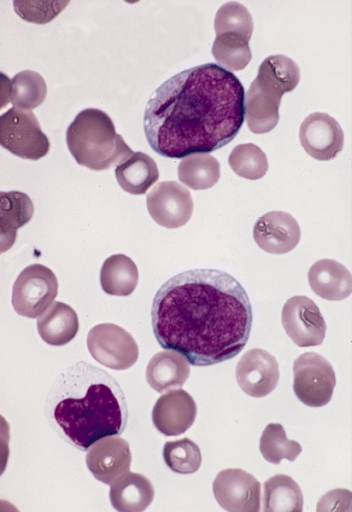
Two Myeloblasts With Auer Rods
2 (two) is a number, numeral and digit. It is the natural number following 1 and preceding 3. It is the smallest and the only even prime number. Because it forms the basis of a duality, it has religious and spiritual significance in many cultures. Mathematics The number 2 is the second natural number after 1. Each natural number, including 2, is constructed by succession, that is, by adding 1 to the previous natural number. 2 is the smallest and the only even prime number, and the first Ramanujan prime. It is also the first superior highly composite number, and the first colossally abundant number. An integer is determined to be even if it is divisible by two. When written in base 10, all multiples of 2 will end in 0, 2, 4, 6, or 8; more generally, in any even base, even numbers will end with an even digit. A digon is a polygon with two sides (or edges) and two vertices. Two distinct points in a plane are always sufficient to define a unique line in a nontr ... [...More Info...] [...Related Items...] OR: [Wikipedia] [Google] [Baidu] |
Endothelial
The endothelium (: endothelia) is a single layer of squamous endothelial cells that line the interior surface of blood vessels and lymphatic vessels. The endothelium forms an interface between circulating blood or lymph in the lumen and the rest of the vessel wall. Endothelial cells in direct contact with blood are called vascular endothelial cells whereas those in direct contact with lymph are known as lymphatic endothelial cells. Vascular endothelial cells line the entire circulatory system, from the heart to the smallest capillaries. These cells have unique functions that include fluid filtration, such as in the glomerulus of the kidney, blood vessel tone, hemostasis, neutrophil recruitment, and hormone trafficking. Endothelium of the interior surfaces of the heart chambers is called endocardium. An impaired function can lead to serious health issues throughout the body. Structure The endothelium is a thin layer of single flat (squamous) cells that line the interior su ... [...More Info...] [...Related Items...] OR: [Wikipedia] [Google] [Baidu] |
Neuroscience Information Framework
The Neuroscience Information Framework is a repository of global neuroscience web resources, including experimental, clinical, and translational neuroscience databases, knowledge bases, atlases, and genetic/ genomic resources and provides many authoritative links throughout the neuroscience portal of Wikipedia. Description The Neuroscience Information Framework (NIF) is an initiative of the NIH Blueprint for Neuroscience Research, which was established in 2004 by the National Institutes of Health. Development of the NIF started in 2008, when the University of California, San Diego School of Medicine obtained an NIH contract to create and maintain "a dynamic inventory of web-based neurosciences data, resources, and tools that scientists and students can access via any computer connected to the Internet". The project is headed by Maryann Martone, co-director of the National Center for Microscopy and Imaging Research (NCMIR), part of the multi-disciplinary Center for Research in Bi ... [...More Info...] [...Related Items...] OR: [Wikipedia] [Google] [Baidu] |
Retinal Degeneration (rhodopsin Mutation)
Retinal degeneration may refer to: * Retinopathy, one of several eye diseases or eye disorders in humans ** Retinal degeneration (rhodopsin mutation) * Progressive retinal atrophy Progressive retinal atrophy (PRA) is a group of genetic diseases seen in certain breeds of dogs and, more rarely, cats. Similar to retinitis pigmentosa in humans, it is characterized by the bilateral degeneration of the retina, causing progressi ..., an eye disease in dogs See also * List of systemic diseases with ocular manifestations, in humans {{disambiguation ... [...More Info...] [...Related Items...] OR: [Wikipedia] [Google] [Baidu] |
Bone Marrow
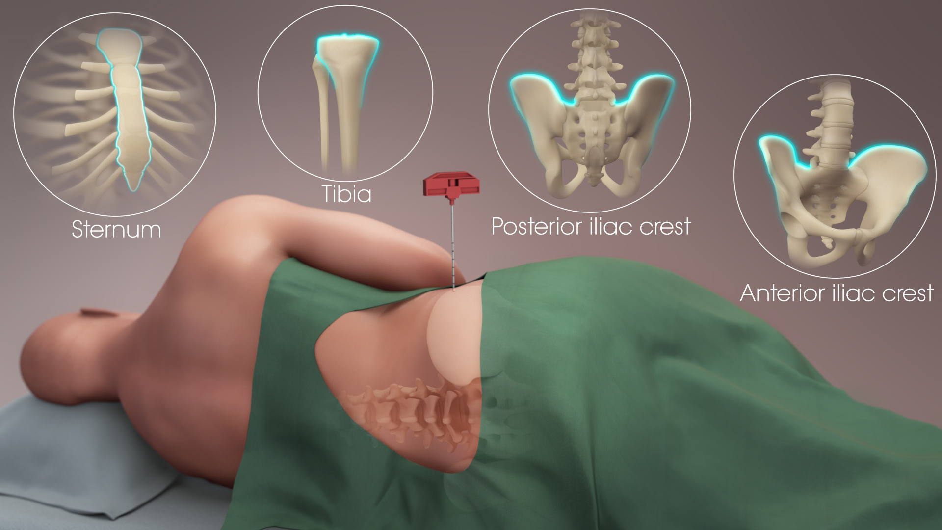
Bone marrow is a semi-solid biological tissue, tissue found within the Spongy bone, spongy (also known as cancellous) portions of bones. In birds and mammals, bone marrow is the primary site of new blood cell production (or haematopoiesis). It is composed of Blood cell, hematopoietic cells, marrow adipose tissue, and supportive stromal cells. In adult humans, bone marrow is primarily located in the Rib cage, ribs, vertebrae, sternum, and Pelvis, bones of the pelvis. Bone marrow comprises approximately 5% of total body mass in healthy adult humans, such that a person weighing 73 kg (161 lbs) will have around 3.7 kg (8 lbs) of bone marrow. Human marrow produces approximately 500 billion blood cells per day, which join the Circulatory system, systemic circulation via permeable vasculature sinusoids within the medullary cavity. All types of Hematopoietic cell, hematopoietic cells, including both Myeloid tissue, myeloid and Lymphocyte, lymphoid lineages, are create ... [...More Info...] [...Related Items...] OR: [Wikipedia] [Google] [Baidu] |
Ischemia
Ischemia or ischaemia is a restriction in blood supply to any tissue, muscle group, or organ of the body, causing a shortage of oxygen that is needed for cellular metabolism (to keep tissue alive). Ischemia is generally caused by problems with blood vessels, with resultant damage to or dysfunction of tissue, i.e., hypoxia and microvascular dysfunction. It also implies local hypoxia in a part of a body resulting from constriction (such as vasoconstriction, thrombosis, or embolism). Ischemia causes not only insufficiency of oxygen but also reduced availability of nutrients and inadequate removal of metabolic wastes. Ischemia can be partial (poor perfusion) or total blockage. The inadequate delivery of oxygenated blood to the organs must be resolved either by treating the cause of the inadequate delivery or reducing the oxygen demand of the system that needs it. For example, patients with myocardial ischemia have a decreased blood flow to the heart and are prescribe ... [...More Info...] [...Related Items...] OR: [Wikipedia] [Google] [Baidu] |
Neovascularization
Neovascularization is the natural formation of new blood vessels ('' neo-'' + ''vascular'' + '' -ization''), usually in the form of functional microvascular networks, capable of perfusion by red blood cells, that form to serve as collateral circulation in response to local poor perfusion or ischemia. Growth factors that inhibit neovascularization include those that affect endothelial cell division and differentiation. These growth factors often act in a paracrine or autocrine fashion; they include fibroblast growth factor, placental growth factor, insulin-like growth factor, hepatocyte growth factor, and platelet-derived endothelial growth factor. There are three different pathways that comprise neovascularization: (1) vasculogenesis, (2) angiogenesis, and (3) arteriogenesis. Three pathways of neovascularization Vasculogenesis Vasculogenesis is the de novo formation of blood vessels. This primarily occurs in the developing embryo with the development of the first primitive ... [...More Info...] [...Related Items...] OR: [Wikipedia] [Google] [Baidu] |
Angiogenesis
Angiogenesis is the physiological process through which new blood vessels form from pre-existing vessels, formed in the earlier stage of vasculogenesis. Angiogenesis continues the growth of the vasculature mainly by processes of sprouting and splitting, but processes such as coalescent angiogenesis, vessel elongation and vessel cooption also play a role. Vasculogenesis is the embryonic formation of endothelial cells from mesoderm cell precursors, and from neovascularization, although discussions are not always precise (especially in older texts). The first vessels in the developing embryo form through vasculogenesis, after which angiogenesis is responsible for most, if not all, blood vessel growth during development and in disease. Angiogenesis is a normal and vital process in growth and development, as well as in wound healing and in the formation of granulation tissue. However, it is also a fundamental step in the transition of tumors from a benign state to a malign ... [...More Info...] [...Related Items...] OR: [Wikipedia] [Google] [Baidu] |
Vasculogenesis
Vasculogenesis is the process of blood vessel formation, occurring by a ''De novo synthesis, de novo'' production of endothelial cells. It is the first stage of the formation of the vascular network, closely followed by angiogenesis. Process In the word sense, sense distinguished from angiogenesis, vasculogenesis is different in one aspect: whereas angiogenesis is the formation of new blood vessels from pre-existing ones, vasculogenesis is the formation of new blood vessels, in blood islands, when there are no pre-existing ones. For example, if a monolayer of endothelial cells begins sprouting to form capillary, capillaries, angiogenesis is occurring. Vasculogenesis, in contrast, is when endothelial precursor cells (angioblasts) migrate and differentiate in response to local cues (such as growth factors and extracellular matrices) to form new blood vessels. These vascular trees are then pruned and extended through angiogenesis. Occurrences Vasculogenesis occurs during embryon ... [...More Info...] [...Related Items...] OR: [Wikipedia] [Google] [Baidu] |
Angioblast
Angioblasts (or vasoformative cells) are embryonic cells from which the endothelium of blood vessels arises. They are derived from embryonic mesoderm. Blood vessels first make their appearance in several scattered vascular areas ( blood islands) that are developed simultaneously between the endoderm and the mesoderm of the yolk-sac, i. e., outside the body of the embryo. Here a new type of cell, the angioblast, is differentiated from the mesoderm. These cells as they divide form small, dense syncytial masses, which soon join with similar masses by means of fine processes to form plexuses. They form capillaries through vasculogenesis and angiogenesis. Angioblasts are one of the two products formed from hemangioblasts (the other being multipotential hemopoietic stem cells). See also *List of human cell types derived from the germ layers This is a list of Cell (biology), cells in humans derived from the three embryonic germ layers – ectoderm, mesoderm, and endoderm. Cells d ... [...More Info...] [...Related Items...] OR: [Wikipedia] [Google] [Baidu] |
Lysosomal Storage Disease
Lysosomal storage diseases (LSDs; ) are a group of over 70 rare inherited metabolic disorders that result from defects in lysosomal function. Lysosomes are sacs of enzymes within cells that digest large molecules and pass the fragments on to other parts of the cell for recycling. This process requires several critical enzymes. If one of these enzymes is defective due to a mutation, the large molecules accumulate within the cell, eventually killing it. Lysosomal storage disorders are caused by lysosomal dysfunction usually as a consequence of deficiency of a single enzyme required for the metabolism of lipids, glycoproteins (sugar-containing proteins), or mucopolysaccharides. Individually, lysosomal storage diseases occur with incidences of less than 1:100,000; however, as a group, the incidence is about 1:5,000 – 1:10,000. Most of these disorders are autosomal recessively inherited such as Niemann–Pick disease, type C, but a few are X-linked recessively inherited, such as Fa ... [...More Info...] [...Related Items...] OR: [Wikipedia] [Google] [Baidu] |
Oligodendrocyte Progenitor Cell
Oligodendrocyte progenitor cells (OPCs), also known as oligodendrocyte precursor cells, NG2-glia, O2A cells, or polydendrocytes, are a subtype of glia in the central nervous system named for their essential role as precursors to oligodendrocytes and myelin. They are typically identified in the human by co-expression of PDGFRA and CSPG4. OPCs play a critical role in developmental and adult myelinogenesis. They give rise to oligodendrocytes, which then wrap around axons and provide electrical insulation by forming a myelin sheath. This enables faster action potential propagation and high fidelity transmission without a need for an increase in axonal diameter. The loss or lack of OPCs, and consequent lack of differentiated oligodendrocytes, is associated with a loss of myelination and subsequent impairment of neurological functions. In addition, OPCs express receptors for various neurotransmitters and undergo membrane depolarization when they receive synaptic inputs from neurons. ... [...More Info...] [...Related Items...] OR: [Wikipedia] [Google] [Baidu] |


