|
Yolk Sac
The yolk sac is a membranous wikt:sac, sac attached to an embryo, formed by cells of the hypoblast layer of the bilaminar embryonic disc. This is alternatively called the umbilical vesicle by the Terminologia Embryologica (TE), though ''yolk sac'' is far more widely used. The yolk sac is one of the fetal membranes and is important in early embryonic blood supply. In humans much of it is incorporated into the primordial gut (anatomy), gut during the fourth week of embryonic development. In humans The yolk sac is the first element seen within the gestational sac during pregnancy, usually at three days gestation. The yolk sac is situated on the front (ventral) part of the embryo; it is lined by extra-embryonic endoderm, outside of which is a layer of extra-embryonic mesenchyme, derived from the epiblast. Blood is conveyed to the wall of the yolk sac by the primitive aorta and after circulating through a wide-meshed capillary plexus, is returned by the vitelline veins to the tubul ... [...More Info...] [...Related Items...] OR: [Wikipedia] [Google] [Baidu] |
Embryo
An embryo ( ) is the initial stage of development for a multicellular organism. In organisms that reproduce sexually, embryonic development is the part of the life cycle that begins just after fertilization of the female egg cell by the male sperm cell. The resulting fusion of these two cells produces a single-celled zygote that undergoes many cell divisions that produce cells known as blastomeres. The blastomeres (4-cell stage) are arranged as a solid ball that when reaching a certain size, called a morula, (16-cell stage) takes in fluid to create a cavity called a blastocoel. The structure is then termed a blastula, or a blastocyst in mammals. The mammalian blastocyst hatches before implantating into the endometrial lining of the womb. Once implanted the embryo will continue its development through the next stages of gastrulation, neurulation, and organogenesis. Gastrulation is the formation of the three germ layers that will form all of the different parts of t ... [...More Info...] [...Related Items...] OR: [Wikipedia] [Google] [Baidu] |
Vitelline Veins
The vitelline veins are veins that drain blood from the yolk sac and the gut tube during gestation. Path They run upward at first in front, and subsequently on either side of the intestinal canal. They unite on the ventral aspect of the canal. Beyond this, they are connected to one another by two anastomotic branches, one on the dorsal, and the other on the ventral aspect of the duodenal portion of the intestine. This is encircled by two venous rings; into the middle or dorsal anastomosis the superior mesenteric vein opens. The portions of the veins above the upper ring become interrupted by the developing liver and broken up by it into a plexus of small capillary-like vessels termed sinusoids. Derivatives The vitelline veins give rise to: * Hepatic veins * Inferior portion of Inferior vena cava * Portal vein * Superior mesenteric vein * Inferior mesenteric vein The branches conveying the blood to the plexus are named the venae advehentes, and become the branches of ... [...More Info...] [...Related Items...] OR: [Wikipedia] [Google] [Baidu] |
Mesoderm
The mesoderm is the middle layer of the three germ layers that develops during gastrulation in the very early development of the embryo of most animals. The outer layer is the ectoderm, and the inner layer is the endoderm.Langman's Medical Embryology, 11th edition. 2010. The mesoderm forms mesenchyme, mesothelium and coelomocytes. Mesothelium lines coeloms. Mesoderm forms the muscles in a process known as myogenesis, septa (cross-wise partitions) and mesenteries (length-wise partitions); and forms part of the gonads (the rest being the gametes). Myogenesis is specifically a function of mesenchyme. The mesoderm differentiates from the rest of the embryo through intercellular signaling, after which the mesoderm is polarized by an organizing center. The position of the organizing center is in turn determined by the regions in which beta-catenin is protected from degradation by GSK-3. Beta-catenin acts as a co-factor that alters the activity of the transcription facto ... [...More Info...] [...Related Items...] OR: [Wikipedia] [Google] [Baidu] |
Heuser's Membrane
Heuser's membrane (or the exocoelomic membrane) is a short lived combination of hypoblast cells and extracellular matrix. At day 9-10 of embryonic development, cells from the hypoblast begin to migrate to the embryonic pole, forming a layer of cells just beneath the cytotrophoblast, called Heuser's membrane. It surrounds the exocoelomic cavity (primary yolk sac), i.e. it lines the inner surface of the cytotrophoblast. At this point, the exocoelomic cavity replaces the blastocyst cavity. At days 11 to 12, there is further delineation of the trophoblastic cells giving rise to a layer of loosely arranged cells that inserts between Heuser's membrane and both syncytiotrophoblast and cytotrophoblast. The Heuser's membrane cells (hypoblast cells) that migrated along the inner cytotrophoblast lining of the blastocoel, secrete an extracellular matrix along the way. Cells of the hypoblast migrate along the outer edges of this reticulum and form the extraembryonic mesoderm (splanchic ... [...More Info...] [...Related Items...] OR: [Wikipedia] [Google] [Baidu] |
Ileum
The ileum () is the final section of the small intestine in most higher vertebrates, including mammals, reptiles, and birds. In fish, the divisions of the small intestine are not as clear and the terms posterior intestine or distal intestine may be used instead of ileum. Its main function is to absorb vitamin B12, vitamin B12, bile salts, and whatever products of digestion that were not absorbed by the jejunum. The ileum follows the duodenum and jejunum and is separated from the cecum by the ileocecal valve (ICV). In humans, the ileum is about 2–4 m long, and the pH is usually between 7 and 8 (neutral or slightly base (chemistry), basic). ''Ileum'' is derived from the Greek word εἰλεός (eileós), referring to a medical condition known as ileus. Structure The ileum is the third and final part of the small intestine. It follows the jejunum and ends at the ileocecal junction, where the wikt:terminal, terminal ileum communicates with the cecum of the large intestine thro ... [...More Info...] [...Related Items...] OR: [Wikipedia] [Google] [Baidu] |
Navel
The navel (clinically known as the umbilicus; : umbilici or umbilicuses; also known as the belly button or tummy button) is a protruding, flat, or hollowed area on the abdomen at the attachment site of the umbilical cord. Structure The umbilicus is used to visually separate the abdomen into quadrants. The umbilicus is a prominent Scar#Umbilical, scar on the abdomen, with its position being relatively consistent among humans. The skin around the waist at the level of the umbilicus is supplied by the tenth thoracic spinal nerve (T10 dermatome (anatomy), dermatome). The umbilicus itself typically lies at a vertical level corresponding to the junction between the L3 and L4 vertebrae, with a normal variation among people between the L3 and L5 vertebrae. Parts of the adult navel include the "umbilical cord remnant" or "umbilical tip", which is the often protruding scar left by the detachment of the umbilical cord. This is located in the center of the navel, sometimes described ... [...More Info...] [...Related Items...] OR: [Wikipedia] [Google] [Baidu] |
Ileocecal Valve
In many Animalia, including humans, an ileocolic structure or problem is something that concerns the region of the gastrointestinal tract from the ileum to the large intestine, colon. In Animalia that have cecum, ceca, the ileocecal region is a subset of the ileocolic region, and the entire range can also be described as ileocecocolic, whereas in some Animalia, the ileocolic region contains no cecum, as the ileum joins the colon directly. Things that are ileocolic, ileocecal, or both include the following: * Ileocecal fold * Ileocecal/ileocolic intussusception (medical disorder), intussusception * Ileocecal valve * Ileocolic artery * Ileocolic lymph nodes * Ileocolic vein {{SIA, animals ... [...More Info...] [...Related Items...] OR: [Wikipedia] [Google] [Baidu] |
Meckel's Diverticulum
A Meckel's diverticulum, a true congenital diverticulum, is a slight bulge in the small intestine present at birth and a vestigial remnant of the vitelline duct. It is the most common malformation of the Human gastrointestinal tract, gastrointestinal tract and is present in approximately 2% of the population, with males more frequently experiencing symptoms. Meckel's diverticulum was first explained by Fabricius Hildanus in the sixteenth century and later named after Johann Friedrich Meckel, who described the embryological origin of this type of diverticulum in 1809. Signs and symptoms The majority of people with a Meckel's diverticulum are asymptomatic. An asymptomatic Meckel's diverticulum is called a ''silent'' Meckel's diverticulum. If symptoms do occur, they typically appear before the age of two years. The most common presenting symptom is painless rectal bleeding such as melaena-like black offensive stools, followed by intestinal obstruction, volvulus and intussusception (m ... [...More Info...] [...Related Items...] OR: [Wikipedia] [Google] [Baidu] |
Placenta
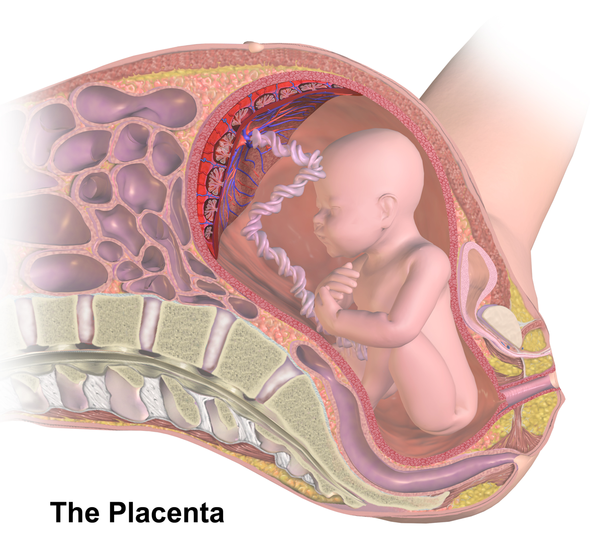
The placenta (: placentas or placentae) is a temporary embryonic and later fetal organ that begins developing from the blastocyst shortly after implantation. It plays critical roles in facilitating nutrient, gas, and waste exchange between the physically separate maternal and fetal circulations, and is an important endocrine organ, producing hormones that regulate both maternal and fetal physiology during pregnancy. The placenta connects to the fetus via the umbilical cord, and on the opposite aspect to the maternal uterus in a species-dependent manner. In humans, a thin layer of maternal decidual ( endometrial) tissue comes away with the placenta when it is expelled from the uterus following birth (sometimes incorrectly referred to as the 'maternal part' of the placenta). Placentas are a defining characteristic of placental mammals, but are also found in marsupials and some non-mammals with varying levels of development. Mammalian placentas probably first evolved abou ... [...More Info...] [...Related Items...] OR: [Wikipedia] [Google] [Baidu] |
Chorion
The chorion is the outermost fetal membrane around the embryo in mammals, birds and reptiles (amniotes). It is also present around the embryo of other animals, like insects and molluscs. Structure In humans and other therian mammals, the chorion is one of the fetal membranes that exist during pregnancy between the developing fetus and mother. The chorion and the amnion together form the amniotic sac. In humans it is formed by extraembryonic mesoderm and the two layers of trophoblast that surround the embryo and other membranes; the chorionic villi emerge from the chorion, invade the endometrium, and allow the transfer of nutrients from maternal blood to fetal blood. Layers The chorion consists of two layers: an outer formed by the trophoblast, and an inner formed by the extra-embryonic mesoderm. The trophoblast is made up of an internal layer of cubical or prismatic cells, the cytotrophoblast or layer of Langhans, and an external multinucleated layer, the syncytiotro ... [...More Info...] [...Related Items...] OR: [Wikipedia] [Google] [Baidu] |
Amnion
The amnion (: amnions or amnia) is a membrane that closely covers human and various other embryos when they first form. It fills with amniotic fluid, which causes the amnion to expand and become the amniotic sac that provides a protective environment for the developing embryo. The amnion, along with the chorion, the yolk sac and the allantois protect the embryo. In birds, reptiles and monotremes, the protective sac is enclosed in a shell. In marsupials and placental mammals, it is enclosed in a uterus. The amnion is a feature of the vertebrate clade ''Amniota'', which includes reptiles, birds, and mammals. Amphibians and fish lack the amnion and thus are anamniotes (non-amniotes). The amnion stems from the extra-embryonic somatic mesoderm on the outer side and the extra-embryonic ectoderm or trophoblast on the inner side. Etymology Etymologists have traditionally assumed that the Greek term ἀμνίον (''amnion'') relates to Ancient Greek ἀμνίον : , "little lam ... [...More Info...] [...Related Items...] OR: [Wikipedia] [Google] [Baidu] |


