|
Trendelenburg Test
The Trendelenburg Test or Brodie–Trendelenburg test is a test which can be carried out as part of a physical examination to determine the competency of the valves in the superficial and deep veins of the legs in patients with varicose veins. __TOC__ Procedure With the patient in the supine position, the leg is flexed at the hip and raised above heart level. The veins will empty due to gravity or with the assistance of the examiner's hand squeezing blood towards the heart. A tourniquet is then applied around the upper thigh to compress the superficial veins but not too tight as to occlude the deeper veins. The leg is then lowered by asking the patient to stand. Normally the superficial saphenous vein will fill from below within 30–35 seconds as blood from the capillary beds reaches the veins; if the superficial veins fill more rapidly with the tourniquet in place there is valvular incompetence below the level of the tourniquet in the "deep" or "communicating" veins. After 20 ... [...More Info...] [...Related Items...] OR: [Wikipedia] [Google] [Baidu] |
Physical Examination
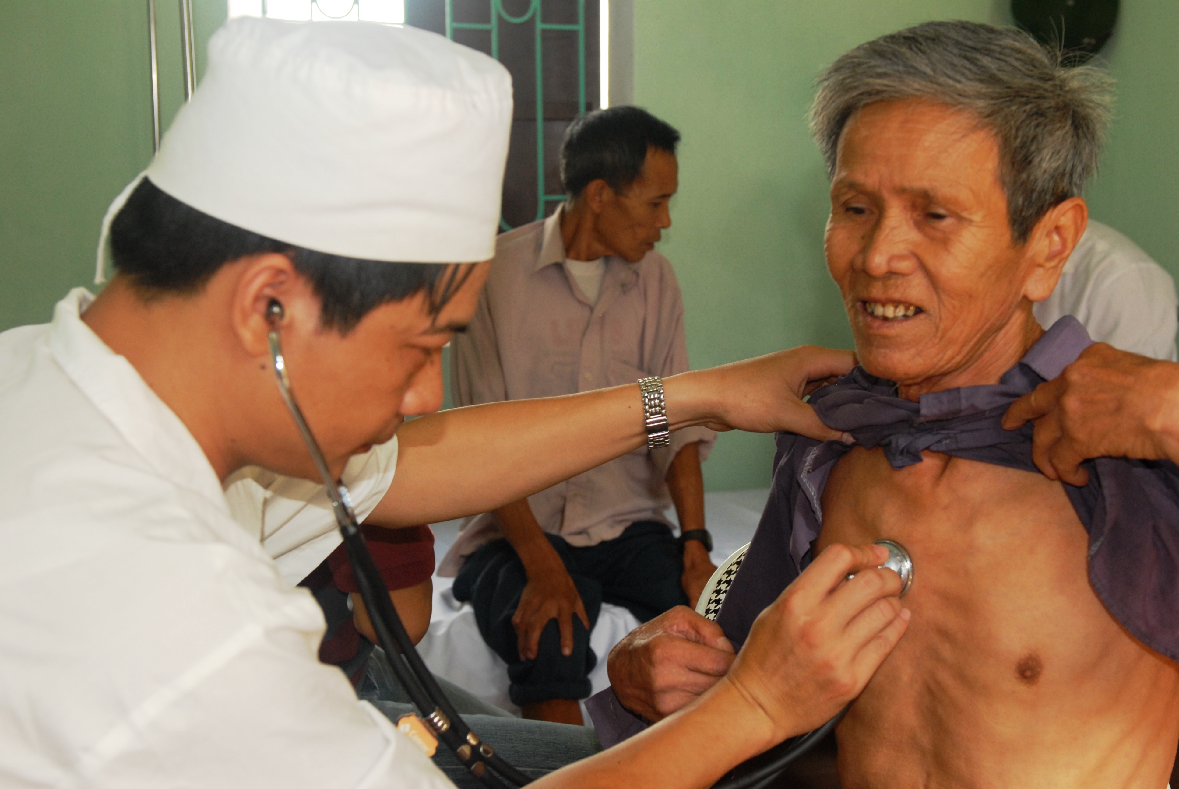
In a physical examination, medical examination, or clinical examination, a medical practitioner examines a patient for any possible medical signs or symptoms of a medical condition. It generally consists of a series of questions about the patient's medical history followed by an examination based on the reported symptoms. Together, the medical history and the physical examination help to determine a diagnosis and devise the treatment plan. These data then become part of the medical record. Types Routine The ''routine physical'', also known as ''general medical examination'', ''periodic health evaluation'', ''annual physical'', ''comprehensive medical exam'', ''general health check'', ''preventive health examination'', ''medical check-up'', or simply ''medical'', is a physical examination performed on an asymptomatic patient for medical screening purposes. These are normally performed by a pediatrician, family practice physician, physician assistant, a certified nurse pr ... [...More Info...] [...Related Items...] OR: [Wikipedia] [Google] [Baidu] |
Vein Valve
Veins are blood vessels in humans and most other animals that carry blood towards the heart. Most veins carry deoxygenated blood from the tissues back to the heart; exceptions are the pulmonary and umbilical veins, both of which carry oxygenated blood to the heart. In contrast to veins, arteries carry blood away from the heart. Veins are less muscular than arteries and are often closer to the skin. There are valves (called ''pocket valves'') in most veins to prevent backflow. Structure Veins are present throughout the body as tubes that carry blood back to the heart. Veins are classified in a number of ways, including superficial vs. deep, pulmonary vs. systemic, and large vs. small. *Superficial veins are those closer to the surface of the body, and have no corresponding arteries. *Deep veins are deeper in the body and have corresponding arteries. *Perforator veins drain from the superficial to the deep veins. These are usually referred to in the lower limbs and feet. *Communica ... [...More Info...] [...Related Items...] OR: [Wikipedia] [Google] [Baidu] |
Superficial Vein
Superficial veins are veins that are close to the surface of the body, as opposed to deep veins, which are far from the surface. Superficial veins are not paired with an artery, unlike the deep veins, which are typically associated with an artery of the same name. Superficial veins are important physiologically for cooling of the body. When the body is too hot, the body shunts blood from the deep veins to the superficial veins to facilitate heat transfer to the body's surroundings. Superficial veins are often visible underneath the skin. Those below the level of the heart tend to bulge out, which can be readily witnessed in the hand, where the veins bulge significantly less after the arm has been raised above the head for a short time. Veins become more visually prominent when lifting heavy weight, especially after a period of proper strength training. Physiologically, the superficial veins are not as important as the deep veins (as they carry less blood) and are sometimes r ... [...More Info...] [...Related Items...] OR: [Wikipedia] [Google] [Baidu] |
Deep Vein
A deep vein is a vein that is deep in the body. This contrasts with superficial veins that are close to the body's surface. Deep veins are almost always beside an artery with the same name (e.g. the femoral vein is beside the femoral artery). Collectively, they carry the vast majority of the blood. Occlusion of a deep vein can be life-threatening and is most often caused by thrombosis. Occlusion of a deep vein by thrombosis is called ''deep vein thrombosis''. Because of their location deep within the body, operation on these veins can be difficult. List *Internal jugular vein Upper limb *Brachial vein *Axillary vein *Subclavian vein Lower limb *Common femoral vein *Femoral vein *Profunda femoris vein *Popliteal vein *Peroneal vein *Anterior tibial vein *Posterior tibial vein The posterior tibial veins are veins of the leg in humans. They drain the posterior compartment of the leg and the plantar surface of the foot to the popliteal vein. Structure The posterior tibi ... [...More Info...] [...Related Items...] OR: [Wikipedia] [Google] [Baidu] |
Varicose Veins
Varicose veins, also known as varicoses, are a medical condition in which superficial veins become enlarged and twisted. These veins typically develop in the legs, just under the skin. Varicose veins usually cause few symptoms. However, some individuals may experience fatigue or pain in the area. Complications can include bleeding or superficial thrombophlebitis. Varices in the scrotum are known as a varicocele, while those around the anus are known as hemorrhoids. Due to the various physical, social, and psychological effects of varicose veins, they can negatively affect one's quality of life. Varicose veins have no specific cause. Risk factors include obesity, lack of exercise, leg trauma, and family history of the condition. They also develop more commonly during pregnancy. Occasionally they result from chronic venous insufficiency. Underlying causes include weak or damaged valves in the veins. They are typically diagnosed by examination, including observation by ultrasound ... [...More Info...] [...Related Items...] OR: [Wikipedia] [Google] [Baidu] |
Small Saphenous Vein
The small saphenous vein (also short saphenous vein or lesser saphenous vein) is a relatively large superficial vein of the posterior leg. Structure The origin of the small saphenous vein, (SSV) is where the dorsal vein from the fifth digit (smallest toe) merges with the dorsal venous arch of the foot, which attaches to the great saphenous vein (GSV). It is a superficial vein, being Subcutaneous tissue, subcutaneous (just under the skin). From its origin, it courses around the lateral aspect of the foot (inferior and posterior to the lateral malleolus) and runs along the posterior aspect of the leg (with the sural nerve), where it passes between the heads of the gastrocnemius muscle. This vein presents a number of different draining points. Usually, it drains into the popliteal vein, at or above the level of the knee joint. Variation Sometimes, the SSV joins the common gastrocnemius vein before draining in the popliteal vein. Sometimes, it does not make contact with the popliteal ... [...More Info...] [...Related Items...] OR: [Wikipedia] [Google] [Baidu] |
Popliteal Vein
The popliteal vein is a vein of the lower limb. It is formed from the anterior tibial vein and the posterior tibial vein. It travels medial to the popliteal artery, and becomes the femoral vein. It drains blood from the leg. It can be assessed using medical ultrasound. It can be affected by popliteal vein entrapment. Structure The popliteal vein is formed by the junction of the venae comitantes of the anterior tibial vein and the posterior tibial vein at the lower border of the popliteus muscle. It travels on the medial side of the popliteal artery. It is superficial to the popliteal artery. As it ascends through the fossa, it crosses behind the popliteal artery so that it comes to lie on its lateral side. It passes through the adductor hiatus (the opening in the adductor magnus muscle) to become the femoral vein.Moore K.L. and Dalley A.F. (2006), Clinically Oriented Anatomy, 5th Edition, Lippincott Williams & Wilkins, Toronto, page 636 Tributaries The tributaries of the p ... [...More Info...] [...Related Items...] OR: [Wikipedia] [Google] [Baidu] |
Friedrich Trendelenburg
Friedrich Trendelenburg (; 24 May 184415 December 1924) was a German surgeon. He was son of the philosophy, philosopher Friedrich Adolf Trendelenburg, father of the pharmacology, pharmacologist Paul Trendelenburg and grandfather of the pharmacologist Ullrich Georg Trendelenburg. Trendelenburg was born in Berlin and studied medicine at the University of Glasgow and the University of Edinburgh. He completed his studies at the Charité, Charité - Universitätsmedizin Berlin under Bernhard von Langenbeck, receiving his doctorate in 1866. He practiced medicine at the University of Rostock and the University of Bonn. In 1895 he became surgeon-in-chief at the University of Leipzig. Trendelenburg was interested in the history of surgery. He founded the German Surgical Society in 1872. Trendelenburg was also interested in the surgical removal of pulmonary embolism, pulmonary emboli. His student Martin Kirschner performed the first successful pulmonary embolectomy in 1924, shortly befor ... [...More Info...] [...Related Items...] OR: [Wikipedia] [Google] [Baidu] |
Brodie–Trendelenburg Percussion Test
The Brodie–Trendelenburg percussion test is a medical test to determine valvular incompetence in superficial veins. A finger is placed over the lower (distal) part of the vein being examined. The upper (proximal) part of the vein is then tapped (percussed). If the impulse is felt by the finger placed at the lower end, it indicates incompetence of valves in that vein. The test is used as part of the examination of varicose veins. It is named after Sir Benjamin Collins Brodie and Friedrich Trendelenburg Friedrich Trendelenburg (; 24 May 184415 December 1924) was a German surgeon. He was son of the philosophy, philosopher Friedrich Adolf Trendelenburg, father of the pharmacology, pharmacologist Paul Trendelenburg and grandfather of the pharmaco .... See also * Trendelenburg test External links Medical signs {{Med-diagnostic-stub ... [...More Info...] [...Related Items...] OR: [Wikipedia] [Google] [Baidu] |
Physical Examination
In a physical examination, medical examination, or clinical examination, a medical practitioner examines a patient for any possible medical signs or symptoms of a medical condition. It generally consists of a series of questions about the patient's medical history followed by an examination based on the reported symptoms. Together, the medical history and the physical examination help to determine a diagnosis and devise the treatment plan. These data then become part of the medical record. Types Routine The ''routine physical'', also known as ''general medical examination'', ''periodic health evaluation'', ''annual physical'', ''comprehensive medical exam'', ''general health check'', ''preventive health examination'', ''medical check-up'', or simply ''medical'', is a physical examination performed on an asymptomatic patient for medical screening purposes. These are normally performed by a pediatrician, family practice physician, physician assistant, a certified nurse pr ... [...More Info...] [...Related Items...] OR: [Wikipedia] [Google] [Baidu] |
Veins
Veins are blood vessels in humans and most other animals that carry blood towards the heart. Most veins carry deoxygenated blood from the tissues back to the heart; exceptions are the pulmonary and umbilical veins, both of which carry oxygenated blood to the heart. In contrast to veins, arteries carry blood away from the heart. Veins are less muscular than arteries and are often closer to the skin. There are valves (called ''pocket valves'') in most veins to prevent backflow. Structure Veins are present throughout the body as tubes that carry blood back to the heart. Veins are classified in a number of ways, including superficial vs. deep, pulmonary vs. systemic, and large vs. small. * Superficial veins are those closer to the surface of the body, and have no corresponding arteries. *Deep veins are deeper in the body and have corresponding arteries. *Perforator veins drain from the superficial to the deep veins. These are usually referred to in the lower limbs and feet. *Communic ... [...More Info...] [...Related Items...] OR: [Wikipedia] [Google] [Baidu] |


