|
Tolosa–Hunt Syndrome
Tolosa–Hunt syndrome is a rare disorder characterized by severe and unilateral headaches with orbital pain, along with weakness and paralysis (ophthalmoplegia) of certain eye muscles ( extraocular palsies). In 2004, the International Headache Society provided a definition of the diagnostic criteria which included granuloma. Signs and symptoms Symptoms are usually limited to one side of the head, and in most cases the individual affected will experience intense, sharp pain and paralysis of muscles around the eye. Symptoms may subside without medical intervention, yet recur without a noticeable pattern. In addition, affected individuals may experience paralysis of various facial nerves and drooping of the upper eyelid ( ptosis). Other signs include double vision, fever, chronic fatigue, vertigo or arthralgia. Occasionally the patient may present with a feeling of protrusion of one or both eyeballs (exophthalmos). Causes The cause of Tolosa–Hunt syndrome is not known, but the d ... [...More Info...] [...Related Items...] OR: [Wikipedia] [Google] [Baidu] |
Ophthalmoplegia
Ophthalmoparesis refers to weakness (-paresis) or paralysis (-plegia) of one or more extraocular muscles which are responsible for eye movements. It is a physical finding in certain neurologic, ophthalmologic, and endocrine disease. Internal ophthalmoplegia means involvement limited to the pupillary sphincter and ciliary muscle. External ophthalmoplegia refers to involvement of only the extraocular muscles. Complete ophthalmoplegia indicates involvement of both. Causes Ophthalmoparesis can result from disorders of various parts of the eye and nervous system: * Infection around the eye. Ophthalmoplegia is an important finding in orbital cellulitis. * The orbit of the eye, including mechanical restrictions of eye movement, as in Graves' disease. * The muscle, as in progressive external ophthalmoplegia or Kearns–Sayre syndrome. * The neuromuscular junction, as in myasthenia gravis. * The relevant cranial nerves (specifically the oculomotor, trochlear, and abducens), as in ca ... [...More Info...] [...Related Items...] OR: [Wikipedia] [Google] [Baidu] |
Fatigue (medical)
Fatigue describes a state of tiredness that does not resolve with rest or sleep. In general usage, fatigue is synonymous with extreme tiredness or exhaustion that normally follows prolonged physical or mental activity. When it does not resolve after rest or sleep, or occurs independently of physical or mental exertion, it may be a symptom of a medical condition that may become severe or progressive. Fatigue can be a feature of a mental disorder such as depression; may be associated with conditions of chronic pain such as fibromyalgia; it may also feature in conditions of chronic low-level inflammation, and be a disease-related symptom in many other conditions. Fatigue often has no known cause, and is recognised as being very complex in nature. Fatigability describes a susceptibility to fatigue. Physical fatigue results from muscle fatigue brought about by intense physical activity. Mental fatigue results from prolonged periods of cognitive activity which impairs cognitive abil ... [...More Info...] [...Related Items...] OR: [Wikipedia] [Google] [Baidu] |
Digital Subtraction Angiography
Digital subtraction angiography (DSA) is a fluoroscopy technique used in interventional radiology to clearly visualize blood vessels in a bony or dense soft tissue environment. Images are produced using contrast medium by subtracting a "pre-contrast image" or ''mask'' from subsequent images, once the contrast medium has been introduced into a structure. Hence the term "digital ''subtraction'' angiography. Subtraction angiography was first described in 1935 and in English sources in 1962 as a manual technique. Digital technology made DSA practical starting in the 1970s. Procedure DSA and fluoroscopy In traditional angiography, images are acquired by exposing an area of interest with time-controlled x-rays while injecting a contrast medium into the blood vessels. The image obtained includes the blood vessels, together with all overlying and underlying structures. The images are useful for determining anatomical position and variations, but unhelpful for visualizing blood vessels acc ... [...More Info...] [...Related Items...] OR: [Wikipedia] [Google] [Baidu] |
Magnetic Resonance Angiography
Magnetic resonance angiography (MRA) is a group of techniques based on magnetic resonance imaging (MRI) to image blood vessels. Magnetic resonance angiography is used to generate images of arteries (and less commonly veins) in order to evaluate them for stenosis (abnormal narrowing), Vascular occlusion, occlusions, aneurysms (vessel wall dilatations, at risk of rupture) or other abnormalities. MRA is often used to evaluate the arteries of the neck and brain, the thoracic and abdominal aorta, the renal arteries, and the legs (the latter exam is often referred to as a "run-off"). Acquisition A variety of techniques can be used to generate the pictures of blood vessels, both artery, arteries and veins, based on flow effects or on contrast (inherent or pharmacologically generated). The most frequently applied MRA methods involve the use intravenous MRI contrast agent, contrast agents, particularly those containing gadolinium to shorten the Spin–lattice relaxation, ''T''1 of blood ... [...More Info...] [...Related Items...] OR: [Wikipedia] [Google] [Baidu] |
Orbit
In celestial mechanics, an orbit is the curved trajectory of an object such as the trajectory of a planet around a star, or of a natural satellite around a planet, or of an artificial satellite around an object or position in space such as a planet, moon, asteroid, or Lagrange point. Normally, orbit refers to a regularly repeating trajectory, although it may also refer to a non-repeating trajectory. To a close approximation, planets and satellites follow elliptic orbits, with the center of mass being orbited at a focal point of the ellipse, as described by Kepler's laws of planetary motion. For most situations, orbital motion is adequately approximated by Newtonian mechanics, which explains gravity as a force obeying an inverse-square law. However, Albert Einstein's general theory of relativity, which accounts for gravity as due to curvature of spacetime, with orbits following geodesics, provides a more accurate calculation and understanding of the exact mechanics of orbi ... [...More Info...] [...Related Items...] OR: [Wikipedia] [Google] [Baidu] |
Cerebrospinal Fluid
Cerebrospinal fluid (CSF) is a clear, colorless body fluid found within the tissue that surrounds the brain and spinal cord of all vertebrates. CSF is produced by specialised ependymal cells in the choroid plexus of the ventricles of the brain, and absorbed in the arachnoid granulations. There is about 125 mL of CSF at any one time, and about 500 mL is generated every day. CSF acts as a shock absorber, cushion or buffer, providing basic mechanical and immunological protection to the brain inside the skull. CSF also serves a vital function in the cerebral autoregulation of cerebral blood flow. CSF occupies the subarachnoid space (between the arachnoid mater and the pia mater) and the ventricular system around and inside the brain and spinal cord. It fills the ventricles of the brain, cisterns, and sulci, as well as the central canal of the spinal cord. There is also a connection from the subarachnoid space to the bony labyrinth of the inner ear via the perilymphat ... [...More Info...] [...Related Items...] OR: [Wikipedia] [Google] [Baidu] |
Serum Protein Electrophoresis
Serum protein electrophoresis (SPEP or SPE) is a laboratory test that examines specific Blood proteins, proteins in the blood called globulins. The most common indications for a serum protein electrophoresis test are to diagnose or monitor multiple myeloma, a monoclonal gammopathy of uncertain significance (MGUS), or further investigate a discrepancy between a low albumin and a relatively high total protein. Unexplained bone pain, anemia, proteinuria, chronic kidney disease, and hypercalcemia are also signs of multiple myeloma, and indications for SPE. Blood must first be collected, usually into an airtight vial or syringe. Electrophoresis is a laboratory technique in which the blood serum (the fluid portion of the blood after the blood has clotted) is applied to either an acetate membrane soaked in a liquid buffer, or to a buffered agarose gel electrophoresis of proteins, gel matrix, or into liquid in a Capillary electrophoresis, capillary tube, and exposed to an electric curren ... [...More Info...] [...Related Items...] OR: [Wikipedia] [Google] [Baidu] |
Thyroid
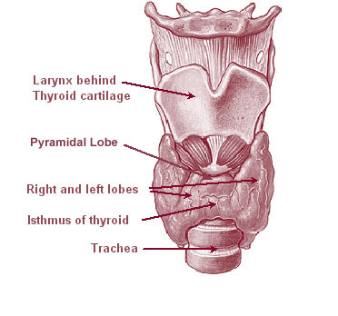
The thyroid, or thyroid gland, is an endocrine gland in vertebrates. In humans it is in the neck and consists of two connected lobes. The lower two thirds of the lobes are connected by a thin band of tissue called the thyroid isthmus. The thyroid is located at the front of the neck, below the Adam's apple. Microscopically, the functional unit of the thyroid gland is the spherical thyroid follicle, lined with follicular cells (thyrocytes), and occasional parafollicular cells that surround a lumen containing colloid. The thyroid gland secretes three hormones: the two thyroid hormones triiodothyronine (T3) and thyroxine (T4)and a peptide hormone, calcitonin. The thyroid hormones influence the metabolic rate and protein synthesis, and in children, growth and development. Calcitonin plays a role in calcium homeostasis. Secretion of the two thyroid hormones is regulated by thyroid-stimulating hormone (TSH), which is secreted from the anterior pituitary gland. TSH is regula ... [...More Info...] [...Related Items...] OR: [Wikipedia] [Google] [Baidu] |
Complete Blood Count
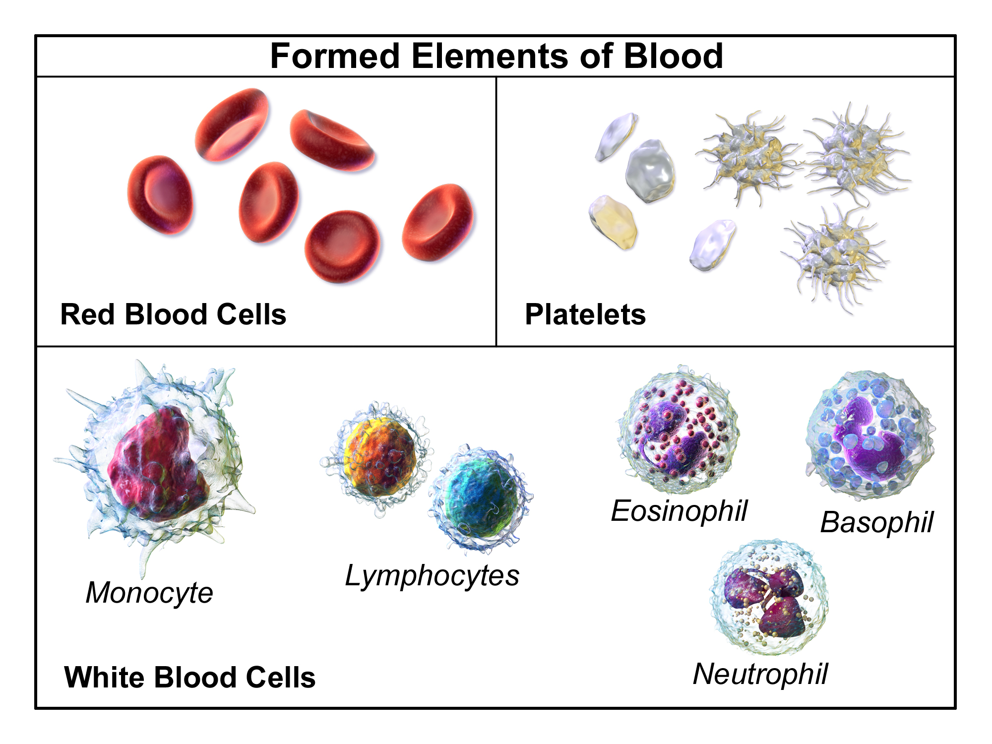
A complete blood count (CBC), also known as a full blood count (FBC), is a set of medical laboratory tests that provide cytometry, information about the cells in a person's blood. The CBC indicates the counts of white blood cells, red blood cells and platelets, the concentration of hemoglobin, and the hematocrit (the volume percentage of red blood cells). The red blood cell indices, which indicate the average size and hemoglobin content of red blood cells, are also reported, and a white blood cell differential, which counts the different types of white blood cells, may be included. The CBC is often carried out as part of a medical assessment and can be used to monitor health or diagnose diseases. The results are interpreted by comparing them to Reference ranges for blood tests, reference ranges, which vary with sex and age. Conditions like anemia and thrombocytopenia are defined by abnormal complete blood count results. The red blood cell indices can provide information about the ... [...More Info...] [...Related Items...] OR: [Wikipedia] [Google] [Baidu] |
Superior Orbital Fissure
The superior orbital fissure is a foramen or cleft of the skull between the lesser and greater wings of the sphenoid bone. It gives passage to multiple structures, including the oculomotor nerve, trochlear nerve, ophthalmic nerve, abducens nerve, ophthalmic veins, and sympathetic fibres from the cavernous plexus. Structure The superior orbital fissure is usually 22 mm wide in adults, and is much larger medially. Its boundaries are formed by the (caudal surface of the) lesser wing of the sphenoid bone, and (medial border of the) greater wing of the sphenoid bone. Contents The superior orbital fissure is traversed by the following structures: * (superior and inferior divisions of the) oculomotor nerve (CN III) * trochlear nerve (CN IV) * lacrimal, frontal, and nasociliary branches of ophthalmic nerve (CN V1) * abducens nerve (CN VI) * superior ophthalmic vein and superior division of the inferior ophthalmic vein * sympathetic fibres from the cavernous nerve pl ... [...More Info...] [...Related Items...] OR: [Wikipedia] [Google] [Baidu] |
Cavernous Sinus
The cavernous sinus within the human head is one of the dural venous sinuses creating a cavity called the lateral sellar compartment bordered by the temporal bone of the skull and the sphenoid bone, lateral to the sella turcica. Structure The cavernous sinus is one of the dural venous sinuses of the head. It is a network of veins that sit in a cavity. It sits on both sides of the sphenoidal bone and pituitary gland, approximately 1 × 2 cm in size in an adult. The carotid siphon of the internal carotid artery, and cranial nerves III, IV, V (branches V1 and V2) and VI all pass through this blood filled space. Both sides of cavernous sinus is connected to each other via intercavernous sinuses. The cavernous sinus lies in between the inner and outer layers of dura mater. Nearby structures * Above: optic tract, optic chiasma, internal carotid artery. * Inferiorly: foramen lacerum, and the junction of the body and greater wing of sphenoid bone. * Medially: pituitary gla ... [...More Info...] [...Related Items...] OR: [Wikipedia] [Google] [Baidu] |

_Phase_Contrast_(PC)_sequence_MRI_of_arterial_dissections.jpg)


