|
Temporal Muscle
In anatomy, the temporalis muscle, also known as the temporal muscle, is one of the muscles of mastication (chewing). It is a broad, fan-shaped convergent muscle on each side of the head that fills the temporal fossa, superior to the zygomatic arch so it covers much of the temporal bone.Illustrated Anatomy of the Head and Neck, Fehrenbach and Herring, Elsevier, 2012, page 98''Temporal'' refers to the head's temples. Structure In humans, the temporalis muscle arises from the temporal fossa and the deep part of temporal fascia. This is a very broad area of attachment. It passes medial to the zygomatic arch. It forms a tendon which inserts onto the coronoid process of the mandible, with its insertion extending into the retromolar fossa posterior to the most distal mandibular molar.Human Anatomy, Jacobs, Elsevier, 2008, page 194 In other mammals, the muscle usually spans the dorsal part of the skull all the way up to the medial line. There, it may be attached to a sagittal crest, a ... [...More Info...] [...Related Items...] OR: [Wikipedia] [Google] [Baidu] |
Zygomatic Arch
In anatomy, the zygomatic arch, or cheek bone, is a part of the skull formed by the zygomatic process of the temporal bone (a bone extending forward from the side of the skull, over the opening of the ear) and the temporal process of the zygomatic bone (the side of the cheekbone), the two being united by an oblique suture (the zygomaticotemporal suture); the tendon of the temporal muscle passes medial to (i.e. through the middle of) the arch, to gain insertion into the coronoid process of the mandible (jawbone). The jugal point is the point at the anterior (towards face) end of the upper border of the zygomatic arch where the masseteric and maxillary edges meet at an angle, and where it meets the process of the zygomatic bone. The arch is typical of '' Synapsida'' (“fused arch”), a clade of amniotes that includes mammals and their extinct relatives, such as ''Moschops'' and '' Dimetrodon''. Structure The zygomatic process of the temporal arises by two roots: * an ''anter ... [...More Info...] [...Related Items...] OR: [Wikipedia] [Google] [Baidu] |
Skeletal Muscle
Skeletal muscles (commonly referred to as muscles) are organs of the vertebrate muscular system and typically are attached by tendons to bones of a skeleton. The muscle cells of skeletal muscles are much longer than in the other types of muscle tissue, and are often known as muscle fibers. The muscle tissue of a skeletal muscle is striated – having a striped appearance due to the arrangement of the sarcomeres. Skeletal muscles are voluntary muscles under the control of the somatic nervous system. The other types of muscle are cardiac muscle which is also striated and smooth muscle which is non-striated; both of these types of muscle tissue are classified as involuntary, or, under the control of the autonomic nervous system. A skeletal muscle contains multiple muscle fascicle, fascicles – bundles of muscle fibers. Each individual fiber, and each muscle is surrounded by a type of connective tissue layer of fascia. Muscle fibers are formed from the cell fusion, fusion of ... [...More Info...] [...Related Items...] OR: [Wikipedia] [Google] [Baidu] |
Sarcomere
A sarcomere (Greek σάρξ ''sarx'' "flesh", μέρος ''meros'' "part") is the smallest functional unit of striated muscle tissue. It is the repeating unit between two Z-lines. Skeletal muscles are composed of tubular muscle cells (called muscle fibers or myofibers) which are formed during embryonic myogenesis. Muscle fibers contain numerous tubular myofibrils. Myofibrils are composed of repeating sections of sarcomeres, which appear under the microscope as alternating dark and light bands. Sarcomeres are composed of long, fibrous proteins as filaments that slide past each other when a muscle contracts or relaxes. The costamere is a different component that connects the sarcomere to the sarcolemma. Two of the important proteins are myosin, which forms the thick filament, and actin, which forms the thin filament. Myosin has a long, fibrous tail and a globular head, which binds to actin. The myosin head also binds to ATP, which is the source of energy for muscle movement. Myos ... [...More Info...] [...Related Items...] OR: [Wikipedia] [Google] [Baidu] |
First Pharyngeal Arch
The pharyngeal arches, also known as visceral arches'','' are structures seen in the embryonic development of vertebrates that are recognisable precursors for many structures. In fish, the arches are known as the branchial arches, or gill arches. In the human embryo, the arches are first seen during the fourth week of development. They appear as a series of outpouchings of mesoderm on both sides of the developing pharynx. The vasculature of the pharyngeal arches is known as the aortic arches. In fish, the branchial arches support the gills. Structure In vertebrates, the pharyngeal arches are derived from all three germ layers (the primary layers of cells that form during embryogenesis). Neural crest cells enter these arches where they contribute to features of the skull and facial skeleton such as bone and cartilage. However, the existence of pharyngeal structures before neural crest cells evolved is indicated by the existence of neural crest-independent mechanisms of pharyng ... [...More Info...] [...Related Items...] OR: [Wikipedia] [Google] [Baidu] |
Trigeminal Nerve
In neuroanatomy, the trigeminal nerve ( lit. ''triplet'' nerve), also known as the fifth cranial nerve, cranial nerve V, or simply CN V, is a cranial nerve responsible for sensation in the face and motor functions such as biting and chewing; it is the most complex of the cranial nerves. Its name ("trigeminal", ) derives from each of the two nerves (one on each side of the pons) having three major branches: the ophthalmic nerve (V), the maxillary nerve (V), and the mandibular nerve (V). The ophthalmic and maxillary nerves are purely sensory, whereas the mandibular nerve supplies motor as well as sensory (or "cutaneous") functions. Adding to the complexity of this nerve is that autonomic nerve fibers as well as special sensory fibers (taste) are contained within it. The motor division of the trigeminal nerve derives from the basal plate of the embryonic pons, and the sensory division originates in the cranial neural crest. Sensory information from the face and body is proc ... [...More Info...] [...Related Items...] OR: [Wikipedia] [Google] [Baidu] |
Middle Temporal Artery
In anatomy, the middle temporal artery is a major artery which arises immediately above the zygomatic arch, and, perforating the temporal fascia, gives branches to the temporalis, anastomosing with the deep temporal branches of the internal maxillary. It occasionally gives off a zygomatico-orbital branch, which runs along the upper border of the zygomatic arch, between the two layers of the temporal fascia, to the lateral angle of the orbit In celestial mechanics, an orbit is the curved trajectory of an object such as the trajectory of a planet around a star, or of a natural satellite around a planet, or of an artificial satellite around an object or position in space such as a p ... (the eye socket). Additional images File:Gray137.png, Left temporal bone. Outer surface. References Arteries of the head and neck {{circulatory-stub ... [...More Info...] [...Related Items...] OR: [Wikipedia] [Google] [Baidu] |
Muscle Contraction
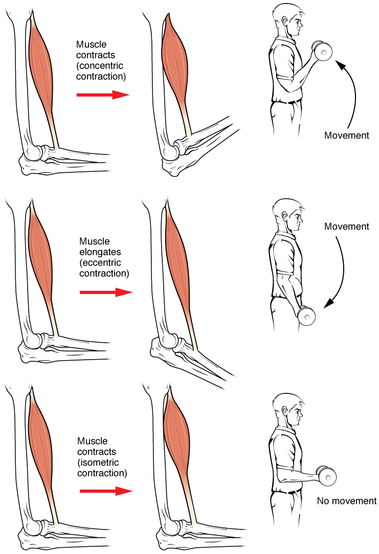
Muscle contraction is the activation of tension-generating sites within muscle cells. In physiology, muscle contraction does not necessarily mean muscle shortening because muscle tension can be produced without changes in muscle length, such as when holding something heavy in the same position. The termination of muscle contraction is followed by muscle relaxation, which is a return of the muscle fibers to their low tension-generating state. For the contractions to happen, the muscle cells must rely on the interaction of two types of filaments which are the thin and thick filaments. Thin filaments are two strands of actin coiled around each, and thick filaments consist of mostly elongated proteins called myosin. Together, these two filaments form myofibrils which are important organelles in the skeletal muscle system. Muscle contraction can also be described based on two variables: length and tension. A muscle contraction is described as isometric if the muscle tension changes ... [...More Info...] [...Related Items...] OR: [Wikipedia] [Google] [Baidu] |
Tympanoplasty
Tympanoplasty is the surgical operation performed to reconstruct hearing mechanism of middle ear Classification Tympanoplasty is classified into five different types, originally described by Horst Ludwig Wullstein (1906–1987) in 1956. # Type 1 involves repair of the tympanic membrane alone, when the middle ear is normal. A type 1 tympanoplasty is synonymous to myringoplasty. # Type 2 involves repair of the tympanic membrane and middle ear in spite of slight defects in the middle ear ossicles. # Type 3 involves removal of ossicles and epitympanum when there are large defects of the malleus and incus. The tympanic membrane is repaired and directly connected to the head of the stapes. # Type 4 describes a repair when the stapes foot plate is movable, but the crura are missing. The resulting middle ear will only consist of the Eustachian tube and hypotympanum. # Type 5 is a repair involving a fixed stapes footplate. Also called fenestration operation. Myringoplasty The term 'myring ... [...More Info...] [...Related Items...] OR: [Wikipedia] [Google] [Baidu] |
Paranthropus Aethiopicus
''Paranthropus aethiopicus'' is an extinct species of robust australopithecine from the Late Pliocene to Early Pleistocene of East Africa about 2.7–2.3 million years ago. However, it is much debated whether or not ''Paranthropus'' is an invalid grouping and is synonymous with ''Australopithecus'', so the species is also often classified as ''Australopithecus aethiopicus''. Whatever the case, it is considered to have been the ancestor of the much more robust '' P. boisei''. It is debated if ''P. aethiopicus'' should be subsumed under ''P. boisei'', and the terms ''P. boisei'' sensu lato ("in the broad sense") and ''P. boisei'' sensu stricto ("in the strict sense") can be used to respectively include and exclude ''P. aethiopicus'' from ''P. boisei''. Like other ''Paranthropus'', ''P. aethiopicus'' had a tall face, thick palate, and especially enlarged cheek teeth. However, likely due to its archaicness, it also diverges from other ''Paranthropus'', with some aspects resembling th ... [...More Info...] [...Related Items...] OR: [Wikipedia] [Google] [Baidu] |
Sagittal Crest
A sagittal crest is a ridge of bone running lengthwise along the midline of the top of the skull (at the sagittal suture) of many mammalian and reptilian skulls, among others. The presence of this ridge of bone indicates that there are exceptionally strong jaw muscles. The sagittal crest serves primarily for attachment of the temporalis muscle, which is one of the main chewing muscles. Development of the sagittal crest is thought to be connected to the development of this muscle. A sagittal crest usually develops during the juvenile stage of an animal in conjunction with the growth of the temporalis muscle, as a result of convergence and gradual heightening of the temporal lines. Function A sagittal crest tends to be present on the skulls of adult animals that rely on powerful biting and clenching of their teeth, usually as a part of their hunting strategy. Skulls of some dinosaur species, including tyrannosaurs, possessed well developed sagittal crests. Among mammals, dogs, cats, ... [...More Info...] [...Related Items...] OR: [Wikipedia] [Google] [Baidu] |
Temporal Fascia
The temporal fascia covers the temporalis muscle. It is a strong, fibrous investment, covered, laterally, by the auricularis anterior and superior, by the galea aponeurotica, and by part of the orbicularis oculi. The superficial temporal vessels and the auriculotemporal nerve cross it from below upward. Superiorly, it is a single layer, attached to the entire extent of the superior temporal line; but inferiorly, where it is fixed to the zygomatic arch, it consists of two layers, one of which is inserted into the lateral, and the other into the medial border of the arch. A small quantity of fat, the orbital branch of the superficial temporal artery, and a filament from the zygomatic branch of the maxillary nerve In neuroanatomy, the maxillary nerve (V) is one of the three branches or divisions of the trigeminal nerve, the fifth (CN V) cranial nerve. It comprises the principal functions of sensation from the maxilla, nasal cavity, sinuses, the palate a ..., are contained be ... [...More Info...] [...Related Items...] OR: [Wikipedia] [Google] [Baidu] |
Temple (anatomy)
The temple is a latch where four skull bones fuse: the frontal, parietal, temporal, and sphenoid. It is located on the side of the head behind the eye between the forehead and the ear. The temporal muscle covers this area and is used during mastication. Cladists classify land vertebrates based on the presence of an upper hole, a lower hole, both, or neither in the cover of dermal bone that formerly covered the temporalis muscle, whose origin is the temple and whose insertion is the jaw. The brain has a lobe called the temporal lobe. Etymology The word "templar" as used in anatomy has a separate etymology from the other meaning of word ''temple'', meaning "place of worship". Both come from Latin, but the word for the place of worship comes from ', whereas the word for the part of the head comes from Vulgar Latin *', modified from ', plural form ("both temples") of ', a word that meant both "time" and the part of the head. Due to the common source with the word for time, ... [...More Info...] [...Related Items...] OR: [Wikipedia] [Google] [Baidu] |


