|
T Wave
In electrocardiography, the T wave represents the repolarization of the ventricles. The interval from the beginning of the QRS complex to the apex of the T wave is referred to as the ''absolute refractory period''. The last half of the T wave is referred to as the ''relative refractory period'' or ''vulnerable period''. The T wave contains more information than the QT interval. The T wave can be described by its symmetry, skewness, slope of ascending and descending limbs, amplitude and subintervals like the Tpeak–Tend interval. In most leads, the T wave is positive. This is due to the repolarization of the membrane. During ventricle contraction (QRS complex), the heart depolarizes. Repolarization of the ventricle happens in the opposite direction of depolarization and is negative current, signifying the relaxation of the cardiac muscle of the ventricles. But this negative flow causes a positive T wave; although the cell becomes more negatively charged, the net effect is in ... [...More Info...] [...Related Items...] OR: [Wikipedia] [Google] [Baidu] |
Tnorm (ECG)
In mathematics, a t-norm (also T-norm or, unabbreviated, triangular norm) is a kind of binary operation used in the framework of probabilistic metric spaces and in multi-valued logic, specifically in fuzzy logic. A t-norm generalizes intersection (set theory), intersection in a lattice (order), lattice and logical conjunction, conjunction in logic. The name ''triangular norm'' refers to the fact that in the framework of probabilistic metric spaces t-norms are used to generalize the triangle inequality of ordinary metric spaces. Definition A t-norm is a function (mathematics), function T: [0, 1] × [0, 1] → [0, 1] that satisfies the following properties: * Commutativity: T(''a'', ''b'') = T(''b'', ''a'') * Monotonicity: T(''a'', ''b'') ≤ T(''c'', ''d'') if ''a'' ≤ ''c'' and ''b'' ≤ ''d'' * Associativity: T(''a'', T(''b'', ''c'')) = T(T(''a'', ''b''), ''c'') * The number 1 acts as identity element: T(''a'', 1) = ''a'' Since a t-norm is a binary operatio ... [...More Info...] [...Related Items...] OR: [Wikipedia] [Google] [Baidu] |
Membrane Potential
Membrane potential (also transmembrane potential or membrane voltage) is the difference in electric potential between the interior and the exterior of a biological cell. It equals the interior potential minus the exterior potential. This is the energy (i.e. work) per charge which is required to move a (very small) positive charge at constant velocity across the cell membrane from the exterior to the interior. (If the charge is allowed to change velocity, the change of kinetic energy and production of radiation must be taken into account.) Typical values of membrane potential, normally given in units of milli volts and denoted as mV, range from −80 mV to −40 mV. For such typical negative membrane potentials, positive work is required to move a positive charge from the interior to the exterior. However, thermal kinetic energy allows ions to overcome the potential difference. For a selectively permeable membrane, this permits a net flow against the gradient. This is a kind ... [...More Info...] [...Related Items...] OR: [Wikipedia] [Google] [Baidu] |
Sinus Tachycardia
Sinus tachycardia is a sinus rhythm of the heart, with an increased rate of electrical discharge from the sinoatrial node, resulting in a tachycardia, a heart rate that is higher than the upper limit of normal (90–100 beats per minute for adult humans). The normal resting heart rate is 60–90 bpm in an average adult. Normal heart rates vary with age and level of fitness, from infants having faster heart rates (110-150 bpm) and the elderly having slower heart rates. Sinus tachycardia is a normal response to physical exercise or other stress, when the heart rate increases to meet the body's higher demand for energy and oxygen, but sinus tachycardia can also be caused by a health problem. Signs and symptoms Tachycardia is often asymptomatic. It is often a resulting symptom of a primary disease state and can be an indication of the severity of a disease. If the heart rate is too high, cardiac output may fall due to the markedly reduced ventricular filling time. Rapid rates, th ... [...More Info...] [...Related Items...] OR: [Wikipedia] [Google] [Baidu] |
Pulmonary Embolism
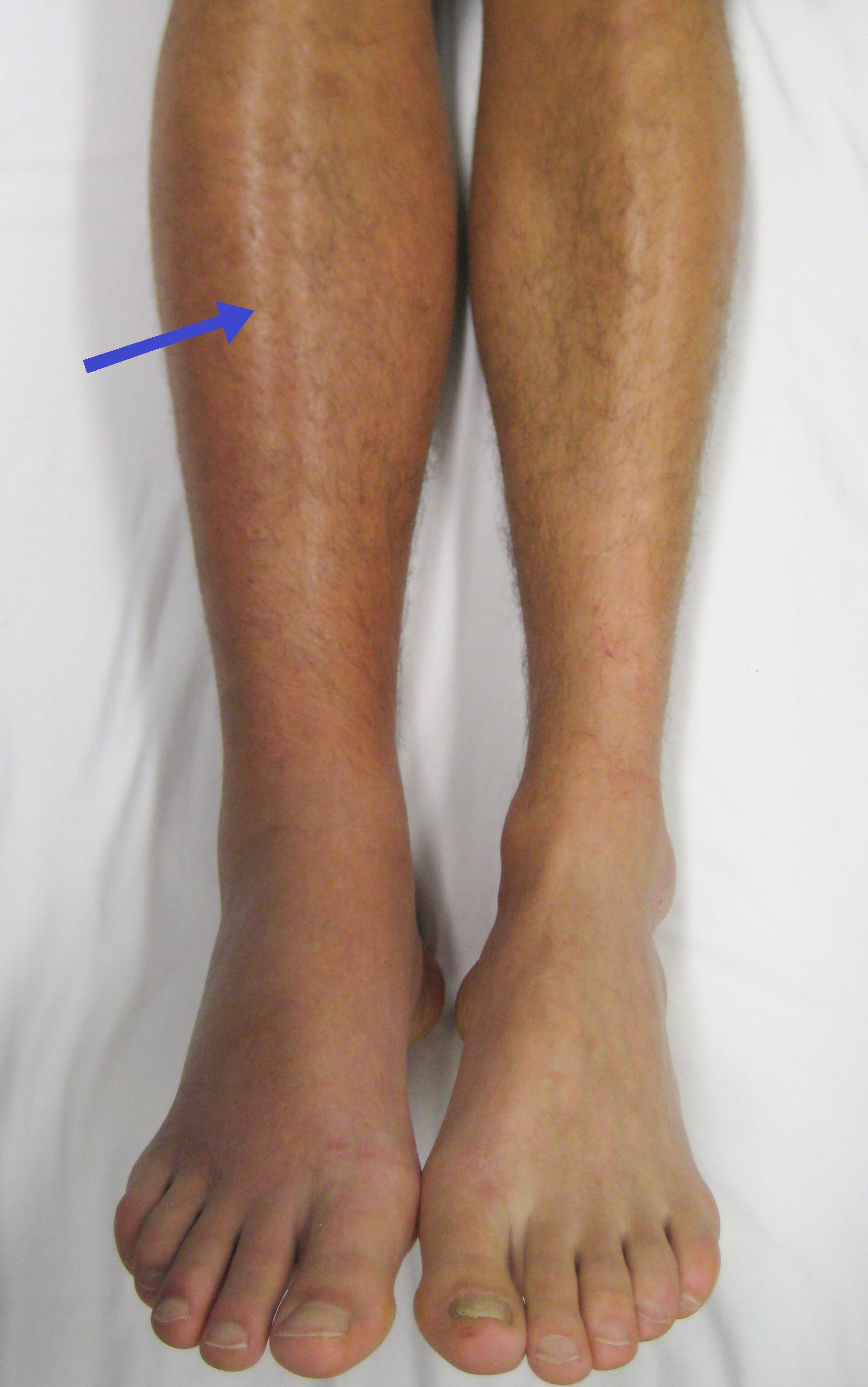
Pulmonary embolism (PE) is a blockage of an pulmonary artery, artery in the lungs by a substance that has moved from elsewhere in the body through the bloodstream (embolism). Symptoms of a PE may include dyspnea, shortness of breath, chest pain particularly upon breathing in, and coughing up blood. Symptoms of a deep vein thrombosis, blood clot in the leg may also be present, such as a erythema, red, warm, swollen, and painful leg. Signs of a PE include low blood oxygen saturation, oxygen levels, tachypnea, rapid breathing, tachycardia, rapid heart rate, and sometimes a mild fever. Severe cases can lead to Syncope (medicine), passing out, shock (circulatory), abnormally low blood pressure, obstructive shock, and cardiac arrest, sudden death. PE usually results from a blood clot in the leg that travels to the lung. The risk of blood clots is increased by advanced age, cancer, prolonged bed rest and immobilization, smoking, stroke, long-haul travel over 4 hours, certain genetics, ... [...More Info...] [...Related Items...] OR: [Wikipedia] [Google] [Baidu] |
Bundle Branch Block
A bundle branch block is a partial or complete interruption in the flow of electrical impulses in either of the bundle branches of the heart's electrical system. Anatomy and physiology The heart's electrical activity begins in the sinoatrial node (the heart's natural pacemaker), which is situated on the upper right atrium. The impulse travels next through the left and right atria and summates at the atrioventricular node. From the AV node the electrical impulse travels down the bundle of His and divides into the right and left bundle branches. The right bundle branch contains one fascicle. The left bundle branch subdivides into two fascicles: the left anterior fascicle, and the left posterior fascicle. Other sources divide the left bundle branch into three fascicles: the left anterior, the left posterior, and the left septal fascicle. The thicker left posterior fascicle bifurcates, with one fascicle being in the septal aspect. Ultimately, the fascicles divide into mill ... [...More Info...] [...Related Items...] OR: [Wikipedia] [Google] [Baidu] |
Right Ventricle
A ventricle is one of two large chambers located toward the bottom of the heart that collect and expel blood towards the peripheral beds within the body and lungs. The blood pumped by a ventricle is supplied by an atrium (heart), atrium, an adjacent chamber in the upper heart that is smaller than a ventricle. Interventricular means between the ventricles (for example the interventricular septum), while intraventricular means within one ventricle (for example an intraventricular block). In a four-chambered heart, such as that in humans, there are two ventricles that operate in a double circulatory system: the right ventricle pumps blood into the pulmonary circulation to the lungs, and the left ventricle pumps blood into the systemic circulation through the aorta. Structure Ventricles have thicker walls than atria and generate higher blood pressures. The physiological load on the ventricles requiring pumping of blood throughout the body and lungs is much greater than the pressure g ... [...More Info...] [...Related Items...] OR: [Wikipedia] [Google] [Baidu] |
Hypertrophic Cardiomyopathy
Hypertrophic cardiomyopathy (HCM, or HOCM when obstructive) is a condition in which muscle tissues of the heart become thickened without an obvious cause. The parts of the heart most commonly affected are the interventricular septum and the ventricles. This results in the heart being less able to pump blood effectively and also may cause electrical conduction problems. Specifically, within the bundle branches that conduct impulses through the interventricular septum and into the Purkinje fibers, as these are responsible for the depolarization of contractile cells of both ventricles. People who have HCM may have a range of symptoms. People may be asymptomatic, or may have fatigue, leg swelling, and shortness of breath. It may also result in chest pain or fainting. Symptoms may be worse when the person is dehydrated. Complications may include heart failure, an irregular heartbeat, and sudden cardiac death. HCM is most commonly inherited in an autosomal dominant pattern. I ... [...More Info...] [...Related Items...] OR: [Wikipedia] [Google] [Baidu] |
Angiography
Angiography or arteriography is a medical imaging technique used to visualize the inside, or lumen, of blood vessels and organs of the body, with particular interest in the arteries, veins, and the heart chambers. Modern angiography is performed by injecting a radio-opaque contrast agent into the blood vessel and imaging using X-ray based techniques such as fluoroscopy. With time-of-flight (TOF) magnetic resonance it is no longer necessary to use a contrast. The word itself comes from the Greek words ἀνγεῖον ''angeion'' 'vessel' and γράφειν ''graphein'' 'to write, record'. The film or image of the blood vessels is called an ''angiograph'', or more commonly an ''angiogram''. Though the word can describe both an arteriogram and a venogram, in everyday usage the terms angiogram and arteriogram are often used synonymously, whereas the term venogram is used more precisely. The term angiography has been applied to radionuclide angiography and newer vascular ima ... [...More Info...] [...Related Items...] OR: [Wikipedia] [Google] [Baidu] |
Left Anterior Descending Artery
The left anterior descending artery (LAD, or anterior descending branch), also called anterior interventricular artery (IVA, or anterior interventricular branch of left coronary artery) is a branch of the left coronary artery. It supplies the anterior portion of the left ventricle. It provides about half of the arterial supply to the left ventricle and is thus considered the most important vessel supplying the left ventricle. Blockage of this artery is often called the ''widow-maker infarction'' due to a high risk of death. Structure Course It first passes at posterior to the pulmonary artery, then passes anteriorward between that pulmonary artery and the left atrium to reach the anterior interventricular sulcus, along which it descends to the notch of cardiac apex. In 78% of cases, it reaches the apex of the heart. Although rare, multiple anomalous courses of the LAD have been described. These include the origin of the artery from the right aortic sinus. Branches The LAD g ... [...More Info...] [...Related Items...] OR: [Wikipedia] [Google] [Baidu] |
Wellens' Syndrome
Wellens' syndrome is an electrocardiographic manifestation of critical proximal left anterior descending (LAD) coronary artery stenosis in people with unstable angina. Originally thought of as two separate types, A and B, it is now considered an evolving wave form, initially of biphasic T wave inversions and later becoming symmetrical, often deep (>2 mm), T wave inversions in the anterior precordial leads. First described by Hein J. J. Wellens and colleagues in 1982 in a subgroup of people with unstable angina, it does not seem to be rare, appearing in 18% of patients in his original study. A subsequent prospective study identified this syndrome in 14% of patients at presentation and 60% of patients within the first 24 hours. The presence of Wellens' syndrome carries significant diagnostic and prognostic value. All people in the De Zwann's study with characteristic findings had more than 50% stenosis of the left anterior descending artery (mean = 85% stenosis) with complete or ... [...More Info...] [...Related Items...] OR: [Wikipedia] [Google] [Baidu] |
Myocardial Ischaemia
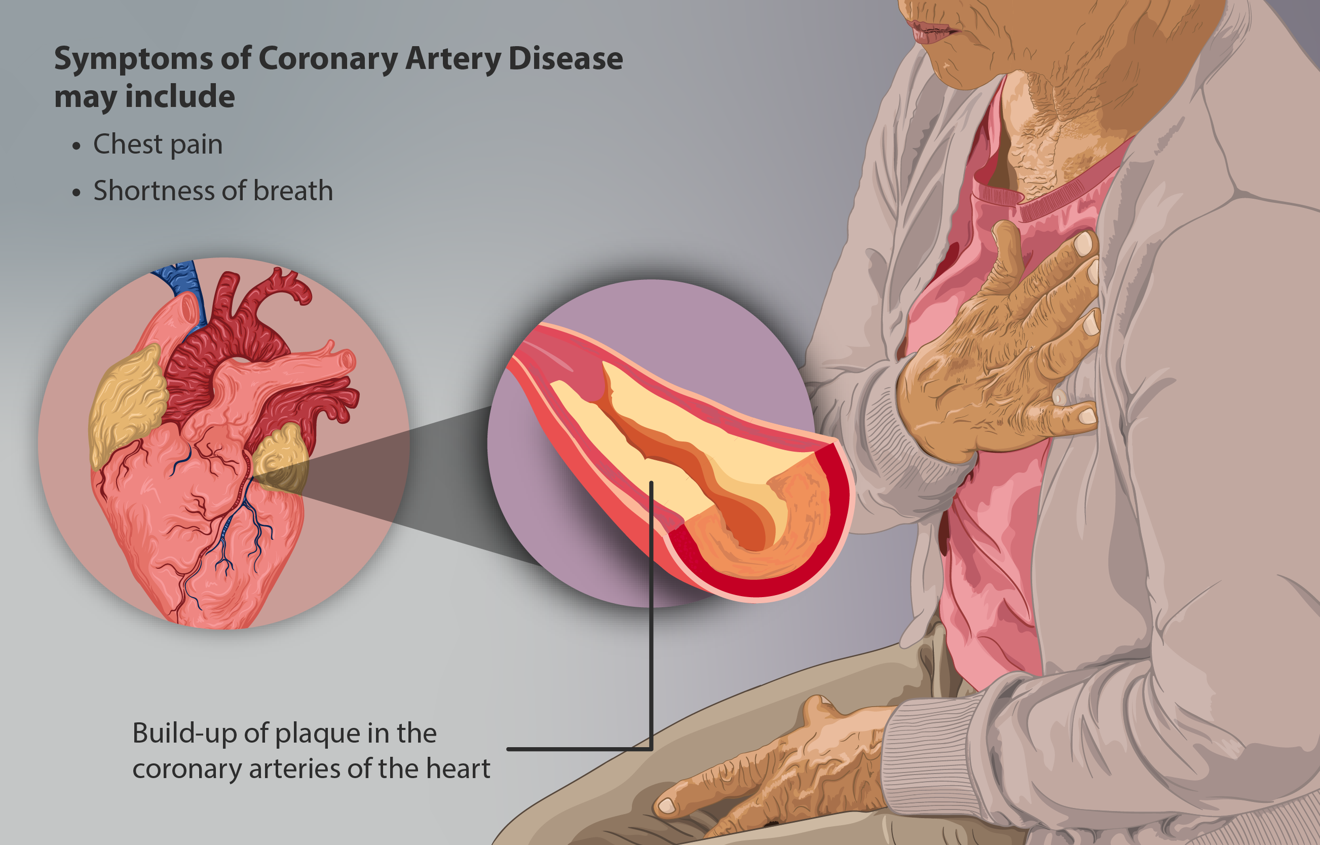
Coronary artery disease (CAD), also called coronary heart disease (CHD), or ischemic heart disease (IHD), is a type of heart disease involving the reduction of blood flow to the cardiac muscle due to a build-up of atheromatous plaque in the arteries of the heart. It is the most common of the cardiovascular diseases. CAD can cause stable angina, unstable angina, myocardial ischemia, and myocardial infarction. A common symptom is angina, which is chest pain or discomfort that may travel into the shoulder, arm, back, neck, or jaw. Occasionally it may feel like heartburn. In stable angina, symptoms occur with exercise or emotional stress, last less than a few minutes, and improve with rest. Shortness of breath may also occur and sometimes no symptoms are present. In many cases, the first sign is a heart attack. Other complications include heart failure or an abnormal heartbeat. Risk factors include high blood pressure, smoking, diabetes mellitus, lack of exercise, obesity, hig ... [...More Info...] [...Related Items...] OR: [Wikipedia] [Google] [Baidu] |


