|
Salpingopharyngeus Muscle
The salpingopharyngeus muscle is a muscle of the pharynx. It arises from cartilage around the Eustachian tube, and inserts into the palatopharyngeus muscle by blending with its posterior fasciculus. It raises the pharynx and larynx during deglutition (swallowing) and laterally draws the pharyngeal walls up. It opens the pharyngeal orifice of the Eustachian tube during swallowing allowing for the equalization of pressure between it and the pharynx. Structure The salpingopharyngeus muscle arises from the superior border of the medial cartilage of the Eustachian tube, in the nasal cavity. This makes the posterior welt of the torus tubarius. It passes downward and blends with the posterior fasciculus of the palatopharyngeus muscle. Nerve supply The salpingopharyngeus is supplied by the vagus nerve (CN X) via the pharyngeal plexus. Blood supply The salpingopharyngeus muscle is supplied by the ascending pharyngeal artery. Function The salpingopharyngeus muscle raises th ... [...More Info...] [...Related Items...] OR: [Wikipedia] [Google] [Baidu] |
Auditory Tube
Auditory means of or relating to the process of hearing: * Auditory system, the neurological structures and pathways of sound perception ** Auditory bulla, part of auditory system found in mammals other than primates ** Auditory nerve, also known as the cochlear nerve is one of two parts of a cranial nerve ** Auditory ossicles, three bones in the middle ear that transmit sounds * Hearing (sense), the auditory sense, the sense by which sound is perceived * Ear, the auditory end organ * Cochlea, the auditory branch of the inner ear * Sound, the physical signal perceived by the auditory system * External auditory meatus, the ear canal * Primary auditory cortex, the part of the higher-level of the brain that serves hearing * Auditory agnosia * Auditory exclusion, a form of temporary hearing loss under high stress * Auditory feedback, an aid to control speech production and singing * Auditory hallucination, perceiving sounds without auditory stimulus * Auditory illusion, sound trick ... [...More Info...] [...Related Items...] OR: [Wikipedia] [Google] [Baidu] |
Muscle Fascicle
A muscle fascicle is a bundle of skeletal muscle fibers surrounded by perimysium, a type of connective tissue. Structure Muscle cells are grouped into muscle fascicles by enveloping perimysium connective tissue. Fascicles are bundled together by epimysium connective tissue. Muscle fascicles typically only contain one type of muscle cell (either type I fibres or type II fibres), but can contain a mixture of both types. Function In the heart specialized cardiac muscle cells transmit electrical impulses from the atrioventricular node (AV node) to the Purkinje fibers – fascicles, also referred to as bundle branches. These start as a single fascicle of fibers at the AV node called the bundle of His that then splits into three bundle branches: the right fascicular branch, left anterior fascicular branch, and left posterior fascicular branch. Clinical significance Myositis may cause thickening of the muscle fascicles. This may be detected with ultrasound scans. Mu ... [...More Info...] [...Related Items...] OR: [Wikipedia] [Google] [Baidu] |
Salpinx
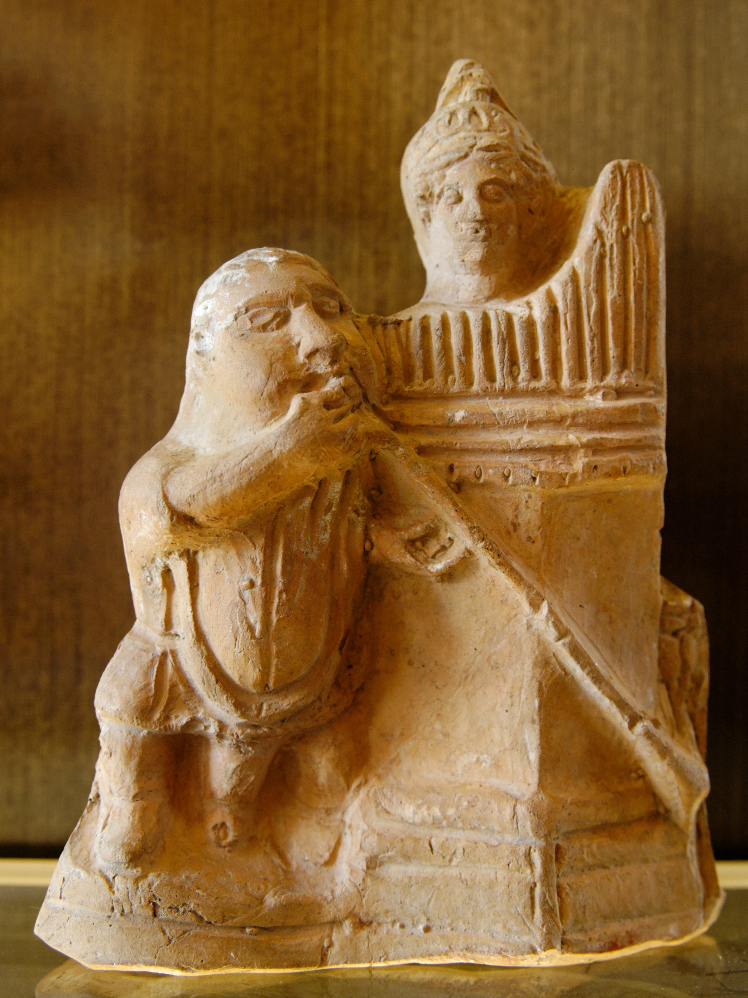
A salpinx (; plural salpinges ; Greek σαλπιγξ) was a trumpet-like instrument of the ancient Greeks. Construction The salpinx consisted of a straight, narrow bronze tube with a mouthpiece of bone and a bell (also constructed of bronze) of variable shape and size; extant descriptions describe conical, bulb-like, and spherical structures. Each type of bell may have had a unique effect on the sound made by the instrument. The instrument has been depicted in some classical era vases as employing the use of a phorbeia, similar to those used by aulos players of the era. Though similar to the Roman tuba, the salpinx was shorter than the approximately 1.5 meter long Roman tuba. A rare example of a salpinx, held at the Museum of Fine Arts, Boston, is unique in that it is constructed from thirteen sections of bone connected using tenons and sockets (with bronze ferrules) rather than the long, bronze tube described elsewhere. This salpinx is over 1.57 m long dwarfing the common sa ... [...More Info...] [...Related Items...] OR: [Wikipedia] [Google] [Baidu] |
Ascending Pharyngeal Artery
The ascending pharyngeal artery is an artery in the neck that supplies the pharynx, developing from the proximal part of the embryonic second aortic arch. It is the smallest branch of the external carotid and is a long, slender vessel, deeply seated in the neck, beneath the other branches of the external carotid and under the stylopharyngeus muscle. It lies just superior to the bifurcation of the common carotid arteries. The artery most typically bifurcates into embryologically distinct pharyngeal and neuromeningeal trunks. The pharyngeal trunk usually consists of several branches which supply the middle and inferior pharyngeal constrictor muscles and the stylopharyngeus, ramifying in their substance and in the mucous membranes lining them. These branches are in hemodynamic equilibrium with contributors from the internal maxillary artery. The neuromeningeal trunk classically consists of jugular and hypoglossal divisions, which enter the jugular and hypoglossal foramina to s ... [...More Info...] [...Related Items...] OR: [Wikipedia] [Google] [Baidu] |
Pharyngeal Plexus Of Vagus Nerve
The pharyngeal plexus is a network of nerve fibers innervating most of the palate and pharynx. (The larynx, which is innervated by the superior and recurrent laryngeal nerves from vagus nerve (CN X), is not included.) It is located on the surface of the middle pharyngeal constrictor muscle. Sources Although the '' Terminologia Anatomica'' name of the plexus has "vagus nerve" in the title, other nerves make contributions to the plexus. It has the following sources: * CN IX – pharyngeal branches of glossopharyngeal nerve – sensory * CN X – pharyngeal branch of vagus nerve – motor * superior cervical ganglion sympathetic fibers – vasomotor Because the cranial part of accessory nerve (CN XI) leaves the jugular foramen as a part of the CN X, it is sometimes considered part of the plexus as well. Innervation Sensory The pharyngeal plexus provides sensory innervation of the oropharynx and laryngopharynx from CN IX and CN X. (The nasopharynx above the pharyngoty ... [...More Info...] [...Related Items...] OR: [Wikipedia] [Google] [Baidu] |
Deglutition
Swallowing, sometimes called deglutition in scientific contexts, is the process in the human or animal body that allows for a substance to pass from the mouth, to the pharynx, and into the esophagus, while shutting the epiglottis. Swallowing is an important part of eating and drinking. If the process fails and the material (such as food, drink, or medicine) goes through the trachea, then choking or pulmonary aspiration can occur. In the human body the automatic temporary closing of the epiglottis is controlled by the swallowing reflex. The portion of food, drink, or other material that will move through the neck in one swallow is called a bolus. In colloquial English, the term "swallowing" is also used to describe the action of taking in a large mouthful of food without any biting, where the word gulping is more adequate. In humans Swallowing comes so easily to most people that the process rarely prompts much thought. However, from the viewpoints of physiology, of speech–lan ... [...More Info...] [...Related Items...] OR: [Wikipedia] [Google] [Baidu] |
Larynx
The larynx (), commonly called the voice box, is an organ in the top of the neck involved in breathing, producing sound and protecting the trachea against food aspiration. The opening of larynx into pharynx known as the laryngeal inlet is about 4–5 centimeters in diameter. The larynx houses the vocal cords, and manipulates pitch and volume, which is essential for phonation. It is situated just below where the tract of the pharynx splits into the trachea and the esophagus. The word ʻlarynxʼ (plural ʻlaryngesʼ) comes from the Ancient Greek word ''lárunx'' ʻlarynx, gullet, throat.ʼ Structure The triangle-shaped larynx consists largely of cartilages that are attached to one another, and to surrounding structures, by muscles or by fibrous and elastic tissue components. The larynx is lined by a ciliated columnar epithelium except for the vocal folds. The cavity of the larynx extends from its triangle-shaped inlet, to the epiglottis, and to the circular outlet at the ... [...More Info...] [...Related Items...] OR: [Wikipedia] [Google] [Baidu] |
Human Pharynx
The pharynx (plural: pharynges) is the part of the throat behind the mouth and nasal cavity, and above the oesophagus and trachea (the tubes going down to the stomach and the lungs). It is found in vertebrates and invertebrates, though its structure varies across species. The pharynx carries food and air to the esophagus and larynx respectively. The flap of cartilage called the epiglottis stops food from entering the larynx. In humans, the pharynx is part of the digestive system and the conducting zone of the respiratory system. (The conducting zone—which also includes the nostrils of the nose, the larynx, trachea, bronchi, and bronchioles—filters, warms and moistens air and conducts it into the lungs). The human pharynx is conventionally divided into three sections: the nasopharynx, oropharynx, and laryngopharynx. It is also important in vocalization. In humans, two sets of pharyngeal muscles form the pharynx and determine the shape of its lumen. They are arranged a ... [...More Info...] [...Related Items...] OR: [Wikipedia] [Google] [Baidu] |
Eustachian Tube
In anatomy, the Eustachian tube, also known as the auditory tube or pharyngotympanic tube, is a tube that links the nasopharynx to the middle ear, of which it is also a part. In adult humans, the Eustachian tube is approximately long and in diameter. It is named after the sixteenth-century Italian anatomist Bartolomeo Eustachi. In humans and other tetrapods, both the middle ear and the ear canal are normally filled with air. Unlike the air of the ear canal, however, the air of the middle ear is not in direct contact with the atmosphere outside the body; thus, a pressure difference can develop between the atmospheric pressure of the ear canal and the middle ear. Normally, the Eustachian tube is collapsed, but it gapes open with swallowing and with positive pressure, allowing the middle ear's pressure to adjust to the atmospheric pressure. When taking off in an aircraft, the ambient air pressure goes from higher (on the ground) to lower (in the sky). The air in the middle ear Bo ... [...More Info...] [...Related Items...] OR: [Wikipedia] [Google] [Baidu] |
Palatopharyngeus Muscle
The palatopharyngeus (palatopharyngeal or pharyngopalatinus) muscle is a small muscle in the roof of the mouth. It is a long, fleshy fasciculus, narrower in the middle than at either end, forming, with the mucous membrane covering its surface, the palatopharyngeal arch. Structure It is separated from the palatoglossus muscle by an angular interval, in which the palatine tonsil is lodged. It arises from the soft palate, where it is divided into two fasciculi by the levator veli palatini and musculus uvulae. * The ''posterior fasciculus'' lies in contact with the mucous membrane, and joins with that of the opposite muscle in the middle line. * The ''anterior fasciculus'', the thicker, lies in the soft palate between the levator and tensor veli palatini muscles, and joins in the middle line the corresponding part of the opposite muscle. Passing laterally and downward behind the palatine tonsil, the palatopharyngeus joins the stylopharyngeus and is inserted with that muscle into the ... [...More Info...] [...Related Items...] OR: [Wikipedia] [Google] [Baidu] |
Skeletal Muscle
Skeletal muscles (commonly referred to as muscles) are organs of the vertebrate muscular system and typically are attached by tendons to bones of a skeleton. The muscle cells of skeletal muscles are much longer than in the other types of muscle tissue, and are often known as muscle fibers. The muscle tissue of a skeletal muscle is striated – having a striped appearance due to the arrangement of the sarcomeres. Skeletal muscles are voluntary muscles under the control of the somatic nervous system. The other types of muscle are cardiac muscle which is also striated and smooth muscle which is non-striated; both of these types of muscle tissue are classified as involuntary, or, under the control of the autonomic nervous system. A skeletal muscle contains multiple fascicles – bundles of muscle fibers. Each individual fiber, and each muscle is surrounded by a type of connective tissue layer of fascia. Muscle fibers are formed from the fusion of developmental myobla ... [...More Info...] [...Related Items...] OR: [Wikipedia] [Google] [Baidu] |
Middle Ear
The middle ear is the portion of the ear medial to the eardrum, and distal to the oval window of the cochlea (of the inner ear). The mammalian middle ear contains three ossicles, which transfer the vibrations of the eardrum into waves in the fluid and membranes of the inner ear. The hollow space of the middle ear is also known as the tympanic cavity and is surrounded by the tympanic part of the temporal bone. The auditory tube (also known as the Eustachian tube or the pharyngotympanic tube) joins the tympanic cavity with the nasal cavity (nasopharynx), allowing pressure to equalize between the middle ear and throat. The primary function of the middle ear is to efficiently transfer acoustic energy from compression waves in air to fluid–membrane waves within the cochlea. Structure Ossicles The middle ear contains three tiny bones known as the ossicles: '' malleus'', '' incus'', and ''stapes''. The ossicles were given their Latin names for their distinctive shapes; they ar ... [...More Info...] [...Related Items...] OR: [Wikipedia] [Google] [Baidu] |


