|
Superficial Fibular Nerve
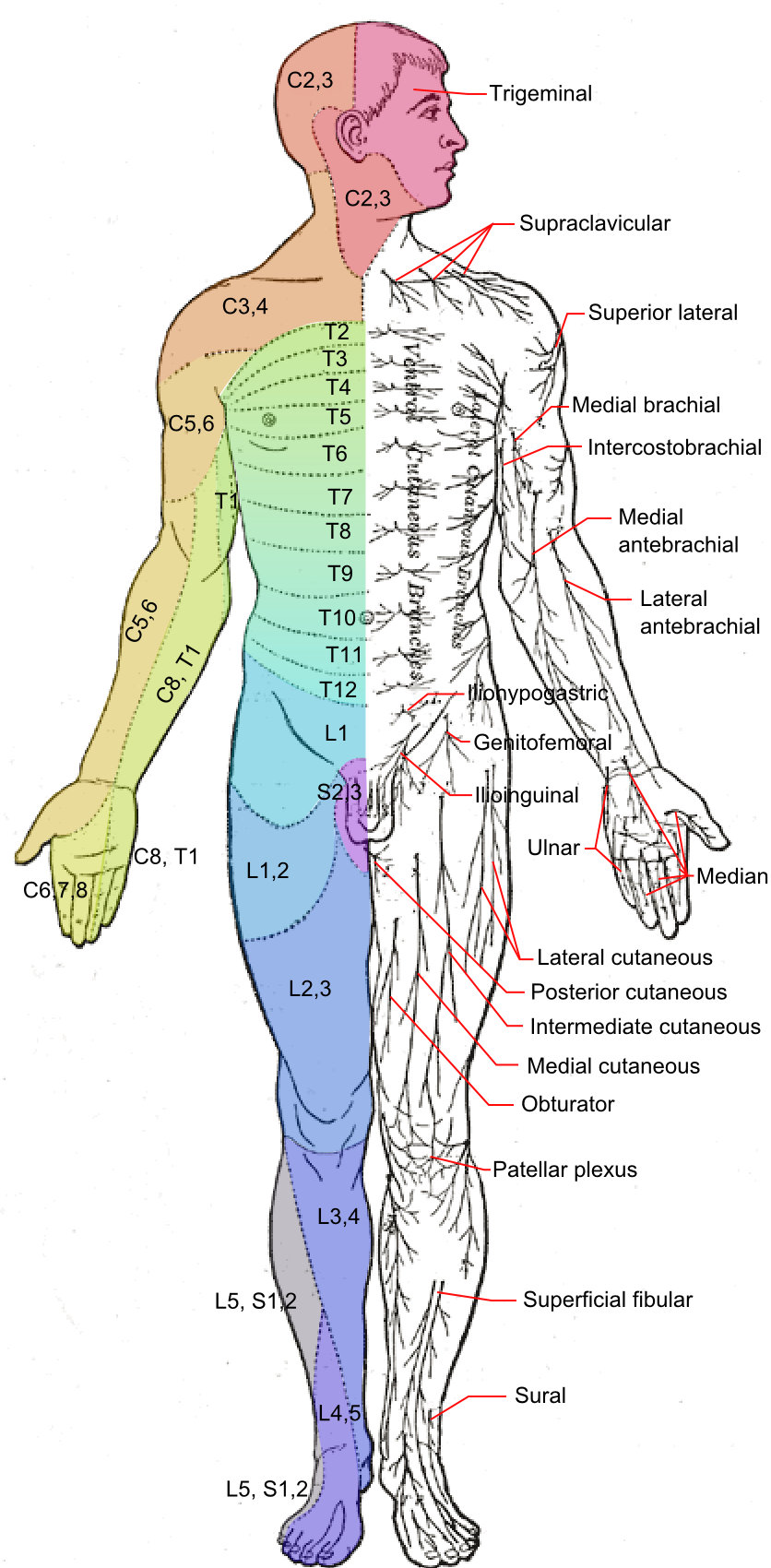
The superficial fibular nerve (also known as superficial peroneal nerve) is a mixed (motor and sensory) nerve that provides motor innervation to the fibularis longus and fibularis brevis muscles, and sensory innervation to skin over the antero-lateral aspect of the leg along with the greater part of the dorsum of the foot (with the exception of the first web space, which is innervated by the deep fibular nerve). Structure Lateral side of the leg The superficial fibular nerve is the main nerve of the lateral compartment of the leg. It begins at the lateral side of the neck of fibula, and runs through the fibularis longus and fibularis brevis muscles. In the middle third of the leg, it descends between the fibularis longus and fibularis brevis, and then reaches the anterior border of the fibularis brevis to enter the groove between the fibularis brevis and the extensor digitorum longus under the deep fascia of leg. It becomes superficial at the junction of upper two-thirds and ... [...More Info...] [...Related Items...] OR: [Wikipedia] [Google] [Baidu] |
Common Fibular Nerve
The common fibular nerve (also known as the common peroneal nerve, external popliteal nerve, or lateral popliteal nerve) is a nerve in the lower leg that provides sensation over the posterolateral part of the leg and the knee joint. It divides at the knee into two terminal branches: the superficial fibular nerve and deep fibular nerve, which innervate the muscles of the lateral and anterior compartments of the leg respectively. When the common fibular nerve is damaged or compressed, foot drop can ensue. Structure The common fibular nerve is the smaller terminal branch of the sciatic nerve. The common fibular nerve has root values of L4, L5, S1, and S2. It arises from the superior angle of the popliteal fossa and extends to the lateral angle of the popliteal fossa, along the medial border of the biceps femoris. It then winds around the neck of the fibula to pierce the fibularis longus and divides into terminal branches of the superficial fibular nerve and the deep fibular nerve ... [...More Info...] [...Related Items...] OR: [Wikipedia] [Google] [Baidu] |
Sural Nerve
The sural nerve ''(L4-S1)'' is generally considered a pure cutaneous nerve of the posterolateral leg to the lateral ankle. The sural nerve originates from a combination of either the sural communicating branch and medial sural cutaneous nerve, or the lateral sural cutaneous nerve. This group of nerves is termed the sural nerve complex. There are eight documented variations of the sural nerve complex. Once formed the sural nerve takes its course midline posterior to posterolateral around the lateral malleolus. The sural nerve terminates as the lateral dorsal cutaneous nerve. Anatomy The sural nerve ''(L4-S1)'' is a cutaneous sensory nerve of the posterolateral calf with cutaneous innervation to the distal one-third of the lower leg. Formation of the ''sural nerve'' is the result of either anastomosis of the medial sural cutaneous nerve and the sural communicating nerve, or it may be found as a continuation of the lateral sural cutaneous nerve traveling parallel to ... [...More Info...] [...Related Items...] OR: [Wikipedia] [Google] [Baidu] |
Great Toe
Toes are the digits of the foot of a tetrapod. Animal species such as cats that walk on their toes are described as being ''digitigrade''. Humans, and other animals that walk on the soles of their feet, are described as being ''plantigrade''; '' unguligrade'' animals are those that walk on hooves at the tips of their toes. Structure There are normally five toes present on each human foot. Each toe consists of three phalanx bones, the proximal, middle, and distal, with the exception of the big toe (). For a minority of people, the little toe also is missing a middle bone. The hallux only contains two phalanx bones, the proximal and distal. The joints between each phalanx are the interphalangeal joints. The proximal phalanx bone of each toe articulates with the metatarsal bone of the foot at the metatarsophalangeal joint. Each toe is surrounded by skin, and present on all five toes is a toenail. The toes are, from medial to lateral: * the first toe, also known as the ... [...More Info...] [...Related Items...] OR: [Wikipedia] [Google] [Baidu] |
Evert
Evert is a Dutch and Swedish short form of the Germanic masculine name "Everhard" (alternative Eberhard). at the Meertens Institute database of given names in the Netherlands. It is also used as surname. Notable people with the name include: Given name * Evert van Aelst (1602–1657), Dutch still life painter * Evert Andersen (1772–1809), Norwegian naval officer * Evert Augustus Duyckinck (1816–1878), American publisher and bi ...[...More Info...] [...Related Items...] OR: [Wikipedia] [Google] [Baidu] |
Cutaneous Nerve
A cutaneous nerve is a nerve that provides nerve supply to the skin. Human anatomy In human anatomy, cutaneous nerves are primarily responsible for providing cutaneous innervation, sensory innervation to the skin. In addition to sympathetic and autonomic afferent (sensory) fibers, most cutaneous nerves also contain sympathetic efferent (visceromotor) fibers, which innervate cutaneous blood vessels, sweat glands, and the arrector pilli muscles of hair follicles. These structures are important to the sympathetic nervous response. There are many cutaneous nerves in the human body, only some of which are named. Some of the larger cutaneous nerves are as follows: Upper body * In the arm (proper) ** Superior lateral cutaneous nerve of arm (Superior LCNOA) ** Inferior lateral cutaneous nerve of arm (Inferior LCNOA) ** Posterior cutaneous nerve of arm (PCNOA) ** Medial cutaneous nerve of arm (MCNOA) * In the forearm ** Lateral cutaneous nerve of forearm (LCNOF) ** Posterior c ... [...More Info...] [...Related Items...] OR: [Wikipedia] [Google] [Baidu] |
Medial Dorsal Cutaneous Nerve
The medial dorsal cutaneous nerve (internal dorsal cutaneous branch) is the more medial one of the two terminal branches of the superficial fibular nerve (the other being the intermediate dorsal cutaneous nerve). Through its branches, it provides innervation to parts of the dorsal aspects of the first, second, and third toes. Anatomy Origin The superficial fibular nerve terminates by bifurcating into the medial dorsal cutaneous nerve and the intermediate dorsal cutaneous nerve immediately after emerging from the deep fascia of leg at the distal two-thirds to three-fourths point of the leg. Branches and distribution The medial dorsal cutaneous nerves trifurcates at the inferior border of the ankle, giving rise to: * a medial branch which passes anteromedially before giving rise to the medial dorsal digital nerves of the first toe; * a middle branch which passes anteriorly superficial to the first intermetatarsal space and anastomoses with the tibial nerve before bifurcati ... [...More Info...] [...Related Items...] OR: [Wikipedia] [Google] [Baidu] |
Lateral Plantar Nerve
The lateral plantar nerve (external plantar nerve) is a branch of the tibial nerve, in turn a branch of the sciatic nerve and supplies the skin of the fifth toe and lateral half of the fourth, as well as most of the deep muscles, its distribution being similar to that of the ulnar nerve in the hand. It passes obliquely forward with the lateral plantar artery to the lateral side of the foot, lying between the flexor digitorum brevis and quadratus plantae and, in the interval between the flexor muscle and the abductor digiti minimi, divides into a superficial and a deep branch. Before its division, it supplies the quadratus plantae and abductor digiti minimi. It divides into deep and superficial branches. Additional images File:Gray357.png, Coronal section through right talocrural and talocalcaneal joint In human anatomy, the subtalar joint, also known as the talocalcaneal joint, is a joint of the foot. It occurs at the meeting point of the talus and the calcaneus. Th ... [...More Info...] [...Related Items...] OR: [Wikipedia] [Google] [Baidu] |
Medial Plantar Nerve
The medial plantar nerve (internal plantar nerve) is the larger of the two terminal divisions of the tibial nerve (medial and lateral plantar nerve), which accompanies the medial plantar artery. From its origin under the laciniate ligament it passes under cover of the abductor hallucis muscle, and, appearing between this muscle and the flexor digitorum brevis, gives off a proper digital plantar nerve and finally divides opposite the bases of the metatarsal bones into three common digital plantar nerves. Branches The branches of the medial plantar nerve are: (1) cutaneous, (2) muscular, (3) articular, (4) a proper digital nerve to the medial side of the great toe, and (5) three common digital nerves. Cutaneous branches The cutaneous branches pierce the plantar aponeurosis between the abductor hallucis and the flexor digitorum brevis and are distributed to the skin of the sole of the foot. Muscular branches The muscular branches supply muscles on the medial side of the sole, in ... [...More Info...] [...Related Items...] OR: [Wikipedia] [Google] [Baidu] |
Saphenous Nerve
The saphenous nerve (long or internal saphenous nerve) is the largest cutaneous branch of the femoral nerve. It is derived from the lumbar plexus (L3-L4). It is a strictly sensory nerve, and has no motor function. It commences in the proximal (upper) thigh and travels along the adductor canal. Upon exiting the adductor canal, the saphenous nerve terminates by splitting into two terminal branches: the sartorial nerve, and the infrapatellar nerve (which together innervate the medial, anteromedial, posteromedial aspects of the distal thigh). The saphenous nerve is responsible for providing sensory innervation to the skin of the anteromedial leg. Structure It is purely a sensory nerve. Origin The saphenous nerve is the largest and terminal branch of the femoral nerve. It is derived from the lumbar plexus (L3-L4). Course Shortly after the femoral nerve passes under the inguinal ligament, it splits into anterior and posterior divisions by the passage of the lateral femoral ci ... [...More Info...] [...Related Items...] OR: [Wikipedia] [Google] [Baidu] |
Medial Dorsal Cutaneous Nerve
The medial dorsal cutaneous nerve (internal dorsal cutaneous branch) is the more medial one of the two terminal branches of the superficial fibular nerve (the other being the intermediate dorsal cutaneous nerve). Through its branches, it provides innervation to parts of the dorsal aspects of the first, second, and third toes. Anatomy Origin The superficial fibular nerve terminates by bifurcating into the medial dorsal cutaneous nerve and the intermediate dorsal cutaneous nerve immediately after emerging from the deep fascia of leg at the distal two-thirds to three-fourths point of the leg. Branches and distribution The medial dorsal cutaneous nerves trifurcates at the inferior border of the ankle, giving rise to: * a medial branch which passes anteromedially before giving rise to the medial dorsal digital nerves of the first toe; * a middle branch which passes anteriorly superficial to the first intermetatarsal space and anastomoses with the tibial nerve before bifurcati ... [...More Info...] [...Related Items...] OR: [Wikipedia] [Google] [Baidu] |
Deep Fascia Of Leg
The deep fascia of leg or crural fascia forms a complete investment to the muscles, and is fused with the periosteum over the subcutaneous surfaces of the bones. The deep fascia of the leg is continuous above with the fascia lata (deep fascia of the thigh), and is attached around the knee to the patella, the patellar ligament, the tuberosity and condyles of the tibia, and the head of the fibula. Behind, it forms the popliteal fascia, covering in the popliteal fossa; here it is strengthened by transverse fibers, and perforated by the small saphenous vein. It receives an expansion from the tendon of the biceps femoris laterally, and from the tendons of the sartorius, gracilis, semitendinosus, and semimembranosus medially; in front, it blends with the periosteum covering the subcutaneous surface of the tibia, and with that covering the head and malleolus of the fibula; below, it is continuous with the transverse crural and laciniate ligaments. It is thick and dense in the upper a ... [...More Info...] [...Related Items...] OR: [Wikipedia] [Google] [Baidu] |
Extensor Digitorum Longus
The extensor digitorum longus is a pennate muscle, situated at the lateral part of the front of the leg. Structure It arises from the lateral condyle of the tibia; from the upper three-quarters of the anterior surface of the body of the fibula; from the upper part of the interosseous membrane; from the deep surface of the fascia; and from the intermuscular septa between it and the tibialis anterior on the medial, and the peroneal muscles on the lateral side. Between it and the tibialis anterior are the upper portions of the anterior tibial vessels and deep peroneal nerve. The muscle passes under the superior and inferior extensor retinaculum of foot in company with the fibularis tertius, and divides into four slips, which run forward on the dorsum of the foot, and are inserted into the second and third phalanges of the four lesser toes. The tendons to the second, third, and fourth toes are each joined, opposite the metatarsophalangeal articulations, on the lateral side by ... [...More Info...] [...Related Items...] OR: [Wikipedia] [Google] [Baidu] |

