|
Striatopallidonigral Bundle
The basal ganglia form a major brain system in all vertebrates, but in primates (including humans) there are special differentiating features. The basal ganglia include the striatum, globus pallidus, substantia nigra and subthalamic nucleus. In primates the pallidus is divided into an external and internal globus pallidus, the external globus pallidus is present in other mammals but not the internal globus pallidus. Also in primates, the dorsal striatum is divided by a large nerve tract called the internal capsule into two masses named the caudate nucleus and the putamen. These differences contribute to a complex circuitry of connections between the striatum and cortex that is specific to primates, reflecting different functions in primate cortical areas. Corticostriatal connection A major output from the cortex, with axons from most of the cortical regions connecting to the striatum, is called the corticostriatal connection, part of the cortico-basal ganglia-thalamo-cortical loo ... [...More Info...] [...Related Items...] OR: [Wikipedia] [Google] [Baidu] |
Basal Ganglia Diagram
Basal or basilar is a term meaning ''base'', ''bottom'', or ''minimum''. Science * Basal (anatomy), an anatomical term of location for features associated with the base of an organism or structure * Basal (medicine), a minimal level that is necessary for health or life, such as a minimum insulin dose * Basal (phylogenetics), a relative position in a phylogenetic tree closer to the root Places * Basal, Hungary, a village in Hungary * Basal, Pakistan, a village in the Attock District Other * Basal plate (other) * Basal sliding, the act of a glacier sliding over the bed before it due to meltwater increasing the water pressure underneath the glacier causing it to be lifted from its bed * Basal conglomerate, see conglomerate (geology) Conglomerate () is a sedimentary rock made up of rounded gravel-sized pieces of rock surrounded by finer-grained sediments (such as sand, silt, or clay). The larger fragments within conglomerate are called clasts, while the finer sediment ... [...More Info...] [...Related Items...] OR: [Wikipedia] [Google] [Baidu] |
Cholinergic
Cholinergic agents are compounds which mimic the action of acetylcholine and/or butyrylcholine. In general, the word " choline" describes the various quaternary ammonium salts containing the ''N'',''N'',''N''-trimethylethanolammonium cation. Found in most animal tissues, choline is a primary component of the neurotransmitter acetylcholine and functions with inositol as a basic constituent of lecithin. Choline also prevents fat deposits in the liver and facilitates the movement of fats into cells. The parasympathetic nervous system, which uses acetylcholine almost exclusively to send its messages, is said to be almost entirely cholinergic. Neuromuscular junctions, preganglionic neurons of the sympathetic nervous system, the basal forebrain, and brain stem complexes are also cholinergic, as are the receptor for the merocrine sweat glands. In neuroscience and related fields, the term cholinergic is used in these related contexts: * A substance (or ligand) is cholinergic if ... [...More Info...] [...Related Items...] OR: [Wikipedia] [Google] [Baidu] |
Connectome
A connectome () is a comprehensive map of neural connections in the brain, and may be thought of as its " wiring diagram". These maps are available in varying levels of detail. A functional connectome shows connections between various brain regions, but not individual neurons. These are available for large animals, including mice and humans, are normally obtained by techniques such as MRI, and have a scale of millimeters. At the other extreme are neural connectomes, which show individual neurons and their interconnections. These are usually obtained by electron microscopy (EM) and have a scale of nanometers. They are only available for small creatures such as the worm ''C. Elegans'' and the fruit fly ''Drosophila melanogaster'', and small regions of mammal brains. Finally there are chemical connectomes, showing which neurons emit, and are sensitive to, a wide variety of neuromodulators. The significance of the connectome stems from the realization that the structure an ... [...More Info...] [...Related Items...] OR: [Wikipedia] [Google] [Baidu] |
Macaque
The macaques () constitute a genus (''Macaca'') of gregarious Old World monkeys of the subfamily Cercopithecinae. The 23 species of macaques inhabit ranges throughout Asia, North Africa, and Europe (in Gibraltar). Macaques are principally frugivorous (preferring fruit), although their diet also includes seeds, leaves, flowers, and tree bark. Some species such as the long-tailed macaque (''M. fascicularis''; also called the crab-eating macaque) will supplement their diets with small amounts of meat from shellfish, insects, and small mammals. On average, a southern pig-tailed macaque (''M. nemestrina'') in Malaysia eats about 70 large rats each year. All macaque social groups are arranged around dominant matriarchs. Macaques are found in a variety of habitats throughout the Asian continent and are highly adaptable. Certain species are synanthropic, having learned to live alongside humans, but they have become problematic in urban areas in Southeast Asia and are not suitabl ... [...More Info...] [...Related Items...] OR: [Wikipedia] [Google] [Baidu] |
Basal Ganglia
The basal ganglia (BG) or basal nuclei are a group of subcortical Nucleus (neuroanatomy), nuclei found in the brains of vertebrates. In humans and other primates, differences exist, primarily in the division of the globus pallidus into external and internal regions, and in the division of the striatum. Positioned at the base of the forebrain and the top of the midbrain, they have strong connections with the cerebral cortex, thalamus, brainstem and other brain areas. The basal ganglia are associated with a variety of functions, including regulating voluntary motor control, motor movements, procedural memory, procedural learning, habituation, habit formation, conditional learning, eye movements, cognition, and emotion. The main functional components of the basal ganglia include the striatum, consisting of both the dorsal striatum (caudate nucleus and putamen) and the ventral striatum (nucleus accumbens and olfactory tubercle), the globus pallidus, the ventral pallidum, the substa ... [...More Info...] [...Related Items...] OR: [Wikipedia] [Google] [Baidu] |
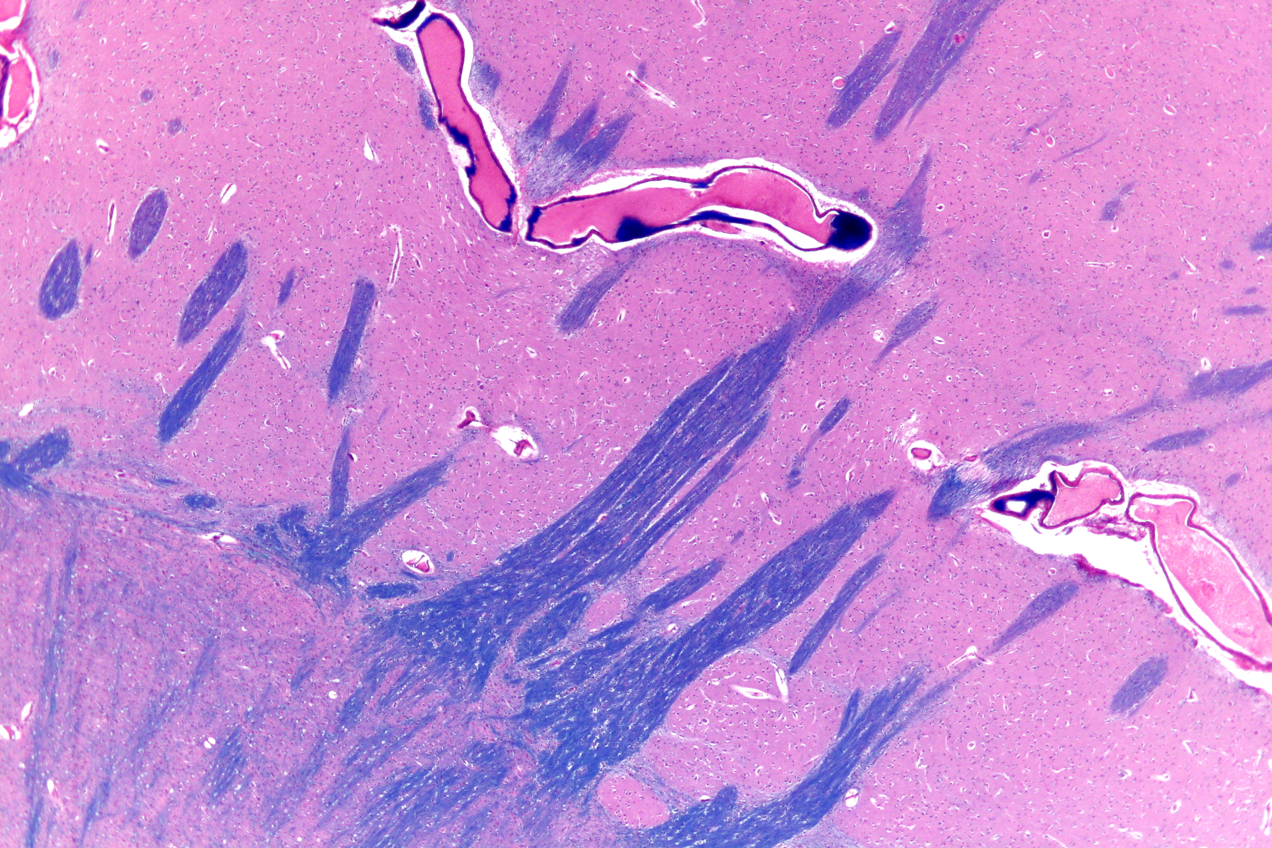
Striatopallidal Fibres
The striatopallidal fibres, also Wilson's pencils, pencil fibres of Wilson, and pencils of Wilson, are prominent myelinated fibres that connect the striatum to the globus pallidus. Their distinctive appearance allows the putamen to be identified on light microscopy. See also *Lentiform nucleus The lentiform nucleus (or lentiform complex, lenticular nucleus, or lenticular complex) are the putamen (laterally) and the globus pallidus (medially), collectively. Due to their proximity, these two structures were formerly considered one, howev ... References {{Reflist, 2 Basal ganglia ... [...More Info...] [...Related Items...] OR: [Wikipedia] [Google] [Baidu] |
Dopamine Receptor D1
Dopamine receptor D1, also known as DRD1. It is one of the two types of D1-like receptor family receptors D1 and D5. It is a protein that in humans is encoded by the DRD1 gene. Tissue distribution D1 receptors are the most abundant kind of dopamine receptor in the central nervous system. Northern blot and in situ hybridization show that the gene expression, mRNA expression of DRD1 is highest in the dorsal striatum (caudate nucleus, caudate and putamen) and ventral striatum (nucleus accumbens and olfactory tubercle). Lower levels occur in the basolateral amygdala, cerebral cortex, septum, thalamus, and hypothalamus. The DRD1 gene expresses primarily in the caudate putamen in humans, and in the caudate putamen, the nucleus accumbens and the olfactory tubercle in mouse. Structure The dopamine receptor D1 (D1R) is a Gs-coupled GPCR characterized by a canonical seven-transmembrane (TM) helical domain, with a ligand-binding pocket located Extracellular space, extracellularl ... [...More Info...] [...Related Items...] OR: [Wikipedia] [Google] [Baidu] |
μ-opioid Receptor
The μ-opioid receptors (MOR) are a class of opioid receptors with a high affinity for enkephalins and beta-endorphin, but a low affinity for dynorphins. They are also referred to as μ(''mu'')-opioid peptide (MOP) receptors. The prototypical μ-opioid receptor agonist is morphine, the primary psychoactive alkaloid in opium and for which the receptor was named, with Mu (letter), mu being the first letter of Morpheus, the compound's namesake in the original Greek. It is an inhibitory G-protein coupled receptor that activates the Gi alpha subunit, Gi alpha subunit, inhibiting adenylate cyclase activity, lowering Cyclic adenosine monophosphate, cAMP levels. Structure The structure of the inactive μ-opioid receptor has been determined with the antagonists Beta-Funaltrexamine, β-FNA and alvimopan. Many structures of the active state are also available, with agonists including DAMGO, Β-Endorphin, β-endorphin, fentanyl and morphine. The structure with the agonist BU72 has the h ... [...More Info...] [...Related Items...] OR: [Wikipedia] [Google] [Baidu] |
Striosome
The striosomes (also referred to as striatal patches) are one of two complementary chemical compartments within the striatum (the other compartment is known as the matrix) that can be visualized by staining for immunocytochemical markers such as mu opioid receptors, acetylcholinesterase, enkephalin, substance P, limbic system-associated membrane protein (LAMP), AMPA receptor subunit 1 (GluR1), dopamine receptor subunits, and calcium binding proteins. Striosomal abnormalities have been associated with neurological disorders, such as mood dysfunction in Huntington's disease, though their precise function remains unknown. Recently studies have identified the presence of "exo-patch" neurons that are biochemically and genetically the same as striosomal neurons, but reside in the matrix compartment. This study also characterized the different input and output connections of the striosome and matrix compartments, revealing that both regions have direct inputs to dopamine neurons ( ... [...More Info...] [...Related Items...] OR: [Wikipedia] [Google] [Baidu] |
Staining
Staining is a technique used to enhance contrast in samples, generally at the Microscope, microscopic level. Stains and dyes are frequently used in histology (microscopic study of biological tissue (biology), tissues), in cytology (microscopic study of cell (biology), cells), and in the medical fields of histopathology, hematology, and cytopathology that focus on the study and diagnoses of diseases at the microscopic level. Stains may be used to define biological tissues (highlighting, for example, muscle fibers or connective tissue), cell (biology), cell populations (classifying different blood cells), or organelles within individual cells. In biochemistry, it involves adding a class-specific (DNA, proteins, lipids, carbohydrates) dye to a substrate to qualify or quantify the presence of a specific compound. Staining and fluorescent tagging can serve similar purposes. Biological staining is also used to mark cells in flow cytometry, and to flag proteins or nucleic acids in gel ... [...More Info...] [...Related Items...] OR: [Wikipedia] [Google] [Baidu] |
Cerebral Cortex
The cerebral cortex, also known as the cerebral mantle, is the outer layer of neural tissue of the cerebrum of the brain in humans and other mammals. It is the largest site of Neuron, neural integration in the central nervous system, and plays a key role in attention, perception, awareness, thought, memory, language, and consciousness. The six-layered neocortex makes up approximately 90% of the Cortex (anatomy), cortex, with the allocortex making up the remainder. The cortex is divided into left and right parts by the longitudinal fissure, which separates the two cerebral hemispheres that are joined beneath the cortex by the corpus callosum and other commissural fibers. In most mammals, apart from small mammals that have small brains, the cerebral cortex is folded, providing a greater surface area in the confined volume of the neurocranium, cranium. Apart from minimising brain and cranial volume, gyrification, cortical folding is crucial for the Neural circuit, brain circuitry ... [...More Info...] [...Related Items...] OR: [Wikipedia] [Google] [Baidu] |
Motor Cortex
The motor cortex is the region of the cerebral cortex involved in the planning, motor control, control, and execution of voluntary movements. The motor cortex is an area of the frontal lobe located in the posterior precentral gyrus immediately anterior to the central sulcus. Components The motor cortex can be divided into three areas: 1. The primary motor cortex is the main contributor to generating neural impulses that pass down to the spinal cord and control the execution of movement. However, some of the other motor areas in the brain also play a role in this function. It is located on the anterior paracentral lobule on the medial surface. 2. The premotor cortex is responsible for some aspects of motor control, possibly including the preparation for movement, the sensory guidance of movement, the spatial guidance of reaching, or the direct control of some movements with an emphasis on control of proximal and trunk muscles of the body. Located anterior to the primary mo ... [...More Info...] [...Related Items...] OR: [Wikipedia] [Google] [Baidu] |


