|
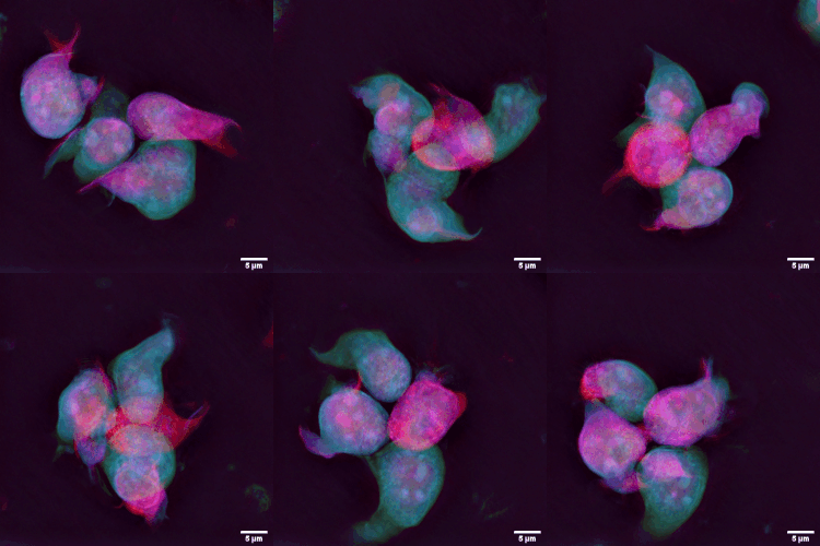
Spermatocytic Tumor
Spermatocytic tumor, previously called spermatocytic seminoma, is a neoplasm of the testis (''i.e.'' a tumour of the testis), and classified as a germ cell tumour. The name of the tumour comes from the similarity (under the microscope) between the small cells of the tumour and secondary spermatocytes. Signs and symptoms Spermatocytic tumor is a rare tumour, making up only one to two percent of all testicular germ cell tumours. Men presenting with this tumour are generally 50 to 60 years old, and its occurrence is rare in men under 30 years old. Most present with slow, painless testicular enlargement, which may involve both testes. Diagnosis Spermatocytic tumors are diagnosed based on tissue from orchiectomy (or partial orchiectomy), done for a lesion suspicious for cancer on medical imaging. The macroscopic appearance of the tumour is of a mutinodular grey-white to tan coloured mass with gelatinous, haemorrhagic and necrotic areas. The tumour may extend beyond the testis. ... [...More Info...] [...Related Items...] OR: [Wikipedia] [Google] [Baidu] |
Micrograph
A micrograph or photomicrograph is a photograph or digital image taken through a microscope or similar device to show a magnify, magnified image of an object. This is opposed to a macrograph or photomacrograph, an image which is also taken on a microscope but is only slightly magnified, usually less than 10 times. Micrography is the practice or art of using microscopes to make photographs. A micrograph contains extensive details of microstructure. A wealth of information can be obtained from a simple micrograph like behavior of the material under different conditions, the phases found in the system, failure analysis, grain size estimation, elemental analysis and so on. Micrographs are widely used in all fields of microscopy. Types Photomicrograph A light micrograph or photomicrograph is a micrograph prepared using an optical microscope, a process referred to as ''photomicroscopy''. At a basic level, photomicroscopy may be performed simply by connecting a camera to a micros ... [...More Info...] [...Related Items...] OR: [Wikipedia] [Google] [Baidu] |
Cell (biology)
The cell is the basic structural and functional unit of life forms. Every cell consists of a cytoplasm enclosed within a membrane, and contains many biomolecules such as proteins, DNA and RNA, as well as many small molecules of nutrients and metabolites.Cell Movements and the Shaping of the Vertebrate Body in Chapter 21 of Molecular Biology of the Cell '' fourth edition, edited by Bruce Alberts (2002) published by Garland Science. The Alberts text discusses how the "cellular building blocks" move to shape developing s. It is also common ... [...More Info...] [...Related Items...] OR: [Wikipedia] [Google] [Baidu] |
Actin
Actin is a protein family, family of Globular protein, globular multi-functional proteins that form microfilaments in the cytoskeleton, and the thin filaments in myofibril, muscle fibrils. It is found in essentially all Eukaryote, eukaryotic cells, where it may be present at a concentration of over 100 micromolar, μM; its mass is roughly 42 kDa, with a diameter of 4 to 7 nm. An actin protein is the monomeric Protein subunit, subunit of two types of filaments in cells: microfilaments, one of the three major components of the cytoskeleton, and thin filaments, part of the Muscle contraction, contractile apparatus in muscle cells. It can be present as either a free monomer called G-actin (globular) or as part of a linear polymer microfilament called F-actin (filamentous), both of which are essential for such important cellular functions as the Motility, mobility and contraction of cell (biology), cells during cell division. Actin participates in many important cellular pr ... [...More Info...] [...Related Items...] OR: [Wikipedia] [Google] [Baidu] |
Vimentin
Vimentin is a structural protein that in humans is encoded by the ''VIM'' gene. Its name comes from the Latin ''vimentum'' which refers to an array of flexible rods. Vimentin is a type III intermediate filament (IF) protein that is expressed in mesenchymal cells. IF proteins are found in all animal cells as well as bacteria. Intermediate filaments, along with tubulin-based microtubules and actin-based microfilaments, comprises the cytoskeleton. All IF proteins are expressed in a highly developmentally-regulated fashion; vimentin is the major cytoskeletal component of mesenchymal cells. Because of this, vimentin is often used as a marker of mesenchymally-derived cells or cells undergoing an epithelial-to-mesenchymal transition (EMT) during both normal development and metastatic progression. Structure A vimentin monomer, like all other intermediate filaments, has a central α-helical domain, capped on each end by non-helical amino (head) and carboxyl (tail) domains. T ... [...More Info...] [...Related Items...] OR: [Wikipedia] [Google] [Baidu] |
Placental Alkaline Phosphatase
Alkaline phosphatase, placental type also known as placental alkaline phosphatase (PLAP) is an allosteric enzyme that in humans is encoded by the ''ALPP'' gene. Gene There are at least four distinct but related alkaline phosphatases: intestinal (ALPI), placental (this enzyme), placental-like ( ALPPL2), and liver/bone/kidney ( ALPL) (tissue-nonspecific). The first three are located together on chromosome 2, whereas the tissue-nonspecific form is located on chromosome 1. The coding sequence for this form of alkaline phosphatase is unique in that the 3' untranslated region contains multiple copies of an Alu family repeat. In addition, this gene is polymorphic and three common alleles (type 1, type 2, and type 3) for this form of alkaline phosphatase have been well-characterized. Function Alkaline phosphatase, placental type is a membrane-bound glycosylated dimeric enzyme, also referred to as the heat-stable form, that is expressed primarily in the placenta, although it is clos ... [...More Info...] [...Related Items...] OR: [Wikipedia] [Google] [Baidu] |
Immunostaining
In biochemistry, immunostaining is any use of an antibody-based method to detect a specific protein in a sample. The term "immunostaining" was originally used to refer to the immunohistochemical staining of tissue sections, as first described by Albert Coons in 1941. However, immunostaining now encompasses a broad range of techniques used in histology, cell biology, and molecular biology that use antibody-based staining methods. Techniques Immunohistochemistry Immunohistochemistry or IHC staining of tissue sections (or immunocytochemistry, which is the staining of cells), is perhaps the most commonly applied immunostaining technique. While the first cases of IHC staining used fluorescent dyes (see '' immunofluorescence''), other non-fluorescent methods using enzymes such as peroxidase (see '' immunoperoxidase staining'') and alkaline phosphatase are now used. These enzymes are capable of catalysing reactions that give a coloured product that is easily detectable by light ... [...More Info...] [...Related Items...] OR: [Wikipedia] [Google] [Baidu] |
Intratubular Germ Cell Neoplasia
Germ cell neoplasia in situ (GCNIS) represents the precursor lesion for many types of testicular germ cell tumors. The term GCNIS was introduced with the 2016 edition of the WHO classification of urological tumours. GCNIS more accurate describes the lesion as it arises between the basement membrane and Sertoli cells (the cells that 'nurse' the developing germ cell). The common, unspecified variant of the entity was once considered to be a carcinoma in situ, although the term "carcinoma ''in situ''" is now largely historical as it is not an accurate description of the process.Mills, S (ed.) 2009.Sternberg's Diagnostic Pathology. 5th Edition. Classification The World Health Organization classification of testicular tumoursEble J.N., Sauter G., Epstein J.I., Sesterhenn I.A. (Eds.): World Health Organization Classification of Tumours. Pathology and Genetics of Tumours of the Urinary System and Male Genital Organs. IARC Press: Lyon 2004. subdivides ITGCN into (1) a more common, uns ... [...More Info...] [...Related Items...] OR: [Wikipedia] [Google] [Baidu] |
Apoptosis
Apoptosis (from grc, ἀπόπτωσις, apóptōsis, 'falling off') is a form of programmed cell death that occurs in multicellular organisms. Biochemical events lead to characteristic cell changes ( morphology) and death. These changes include blebbing, cell shrinkage, nuclear fragmentation, chromatin condensation, DNA fragmentation, and mRNA decay. The average adult human loses between 50 and 70 billion cells each day due to apoptosis. For an average human child between eight and fourteen years old, approximately twenty to thirty billion cells die per day. In contrast to necrosis, which is a form of traumatic cell death that results from acute cellular injury, apoptosis is a highly regulated and controlled process that confers advantages during an organism's life cycle. For example, the separation of fingers and toes in a developing human embryo occurs because cells between the digits undergo apoptosis. Unlike necrosis, apoptosis produces cell fragments called apopt ... [...More Info...] [...Related Items...] OR: [Wikipedia] [Google] [Baidu] |
Mitosis
In cell biology, mitosis () is a part of the cell cycle in which replicated chromosomes are separated into two new nuclei. Cell division by mitosis gives rise to genetically identical cells in which the total number of chromosomes is maintained. Therefore, mitosis is also known as equational division. In general, mitosis is preceded by S phase of interphase (during which DNA replication occurs) and is often followed by telophase and cytokinesis; which divides the cytoplasm, organelles and cell membrane of one cell into two new cells containing roughly equal shares of these cellular components. The different stages of mitosis altogether define the mitotic (M) phase of an animal cell cycle—the division of the mother cell into two daughter cells genetically identical to each other. The process of mitosis is divided into stages corresponding to the completion of one set of activities and the start of the next. These stages are preprophase (specific to plant cells), p ... [...More Info...] [...Related Items...] OR: [Wikipedia] [Google] [Baidu] |
Lymphocyte
A lymphocyte is a type of white blood cell (leukocyte) in the immune system of most vertebrates. Lymphocytes include natural killer cells (which function in cell-mediated, cytotoxic innate immunity), T cells (for cell-mediated, cytotoxic adaptive immunity), and B cells (for humoral, antibody-driven adaptive immunity). They are the main type of cell found in lymph, which prompted the name "lymphocyte". Lymphocytes make up between 18% and 42% of circulating white blood cells. Types The three major types of lymphocyte are T cells, B cells and natural killer (NK) cells. Lymphocytes can be identified by their large nucleus. T cells and B cells T cells ( thymus cells) and B cells (bone marrow- or bursa-derived cells) are the major cellular components of the adaptive immune response. T cells are involved in cell-mediated immunity, whereas B cells are primarily responsible for humoral immunity (relating to antibodies). The function of T cells and B cells is to recognize ... [...More Info...] [...Related Items...] OR: [Wikipedia] [Google] [Baidu] |
Seminoma
A seminoma is a germ cell tumor of the testicle or, more rarely, the mediastinum or other extra-gonadal locations. It is a malignant neoplasm and is one of the most treatable and curable cancers, with a survival rate above 95% if discovered in early stages. Testicular seminoma originates in the germinal epithelium of the seminiferous tubules. About half of germ cell tumors of the testicles are seminomas. Treatment usually requires removal of one testicle. However, fertility usually isn't affected. All other sexual functions will remain intact. Signs and symptoms The average age of diagnosis is between 35 and 50 years. This is about 5 to 10 years older than men with other germ cell tumors of the testes. In most cases, they produce masses that are readily felt on testicular self-examination; however, in up to 11 percent of cases, there may be no mass able to be felt, or there may be testicular atrophy. Testicular pain is reported in up to one fifth of cases. Low back pain m ... [...More Info...] [...Related Items...] OR: [Wikipedia] [Google] [Baidu] |
Nucleoli
The nucleolus (, plural: nucleoli ) is the largest structure in the nucleus of eukaryotic cells. It is best known as the site of ribosome biogenesis, which is the synthesis of ribosomes. The nucleolus also participates in the formation of signal recognition particles and plays a role in the cell's response to stress. Nucleoli are made of proteins, DNA and RNA, and form around specific chromosomal regions called nucleolar organizing regions. Malfunction of nucleoli can be the cause of several human conditions called "nucleolopathies" and the nucleolus is being investigated as a target for cancer chemotherapy. History The nucleolus was identified by bright-field microscopy during the 1830s. Little was known about the function of the nucleolus until 1964, when a study of nucleoli by John Gurdon and Donald Brown in the African clawed frog ''Xenopus laevis'' generated increasing interest in the function and detailed structure of the nucleolus. They found that 25% of the frog ... [...More Info...] [...Related Items...] OR: [Wikipedia] [Google] [Baidu] |


