|
Pemphigus Vulgaris
Pemphigus vulgaris is a rare chronic blistering skin disease and the most common form of pemphigus. Pemphigus was derived from the Greek word ''pemphix'', meaning blister. It is classified as a type II hypersensitivity reaction in which antibodies are formed against desmosomes, components of the skin that function to keep certain layers of skin bound to each other. As desmosomes are attacked, the layers of skin separate and the clinical picture resembles a blister. These blisters are due to acantholysis, or breaking apart of intercellular connections through an autoantibody-mediated response. Over time the condition inevitably progresses without treatment: lesions increase in size and distribution throughout the body, behaving physiologically like a severe burn. Before the advent of modern treatments, mortality for the disease was close to 90%. Today, the mortality rate with treatment is between 5-15% due to the introduction of corticosteroids as primary treatment. Nevertheless ... [...More Info...] [...Related Items...] OR: [Wikipedia] [Google] [Baidu] |
Micrograph
A micrograph or photomicrograph is a photograph or digital image taken through a microscope or similar device to show a magnified image of an object. This is opposed to a macrograph or photomacrograph, an image which is also taken on a microscope but is only slightly magnified, usually less than 10 times. Micrography is the practice or art of using microscopes to make photographs. A micrograph contains extensive details of microstructure. A wealth of information can be obtained from a simple micrograph like behavior of the material under different conditions, the phases found in the system, failure analysis, grain size estimation, elemental analysis and so on. Micrographs are widely used in all fields of microscopy. Types Photomicrograph A light micrograph or photomicrograph is a micrograph prepared using an optical microscope, a process referred to as ''photomicroscopy''. At a basic level, photomicroscopy may be performed simply by connecting a camera to a microscope ... [...More Info...] [...Related Items...] OR: [Wikipedia] [Google] [Baidu] |
Erosion (dermatology)
A skin condition, also known as cutaneous condition, is any medical condition that affects the integumentary system—the organ system that encloses the body and includes skin, nails, and related muscle and glands. The major function of this system is as a barrier against the external environment. Conditions of the human integumentary system constitute a broad spectrum of diseases, also known as dermatoses, as well as many nonpathologic states (like, in certain circumstances, melanonychia and racquet nails). While only a small number of skin diseases account for most visits to the physician, thousands of skin conditions have been described. Classification of these conditions often presents many nosological challenges, since underlying causes and pathogenetics are often not known. Therefore, most current textbooks present a classification based on location (for example, conditions of the mucous membrane), morphology ( chronic blistering conditions), cause (skin conditions r ... [...More Info...] [...Related Items...] OR: [Wikipedia] [Google] [Baidu] |
Skin Biopsy
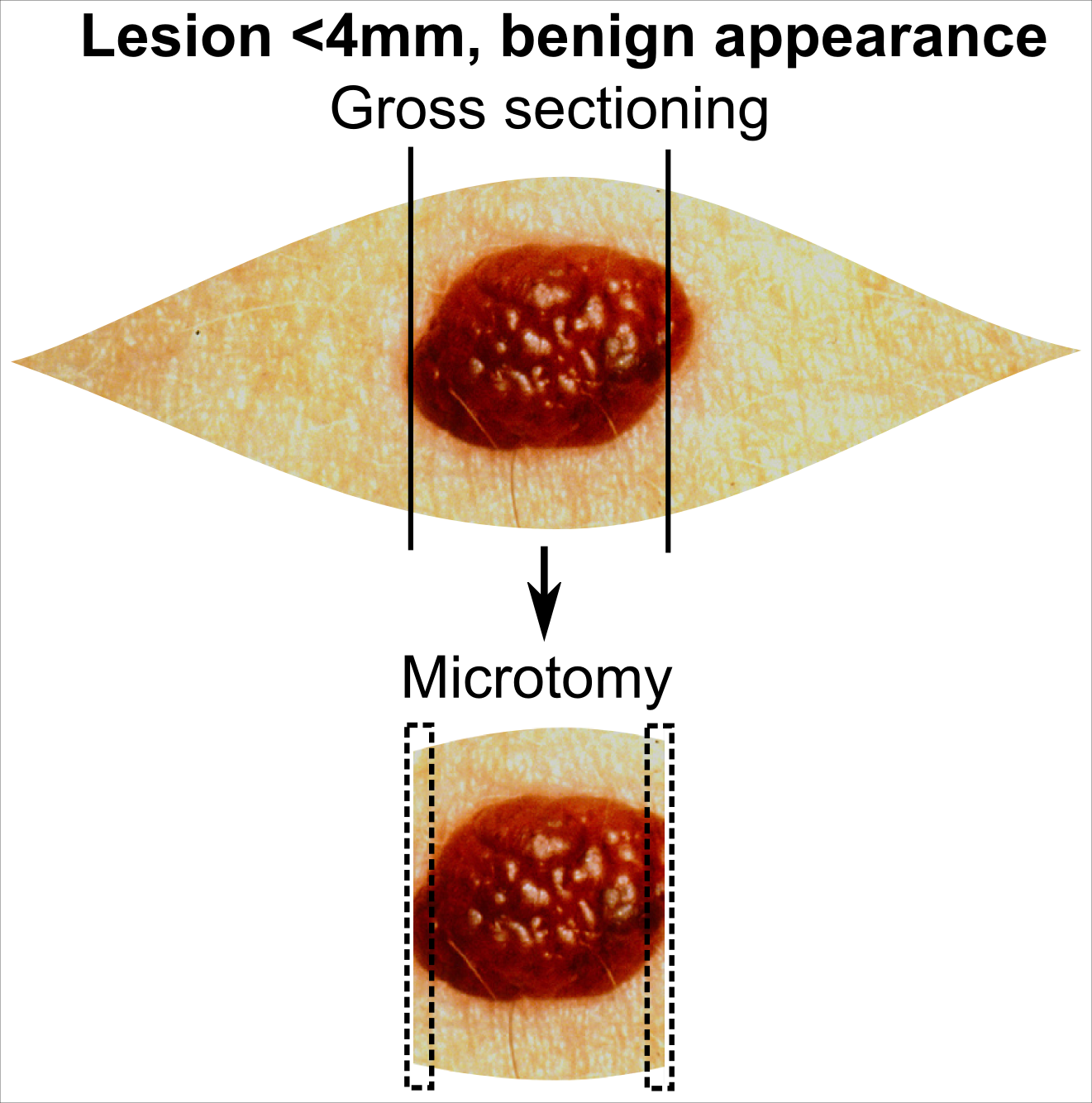
Skin biopsy is a biopsy technique in which a skin lesion is removed to be sent to a pathologist to render a microscopic diagnosis. It is usually done under local anesthetic in a physician's office, and results are often available in 4 to 10 days. It is commonly performed by dermatologists. Skin biopsies are also done by family physicians, internists, surgeons, and other specialties. However, performed incorrectly, and without appropriate clinical information, a pathologist's interpretation of a skin biopsy can be severely limited, and therefore doctors and patients may forgo traditional biopsy techniques and instead choose Mohs surgery. There are four main types of skin biopsies: shave biopsy, punch biopsy, excisional biopsy, and incisional biopsy. The choice of the different skin biopsies is dependent on the suspected diagnosis of the skin lesion. Like most biopsies, patient consent and anesthesia (usually lidocaine injected into the skin) are prerequisites. Types Shave bio ... [...More Info...] [...Related Items...] OR: [Wikipedia] [Google] [Baidu] |
Dermis
The dermis or corium is a layer of skin between the epidermis (with which it makes up the cutis) and subcutaneous tissues, that primarily consists of dense irregular connective tissue and cushions the body from stress and strain. It is divided into two layers, the superficial area adjacent to the epidermis called the papillary region and a deep thicker area known as the reticular dermis.James, William; Berger, Timothy; Elston, Dirk (2005). ''Andrews' Diseases of the Skin: Clinical Dermatology'' (10th ed.). Saunders. Pages 1, 11–12. . The dermis is tightly connected to the epidermis through a basement membrane. Structural components of the dermis are collagen, elastic fibers, and extrafibrillar matrix.Marks, James G; Miller, Jeffery (2006). ''Lookingbill and Marks' Principles of Dermatology'' (4th ed.). Elsevier Inc. Page 8–9. . It also contains mechanoreceptors that provide the sense of touch and thermoreceptors that provide the sense of heat. In addition, hair foll ... [...More Info...] [...Related Items...] OR: [Wikipedia] [Google] [Baidu] |
Bullous Pemphigoid
Bullous pemphigoid (type of pemphigoid) is an autoimmune pruritic skin disease which typically occurs in people aged over 60, that may involve the formation of blisters ( bullae) in the space between the epidermal and dermal skin layers. It is classified as a type II hypersensitivity reaction, which involves formation of anti- hemidesmosome antibodies, causing a loss of keratinocytes to basement membrane adhesion. Signs and symptoms Clinically, the earliest lesions may appear as a hives-like red raised rash, but could also appear dermatitic, targetoid, lichenoid, nodular, or even without a rash ( essential pruritus). Tense bullae eventually erupt, most commonly at the inner thighs and upper arms, but the trunk and extremities are frequently both involved. Any part of the skin surface can be involved. Oral lesions are present in a minority of cases. The disease may be acute, but can last from months to years with periods of exacerbation and remission. Several other skin ... [...More Info...] [...Related Items...] OR: [Wikipedia] [Google] [Baidu] |
Transudate
Transudate is extravascular fluid with low protein content and a low specific gravity (< 1.012). It has low nucleated cell counts (less than 500 to 1000 /microliter) and the primary cell types are mononuclear cells: s, s and l cells. For instance, an of |
Basement Membrane
The basement membrane is a thin, pliable sheet-like type of extracellular matrix that provides cell and tissue support and acts as a platform for complex signalling. The basement membrane sits between epithelial tissues including mesothelium and endothelium, and the underlying connective tissue. Structure As seen with the electron microscope, the basement membrane is composed of two layers, the basal lamina and the reticular lamina. The underlying connective tissue attaches to the basal lamina with collagen VII anchoring fibrils and fibrillin microfibrils. The basal lamina layer can further be subdivided into two layers based on their visual appearance in electron microscopy. The lighter-colored layer closer to the epithelium is called the lamina lucida, while the denser-colored layer closer to the connective tissue is called the lamina densa. The electron-dense lamina densa layer is about 30–70 nanometers thick and consists of an underlying network of reticular col ... [...More Info...] [...Related Items...] OR: [Wikipedia] [Google] [Baidu] |
Epidermis (skin)
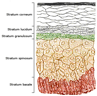
The epidermis is the outermost of the three layers that comprise the skin, the inner layers being the dermis and hypodermis. The epidermis layer provides a barrier to infection from environmental pathogens and regulates the amount of water released from the body into the atmosphere through transepidermal water loss. The epidermis is composed of multiple layers of flattened cells that overlie a base layer ( stratum basale) composed of columnar cells arranged perpendicularly. The layers of cells develop from stem cells in the basal layer. The human epidermis is a familiar example of epithelium, particularly a stratified squamous epithelium. The word epidermis is derived through Latin , itself and . Something related to or part of the epidermis is termed epidermal. Structure Cellular components The epidermis primarily consists of keratinocytes ( proliferating basal and differentiated suprabasal), which comprise 90% of its cells, but also contains melanocytes, Langerha ... [...More Info...] [...Related Items...] OR: [Wikipedia] [Google] [Baidu] |
Keratinocyte
Keratinocytes are the primary type of cell found in the epidermis, the outermost layer of the skin. In humans, they constitute 90% of epidermal skin cells. Basal cells in the basal layer (''stratum basale'') of the skin are sometimes referred to as basal keratinocytes. Keratinocytes form a barrier against environmental damage by heat, UV radiation, water loss, pathogenic bacteria, fungi, parasites, and viruses. A number of structural proteins, enzymes, lipids, and antimicrobial peptides contribute to maintain the important barrier function of the skin. Keratinocytes differentiate from epidermal stem cells in the lower part of the epidermis and migrate towards the surface, finally becoming corneocytes and eventually be shed off, which happens every 40 to 56 days in humans. Function The primary function of keratinocytes is the formation of a barrier against environmental damage by heat, UV radiation, water loss, pathogenic bacteria, fungi, parasites, and viruses. Pathogens ... [...More Info...] [...Related Items...] OR: [Wikipedia] [Google] [Baidu] |
Desmosomes
A desmosome (; "binding body"), also known as a macula adherens (plural: maculae adherentes) (Latin for ''adhering spot''), is a cell structure specialized for cell-to- cell adhesion. A type of junctional complex, they are localized spot-like adhesions randomly arranged on the lateral sides of plasma membranes. Desmosomes are one of the stronger cell-to-cell adhesion types and are found in tissue that experience intense mechanical stress, such as cardiac muscle tissue, bladder tissue, gastrointestinal mucosa, and epithelia. Structure Desmosomes are composed of desmosome-intermediate filament complexes (DIFC), which is a network of cadherin proteins, linker proteins and intermediate filaments. The DIFCs can be broken into three regions: the extracellular core region, or desmoglea, the outer dense plaque, or ODP, and the inner dense plaque, or IDP. The extracellular core region, approximately 34 nm in length, contains desmoglein and desmocollin, which are in the cadherin ... [...More Info...] [...Related Items...] OR: [Wikipedia] [Google] [Baidu] |
Desmoglein 3
Desmoglein-3 is a protein that in humans is encoded by the ''DSG3'' gene. In the skin epidermis Desmoglein-3 is expressed in the basal lower layers of the epidermis, and dominates in terms of expression on mucosal surfaces compared to Desmoglein-1. Function Desmosomes are cell-cell junctions between epithelial, myocardial, and certain other cell types. Desmoglein 3 is a calcium-binding transmembrane glycoprotein component of desmosomes in vertebrate epithelial cells. Currently, four desmoglein subfamily members have been identified and all are members of the cadherin cell adhesion molecule superfamily. These desmoglein gene family members are located in a cluster on chromosome 18. This protein, along with Desmoglein-1, has been identified as the autoantigen of the autoimmune skin blistering disease pemphigus vulgaris. The mucosal dominant form of pemphigus vulgaris only involves antibodies against Desmoglein-3 and causes mucosal erosions, but no skin lesions. Desmoglein-3 serve ... [...More Info...] [...Related Items...] OR: [Wikipedia] [Google] [Baidu] |
Desmoglein 1
Desmoglein-1 is a protein that in humans is encoded by the ''DSG1'' gene. Desmoglein-1 is expressed everywhere in the skin epidermis, but mainly it is expressed in the superficial upper layers of the skin epidermis. Function Desmosomes are Cell junction, cell-cell junctions between epithelial, myocardial and certain other cell types. Desmoglein-1 is a calcium-binding transmembrane glycoprotein component of desmosomes in vertebrate epithelial cells. Currently, four desmoglein subfamily members have been identified and all are members of the cadherin cell adhesion molecule Protein superfamily, superfamily. These desmoglein gene family members are located in a gene cluster, cluster on Chromosome 18 (human), chromosome 18. The protein encoded by this gene has been identified as the autoantigen of the autoimmune disease, autoimmune skin blistering disease pemphigus foliaceus. It has been found that desmoglein-1 is the target antigen in majority of the cases linked to IgG/IgA pemphigus ... [...More Info...] [...Related Items...] OR: [Wikipedia] [Google] [Baidu] |


