|
Orbital Septum
In anatomy, the orbital septum (palpebral fascia) is a membranous sheet that acts as the anterior (frontal) boundary of the orbit. It extends from the orbital rims to the eyelids. It forms the fibrous portion of the eyelids. Structure In the upper eyelid, the orbital septum blends with the tendon of the levator palpebrae superioris, and in the lower eyelid with the tarsal plate. When the eyes are closed, the whole orbital opening is covered by the septum and tarsi. Medially it is thin, and, becoming separated from the medial palpebral ligament, attaches to the lacrimal bone at its posterior crest. The medial ligament and its much weaker lateral counterpart, attached to the septum and orbit, keep the lids stable as the eye moves. The septum is perforated by the vessels and nerves which pass from the orbital cavity to the face and scalp. Clinical significance Orbital septum supports the orbital contents located posterior to it, especially orbital fat. The septum can be w ... [...More Info...] [...Related Items...] OR: [Wikipedia] [Google] [Baidu] |
Anatomy
Anatomy () is the branch of morphology concerned with the study of the internal structure of organisms and their parts. Anatomy is a branch of natural science that deals with the structural organization of living things. It is an old science, having its beginnings in prehistoric times. Anatomy is inherently tied to developmental biology, embryology, comparative anatomy, evolutionary biology, and phylogeny, as these are the processes by which anatomy is generated, both over immediate and long-term timescales. Anatomy and physiology, which study the structure and function of organisms and their parts respectively, make a natural pair of related disciplines, and are often studied together. Human anatomy is one of the essential basic sciences that are applied in medicine, and is often studied alongside physiology. Anatomy is a complex and dynamic field that is constantly evolving as discoveries are made. In recent years, there has been a significant increase in the use of ... [...More Info...] [...Related Items...] OR: [Wikipedia] [Google] [Baidu] |
Blepharoplasty
Blepharoplasty (Greek: ''blepharon'', "eyelid" + ''plassein'' "to form") is the plastic surgery operation for correcting defects, deformities, and disfigurations of the eyelids; and for aesthetically modifying the eye region of the face. With the excision and the removal, or the repositioning (or both) of excess tissues, such as skin and adipocyte fat, and the reinforcement of the corresponding muscle and tendon tissues, the blepharoplasty procedure resolves functional and cosmetic problems of the periorbita, which is the area from the eyebrow to the upper portion of the cheek. The procedure is more common among women, who accounted for approximately 85% of blepharoplasty procedures in 2014 in the US and 88% of such procedures in the UK. The operative goals of a blepharoplastic procedure are the restoration of the correct functioning to the affected eyelid(s) and the restoration of the aesthetics of the eye-region of the face, which are achieved by eliminating excess skin from ... [...More Info...] [...Related Items...] OR: [Wikipedia] [Google] [Baidu] |
Periosteum
The periosteum is a membrane that covers the outer surface of all bones, except at the articular surfaces (i.e. the parts within a joint space) of long bones. (At the joints of long bones the bone's outer surface is lined with "articular cartilage", a type of hyaline cartilage.) Endosteum lines the inner surface of the medullary cavity of all long bones. Structure The periosteum consists of an outer fibrous layer, and an inner ''cambium layer'' (or osteogenic layer). The fibrous layer is of dense irregular connective tissue, containing fibroblasts, while the cambium layer is highly cellular containing progenitor cells that develop into osteoblasts. These osteoblasts are responsible for increasing the width of a long bone (the length of a long bone is controlled by the epiphyseal plate) and the overall size of the other bone types. After a bone fracture A bone fracture (abbreviated FRX or Fx, Fx, or #) is a medical condition in which there is a partial or complete break ... [...More Info...] [...Related Items...] OR: [Wikipedia] [Google] [Baidu] |
Cerebral Abscess
Cerebral may refer to: * Of or relating to the brain * Cerebrum, the largest and uppermost part of the brain * Cerebral cortex, the outer layer of the cerebrum * Retroflex consonant, also referred to as a cerebral consonant, a type of consonant sound used in some languages * Intellectual An intellectual is a person who engages in critical thinking, research, and Human self-reflection, reflection about the nature of reality, especially the nature of society and proposed solutions for its normative problems. Coming from the wor ..., rather than emotional See also * {{Disambiguation ... [...More Info...] [...Related Items...] OR: [Wikipedia] [Google] [Baidu] |
Meningitis
Meningitis is acute or chronic inflammation of the protective membranes covering the brain and spinal cord, collectively called the meninges. The most common symptoms are fever, intense headache, vomiting and neck stiffness and occasionally photophobia. Other symptoms include confusion or altered consciousness, nausea, and an inability to tolerate loud noises. Young children often exhibit only nonspecific symptoms, such as irritability, drowsiness, or poor feeding. A non-blanching rash (a rash that does not fade when a glass is rolled over it) may also be present. The inflammation may be caused by infection with viruses, bacteria, fungi or parasites. Non-infectious causes include malignancy (cancer), subarachnoid hemorrhage, chronic inflammatory disease ( sarcoidosis) and certain drugs. Meningitis can be life-threatening because of the inflammation's proximity to the brain and spinal cord; therefore, the condition is classified as a medical emergency. A lumba ... [...More Info...] [...Related Items...] OR: [Wikipedia] [Google] [Baidu] |
Lamina Papyracea
The orbital lamina of ethmoid bone (or lamina papyracea or orbital lamina) is a smooth, oblong, paper-thin bone plate which forms the lateral wall of the labyrinth of the ethmoid bone. It covers the middle and posterior ethmoidal cells, and forms a large part of the medial wall of the orbit. It articulates above with the orbital plate of the frontal bone, below with the maxilla In vertebrates, the maxilla (: maxillae ) is the upper fixed (not fixed in Neopterygii) bone of the jaw formed from the fusion of two maxillary bones. In humans, the upper jaw includes the hard palate in the front of the mouth. The two maxil ... and the orbital process of palatine bone, in front with the lacrimal, and behind with the sphenoid. Its name lamina papyracea is an appropriate description, as this part of the ethmoid bone is paper-thin and fractures easily. A fracture here could cause entrapment of the medial rectus muscle. Additional images File:Slide4fen.JPG, Orbital lamina of et ... [...More Info...] [...Related Items...] OR: [Wikipedia] [Google] [Baidu] |
Ethmoidal Sinus
The ethmoid sinuses or ethmoid air cells of the ethmoid bone are one of the four paired paranasal sinuses. Unlike the other three pairs of paranasal sinuses which consist of one or two large cavities, the ethmoidal sinuses entail a number of small air-filled cavities ("air cells"). The cells are located within the labyrinth of ethmoid, lateral mass (labyrinth) of each ethmoid bone and are variable in both size and number.Illustrated Anatomy of the Head and Neck, Fehrenbach and Herring, Elsevier, 2012, page 64 The cells are grouped into anterior, middle, and posterior groups; the groups differ in their drainage modalities, though all ultimately drain into either the Superior nasal meatus, superior or the middle nasal meatus of the lateral wall of the nasal cavity. Structure The ethmoid air cells consist of numerous thin-walled cavities in the ethmoidal labyrinthOtorhinolaryngology, Head and Neck Surgery, Anniko, Springer, 2010, page 188 that represent invaginations of the mucous mem ... [...More Info...] [...Related Items...] OR: [Wikipedia] [Google] [Baidu] |
Orbital Cellulitis
Orbital cellulitis is inflammation of eye tissues behind the orbital septum. It is most commonly caused by an acute spread of infection into the eye socket from either the adjacent sinuses or through the blood. It may also occur after trauma. When it affects the rear of the eye, it is known as retro-orbital cellulitis. Without proper treatment, orbital cellulitis may lead to serious consequences, including permanent loss of vision or even death. Signs and symptoms Orbital cellulitis commonly presents with painful eye movement, sudden vision loss, chemosis, bulging of the infected eye, and limited eye movement. Along with these symptoms, patients typically have redness and swelling of the eyelid, pain, discharge, inability to open the eye, occasional fever and lethargy. Complications Complications include hearing loss, blood infection, meningitis, cavernous sinus thrombosis, cerebral abscess, and blindness. It is possible that children experience more severe complications d ... [...More Info...] [...Related Items...] OR: [Wikipedia] [Google] [Baidu] |
Preseptal Cellulitis
Periorbital cellulitis, or preseptal cellulitis, is an inflammation and infection of the eyelid and portions of skin around the eye anterior to the orbital septum. It may be caused by breaks in the skin around the eye, and subsequent spread to the eyelid; infection of the Paranasal sinuses, sinuses around the nose (sinusitis); or from spread of an infection elsewhere through the blood. Signs and symptoms Periorbital cellulitis must be differentiated from orbital cellulitis, which is an emergency and requires intravenous (IV) antibiotics. In contrast to orbital cellulitis, patients with periorbital cellulitis do not have bulging of the eye (proptosis), limited eye movement (ophthalmoplegia), pain on eye movement, or loss of vision. If any of these features is present, one must assume that the patient has orbital cellulitis and begin treatment with IV antibiotics. computed tomography, CT scan may be done to delineate the extension of the infection. Affected individuals may experien ... [...More Info...] [...Related Items...] OR: [Wikipedia] [Google] [Baidu] |
Scalp
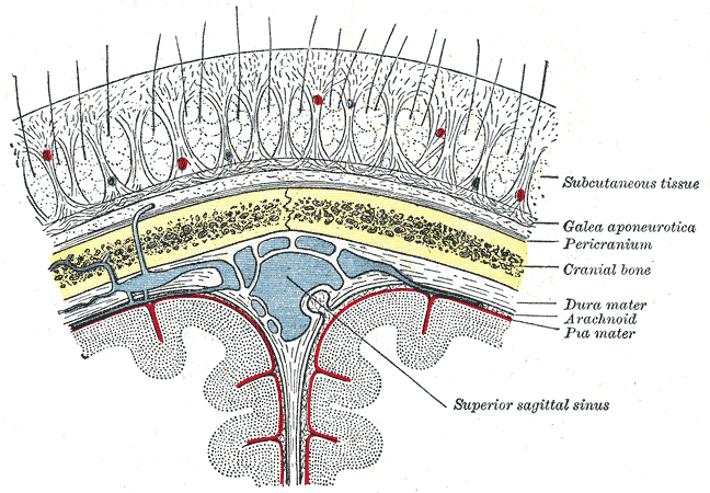
The scalp is the area of the head where head hair grows. It is made up of skin, layers of connective and fibrous tissues, and the membrane of the skull. Anatomically, the scalp is part of the epicranium, a collection of structures covering the cranium. The scalp is bordered by the face at the front, and by the neck at the sides and back. The scientific study of hair and scalp is called trichology. Structure Layers The scalp is usually described as having five layers, which can be remembered using the mnemonic 'SCALP': * S: Skin. The skin of the scalp contains numerous hair follicles and sebaceous glands. * C: Connective tissue. A dense subcutaneous layer of fat and fibrous tissue that lies beneath the skin, containing the nerves and vessels of the scalp. * A: Aponeurosis. The epicranial aponeurosis or galea aponeurotica is a tough layer of dense fibrous tissue which anchors the above layers in place. It runs from the frontalis muscle anteriorly to the occipitalis ... [...More Info...] [...Related Items...] OR: [Wikipedia] [Google] [Baidu] |
Biological Membrane
A biological membrane, biomembrane or cell membrane is a selectively permeable membrane that separates the interior of a cell from the external environment or creates intracellular compartments by serving as a boundary between one part of the cell and another. Biological membranes, in the form of eukaryotic cell membranes, consist of a phospholipid bilayer with embedded, integral and peripheral proteins used in communication and transportation of chemicals and ions. The bulk of lipids in a cell membrane provides a fluid matrix for proteins to rotate and laterally diffuse for physiological functioning. Proteins are adapted to high membrane fluidity environment of the lipid bilayer with the presence of an annular lipid shell, consisting of lipid molecules bound tightly to the surface of integral membrane proteins. The cell membranes are different from the isolating tissues formed by layers of cells, such as mucous membranes, basement membranes, and serous membranes. ... [...More Info...] [...Related Items...] OR: [Wikipedia] [Google] [Baidu] |
Face
The face is the front of the head that features the eyes, nose and mouth, and through which animals express many of their emotions. The face is crucial for human identity, and damage such as scarring or developmental deformities may affect the psyche adversely. Structure The front of the human head is called the face. It includes several distinct areas, of which the main features are: *The forehead, comprising the skin beneath the hairline, bordered laterally by the temples and inferiorly by eyebrows and ears *The eyes, sitting in the orbit and protected by eyelids and eyelashes * The distinctive human nose shape, nostrils, and nasal septum *The cheeks, covering the maxilla and mandible (or jaw), the extremity of which is the chin *The mouth, with the upper lip divided by the philtrum, sometimes revealing the teeth Facial appearance is vital for human recognition and communication. Facial muscles in humans allow expression of emotions. The face is itself a highly s ... [...More Info...] [...Related Items...] OR: [Wikipedia] [Google] [Baidu] |


