|
Node Of Ranvier
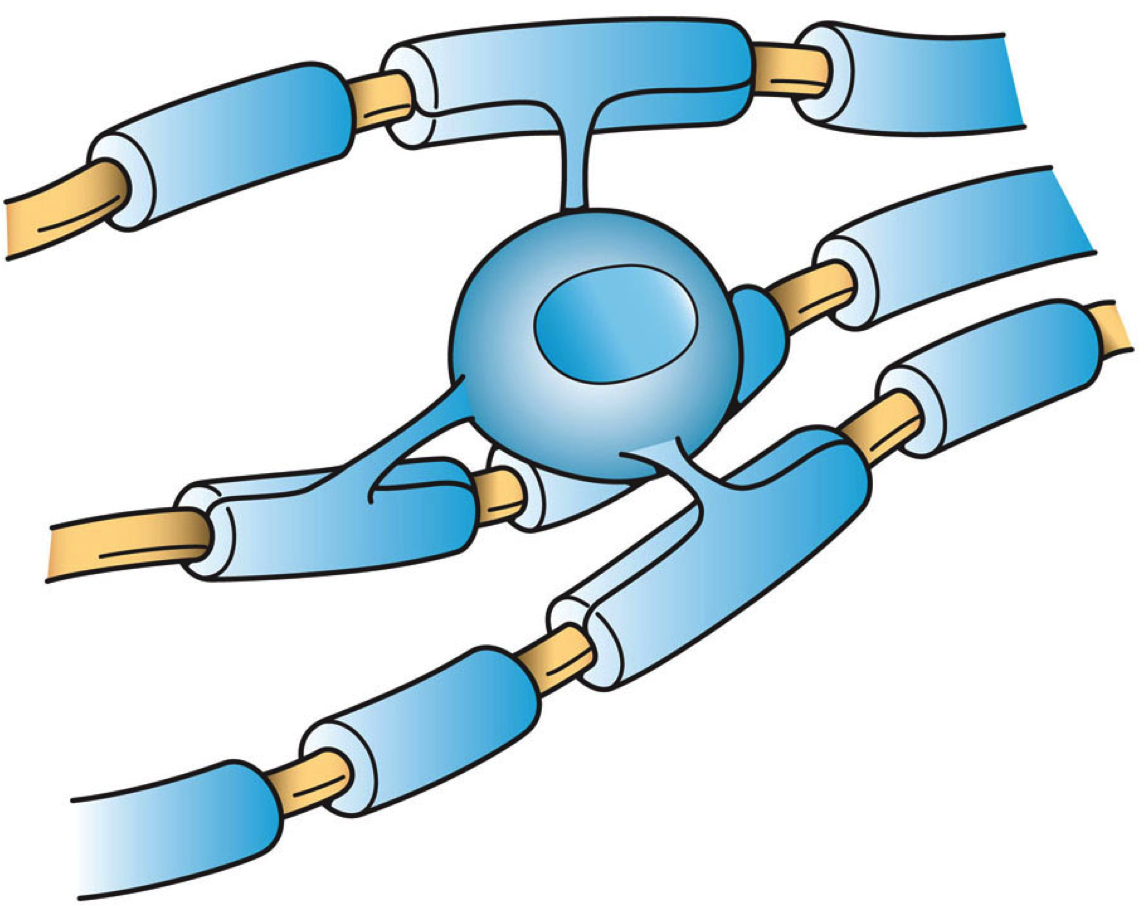
Nodes of Ranvier ( ), also known as myelin-sheath gaps, occur along a myelinated axon where the axolemma is exposed to the extracellular space. Nodes of Ranvier are uninsulated axonal domains that are high in sodium and potassium ion channels complexed with cell adhesion molecules, allowing them to participate in the exchange of ions required to regenerate the action potential. Nerve conduction in myelinated axons is referred to as saltatory conduction () due to the manner in which the action potential seems to "jump" from one node to the next along the axon. This results in faster conduction of the action potential. The nodes of Ranvier are present in both the Peripheral nervous system, peripheral and Central nervous system, central nervous systems. Overview The nodes are primarily composed of sodium and potassium voltage-gated ion channels; Cell adhesion molecule, CAMs such as neurofascin-186 and NRCAM, NrCAM; and cytoskeletal adaptor proteins such as Ankyrin, ankyrin-G and Sp ... [...More Info...] [...Related Items...] OR: [Wikipedia] [Google] [Baidu] |
Nervous System
In biology, the nervous system is the complex system, highly complex part of an animal that coordinates its behavior, actions and sense, sensory information by transmitting action potential, signals to and from different parts of its body. The nervous system detects environmental changes that impact the body, then works in tandem with the endocrine system to respond to such events. Nervous tissue first arose in Ediacara biota, wormlike organisms about 550 to 600 million years ago. In Vertebrate, vertebrates, it consists of two main parts, the central nervous system (CNS) and the peripheral nervous system (PNS). The CNS consists of the brain and spinal cord. The PNS consists mainly of nerves, which are enclosed bundles of the long fibers, or axons, that connect the CNS to every other part of the body. Nerves that transmit signals from the brain are called motor nerves (efferent), while those nerves that transmit information from the body to the CNS are called sensory nerves (aff ... [...More Info...] [...Related Items...] OR: [Wikipedia] [Google] [Baidu] |
NRCAM
Neuronal cell adhesion molecule is a protein that in humans is encoded by the ''NRCAM'' gene. Cell adhesion molecules Cell adhesion molecules (CAMs) are a subset of cell surface proteins that are involved in the binding of cells with other cells or with the extracellular matrix (ECM), in a process called cell adhesion. In essence, CAMs help cells stick to each ... (CAMs) are members of the immunoglobulin superfamily. This gene encodes a neuronal cell adhesion molecule with multiple immunoglobulin-like C2-type domains and fibronectin type-III domains. This ankyrin-binding protein is involved in neuron-neuron adhesion and promotes directional signaling during axonal cone growth. This gene is also expressed in non-neural tissues and may play a general role in cell-cell communication via signaling from its intracellular domain to the actin cytoskeleton during directional cell migration. Allelic variants of this gene have been associated with autism and addiction vulnerability. Alt ... [...More Info...] [...Related Items...] OR: [Wikipedia] [Google] [Baidu] |
Neuron (journal)
''Neuron'' is a biweekly peer-reviewed scientific journal published by Cell Press, an imprint of Elsevier. Established in 1988, it covers neuroscience and related biological processes. The current editor-in-chief An editor-in-chief (EIC), also known as lead editor or chief editor, is a publication's editorial leader who has final responsibility for its operations and policies. The editor-in-chief heads all departments of the organization and is held accoun ... is Mariela Zirlinger. The founding editors were Lily Jan, A. James Hudspeth, Louis Reichardt, Roger Nicoll, and Zach Hall. A past editor-in-chief was Katja Brose. Transcript and video available. Click on "Transcript" for text. * See alsoA Career in Science Editing: Katja BroseEditor in Chief, Neuron References Neuroscience journals Cell Press academic journals Academic journals established in 1988 English-language journals Biweekly journals {{neuroscience-journal-stub ... [...More Info...] [...Related Items...] OR: [Wikipedia] [Google] [Baidu] |
Astrocytes
Astrocytes (from Ancient Greek , , "star" and , , "cavity", "cell"), also known collectively as astroglia, are characteristic star-shaped glial cells in the brain and spinal cord. They perform many functions, including biochemical control of endothelial cells that form the blood–brain barrier, provision of nutrients to the nervous tissue, maintenance of extracellular ion balance, regulation of cerebral blood flow, and a role in the repair and Glial scar, scarring process of the brain and spinal cord following infection and traumatic injuries. The proportion of astrocytes in the brain is not well defined; depending on the counting technique used, studies have found that the astrocyte proportion varies by region and ranges from 20% to around 40% of all glia. Another study reports that astrocytes are the most numerous cell type in the brain. Astrocytes are the major source of cholesterol in the central nervous system. Apolipoprotein E transports cholesterol from astrocytes to neuro ... [...More Info...] [...Related Items...] OR: [Wikipedia] [Google] [Baidu] |
Schwann Cell
Schwann cells or neurolemmocytes (named after German physiologist Theodor Schwann) are the principal glia of the peripheral nervous system (PNS). Glial cells function to support neurons and in the PNS, also include Satellite glial cell, satellite cells, olfactory ensheathing cells, enteric glia and glia that reside at sensory nerve endings, such as the Pacinian corpuscle. The two types of Schwann cells are Myelin, myelinating and Nonmyelinating Schwann cell, nonmyelinating. Myelinating Schwann cells wrap around axons of motor and sensory neurons to form the myelin sheath. The Schwann cell promoter is present in the Upstream and downstream (DNA), downstream region of the human dystrophin gene that gives shortened Transcription (biology), transcript that are again synthesized in a tissue-specific manner. During the development of the PNS, the regulatory mechanisms of myelination are controlled by feedforward interaction of specific genes, influencing transcriptional cascades and sh ... [...More Info...] [...Related Items...] OR: [Wikipedia] [Google] [Baidu] |
Microvillus
Microvilli (: microvillus) are microscopic cellular membrane protrusions that increase the surface area for diffusion and minimize any increase in volume, and are involved in a wide variety of functions, including absorption, secretion, cellular adhesion, and mechanotransduction. Structure Microvilli are covered in plasma membrane, which encloses cytoplasm and microfilaments. Though these are cellular extensions, there are little or no cellular organelles present in the microvilli. Each microvillus has a dense bundle of cross-linked actin filaments, which serves as its structural core. 20 to 30 tightly bundled actin filaments are cross-linked by bundling proteins fimbrin (or plastin-1), villin and espin to form the core of the microvilli. In the enterocyte microvillus, the structural core is attached to the plasma membrane along its length by lateral arms made of myosin 1a and Ca2+ binding protein calmodulin. Myosin 1a functions through a binding site for filamentous ac ... [...More Info...] [...Related Items...] OR: [Wikipedia] [Google] [Baidu] |
Internodal Segment
An internodal segment (or internode) is the portion of a nerve fiber between two Nodes of Ranvier Nodes of Ranvier ( ), also known as myelin-sheath gaps, occur along a myelinated axon where the axolemma is exposed to the extracellular space. Nodes of Ranvier are uninsulated axonal domains that are high in sodium and potassium ion channels .... The neurilemma or primitive sheath is not interrupted at the nodes, but passes over them as a continuous membrane. References External links Neurohistology {{Neuroanatomy-stub ... [...More Info...] [...Related Items...] OR: [Wikipedia] [Google] [Baidu] |
Peripheral Nervous System
The peripheral nervous system (PNS) is one of two components that make up the nervous system of Bilateria, bilateral animals, with the other part being the central nervous system (CNS). The PNS consists of nerves and ganglia, which lie outside the brain and the spinal cord. The main function of the PNS is to connect the CNS to the Limb (anatomy), limbs and Organ (anatomy), organs, essentially serving as a relay between the brain and spinal cord and the rest of the body. Unlike the CNS, the PNS is not protected by the vertebral column and skull, or by the blood–brain barrier, which leaves it exposed to toxins. The peripheral nervous system can be divided into a somatic nervous system, somatic division and an autonomic nervous system, autonomic division. Each of these can further be differentiated into a sensory and a motor sector. In the somatic nervous system, the cranial nerves are part of the PNS with the exceptions of the olfactory nerve and epithelia and the optic nerve (c ... [...More Info...] [...Related Items...] OR: [Wikipedia] [Google] [Baidu] |
Schwann Cells
Schwann cells or neurolemmocytes (named after German physiologist Theodor Schwann) are the principal glia of the peripheral nervous system (PNS). Glial cells function to support neurons and in the PNS, also include Satellite glial cell, satellite cells, olfactory ensheathing cells, enteric glia and glia that reside at sensory nerve endings, such as the Pacinian corpuscle. The two types of Schwann cells are Myelin, myelinating and Nonmyelinating Schwann cell, nonmyelinating. Myelinating Schwann cells wrap around axons of motor and sensory neurons to form the myelin sheath. The Schwann cell promoter is present in the Upstream and downstream (DNA), downstream region of the human dystrophin gene that gives shortened Transcription (biology), transcript that are again synthesized in a tissue-specific manner. During the development of the PNS, the regulatory mechanisms of myelination are controlled by feedforward interaction of specific genes, influencing transcriptional cascades and sh ... [...More Info...] [...Related Items...] OR: [Wikipedia] [Google] [Baidu] |
Central Nervous System
The central nervous system (CNS) is the part of the nervous system consisting primarily of the brain, spinal cord and retina. The CNS is so named because the brain integrates the received information and coordinates and influences the activity of all parts of the bodies of bilateria, bilaterally symmetric and triploblastic animals—that is, all multicellular animals except sponges and Coelenterata, diploblasts. It is a structure composed of nervous tissue positioned along the Anatomical_terms_of_location#Rostral,_cranial,_and_caudal, rostral (nose end) to caudal (tail end) axis of the body and may have an enlarged section at the rostral end which is a brain. Only arthropods, cephalopods and vertebrates have a true brain, though precursor structures exist in onychophorans, gastropods and lancelets. The rest of this article exclusively discusses the vertebrate central nervous system, which is radically distinct from all other animals. Overview In vertebrates, the brain and spinal ... [...More Info...] [...Related Items...] OR: [Wikipedia] [Google] [Baidu] |
Oligodendrocytes
Oligodendrocytes (), also known as oligodendroglia, are a type of neuroglia whose main function is to provide the myelin sheath to neuronal axons in the central nervous system (CNS). Myelination gives metabolic support to, and insulates the axons of most vertebrates. A single oligodendrocyte can extend its processes to cover up to 40 axons, that can include multiple adjacent axons. The myelin sheath is segmented along the axon's length at gaps known as the nodes of Ranvier. In the peripheral nervous system the myelination of axons is carried out by Schwann cells. Oligodendrocytes are found exclusively in the CNS, which comprises the brain and spinal cord. They are the most widespread cell lineage, including oligodendrocyte progenitor cells, pre-myelinating cells, and mature myelinating oligodendrocytes in the CNS white matter. Non-myelinating oligodendrocytes are found in the grey matter surrounding and lying next to neuronal cell bodies. They are known as neuronal satellite ce ... [...More Info...] [...Related Items...] OR: [Wikipedia] [Google] [Baidu] |
Glial Cells
Glia, also called glial cells (gliocytes) or neuroglia, are non-neuronal cells in the central nervous system (the brain and the spinal cord) and in the peripheral nervous system that do not produce electrical impulses. The neuroglia make up more than one half the volume of neural tissue in the human body. They maintain homeostasis, form myelin, and provide support and protection for neurons. In the central nervous system, glial cells include oligodendrocytes (that produce myelin), astrocytes, ependymal cells and microglia, and in the peripheral nervous system they include Schwann cells (that produce myelin), and satellite cells. Function They have four main functions: * to surround neurons and hold them in place * to supply nutrients and oxygen to neurons * to insulate one neuron from another * to destroy pathogens and remove dead neurons. They also play a role in neurotransmission and synaptic connections, and in physiological processes such as breathing. While glia wer ... [...More Info...] [...Related Items...] OR: [Wikipedia] [Google] [Baidu] |


