|
Neuronal Tuning
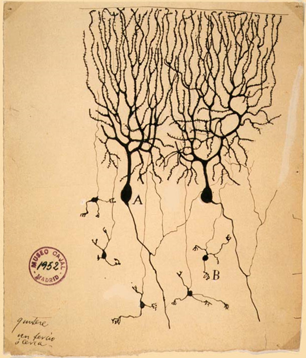
In neuroscience, neuronal tuning refers to the hypothesized property of brain cells by which they selectively represent a particular type of sensory, association, motor, or cognitive information. Some neuronal responses have been hypothesized to be optimally tuned to specific patterns through experience. Neuronal tuning can be strong and sharp, as observed in primary visual cortex (area V1),Matteo Carandini, Jonathan B. Demb, Valerio Mante, David J. Tolhurst, Yang Dan, Bruno A. Olshausen, Jack L. Gallant and Nicole C. Rust. Do we know what the early visual system does? ''Journal of Neuroscience'' 25:10577-10597. or weak and broad, as observed in neural ensembles. Single neurons are hypothesized to be simultaneously tuned to several modalities, such as visual, auditory, and olfactory. Neurons hypothesized to be tuned to different signals are often hypothesized to integrate information from the different sources. In computational models called neural networks, such integratio ... [...More Info...] [...Related Items...] OR: [Wikipedia] [Google] [Baidu] |
Neuroscience
Neuroscience is the scientific study of the nervous system (the brain, spinal cord, and peripheral nervous system), its functions, and its disorders. It is a multidisciplinary science that combines physiology, anatomy, molecular biology, developmental biology, cytology, psychology, physics, computer science, chemistry, medicine, statistics, and mathematical modeling to understand the fundamental and emergent properties of neurons, glia and neural circuits. The understanding of the biological basis of learning, memory, behavior, perception, and consciousness has been described by Eric Kandel as the "epic challenge" of the biological sciences. The scope of neuroscience has broadened over time to include different approaches used to study the nervous system at different scales. The techniques used by neuroscientists have expanded enormously, from molecular and cellular studies of individual neurons to imaging of sensory, motor and cognitive tasks in the brain. Hist ... [...More Info...] [...Related Items...] OR: [Wikipedia] [Google] [Baidu] |
Memory
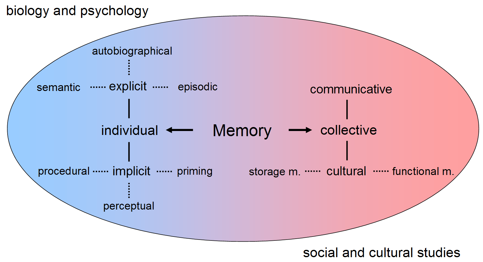
Memory is the faculty of the mind by which data or information is encoded, stored, and retrieved when needed. It is the retention of information over time for the purpose of influencing future action. If past events could not be remembered, it would be impossible for language, relationships, or personal identity to develop. Memory loss is usually described as forgetfulness or amnesia. Memory is often understood as an informational processing system with explicit and implicit functioning that is made up of a sensory processor, short-term (or working) memory, and long-term memory. This can be related to the neuron. The sensory processor allows information from the outside world to be sensed in the form of chemical and physical stimuli and attended to various levels of focus and intent. Working memory serves as an encoding and retrieval processor. Information in the form of stimuli is encoded in accordance with explicit or implicit functions by the working memory p ... [...More Info...] [...Related Items...] OR: [Wikipedia] [Google] [Baidu] |
Superior Temporal Sulcus
The superior temporal sulcus (STS) is the sulcus separating the superior temporal gyrus from the middle temporal gyrus, in the temporal lobe of the mammalian brain. A sulcus (plural sulci) is a deep groove that curves into the largest part of the brain, the cerebrum, and a gyrus (plural gyri) is a ridge that curves outward of the cerebrum. The STS is located under the lateral fissure, which is the fissure that separates the temporal lobe, parietal lobe, and frontal lobe. The STS has an asymmetric structure between the left and right hemisphere, with the STS being longer in the left hemisphere, but deeper in the right hemisphere. This asymmetrical structural organization between hemispheres has only been found to occur in the STS of the human brain. The STS has been shown to produce strong responses when subjects perceive stimuli in research areas that include theory of mind, biological motion, faces, voices, and language. Language processing Spoken language processing ... [...More Info...] [...Related Items...] OR: [Wikipedia] [Google] [Baidu] |
Fusiform Face Area
The fusiform face area (FFA, meaning spindle-shaped face area) is a part of the human visual system (while also activated in people blind from birth) that is specialized for facial recognition. It is located in the inferior temporal cortex (IT), in the fusiform gyrus ( Brodmann area 37). Structure The FFA is located in the ventral stream on the ventral surface of the temporal lobe on the lateral side of the fusiform gyrus. It is lateral to the parahippocampal place area. It displays some lateralization, usually being larger in the right hemisphere. The FFA was discovered and continues to be investigated in humans using positron emission tomography (PET) and functional magnetic resonance imaging (fMRI) studies. Usually, a participant views images of faces, objects, places, bodies, scrambled faces, scrambled objects, scrambled places, and scrambled bodies. This is called a functional localizer. Comparing the neural response between faces and scrambled faces will reveal are ... [...More Info...] [...Related Items...] OR: [Wikipedia] [Google] [Baidu] |
Extrastriate Body Area
The extrastriate body area (EBA) is a subpart of the Extrastriate cortex, extrastriate visual cortex involved in the visual perception of human body and body parts, akin in its respective domain to the fusiform face area, involved in the perception of human faces. The EBA was identified in 2001 by the team of Nancy Kanwisher using fMRI. Function The extrastriate body area is a category-selective region for the visual processing of static and moving images of the human body and parts of it. It is also modulated even in the absence of visual feedback from the limb movement. It is insensitive to faces and stimulus categories unrelated to the human body. The extrastriate cortex responds not only during the perception of other people's body parts but also during goal-directed movement of the participant's body parts. The extrastriate cortex represents the human body in a more integrative and dynamic manner, being able to detect an incongruence of internal body or action representation ... [...More Info...] [...Related Items...] OR: [Wikipedia] [Google] [Baidu] |
Ventral Stream
The two-streams hypothesis is a model of the neural processing of vision as well as hearing. The hypothesis, given its initial characterisation in a paper by David Milner and Melvyn A. Goodale in 1992, argues that humans possess two distinct visual systems. Recently there seems to be evidence of two distinct auditory systems as well. As visual information exits the occipital lobe, and as sound leaves the phonological network, it follows two main pathways, or "streams". The ventral stream (also known as the "what pathway") leads to the temporal lobe, which is involved with object and visual identification and recognition. The dorsal stream (or, "where pathway") leads to the parietal lobe, which is involved with processing the object's spatial location relative to the viewer and with speech repetition. History Several researchers had proposed similar ideas previously. The authors themselves credit the inspiration of work on blindsight by Weiskrantz, and previous neuroscientif ... [...More Info...] [...Related Items...] OR: [Wikipedia] [Google] [Baidu] |
Inferior Temporal Gyrus
The inferior temporal gyrus is one of three gyri of the temporal lobe and is located below the middle temporal gyrus, connected behind with the inferior occipital gyrus; it also extends around the infero-lateral border on to the inferior surface of the temporal lobe, where it is limited by the inferior Sulcus (neuroanatomy), sulcus. This region is one of the higher levels of the ventral stream of visual processing, associated with the representation of objects, places, faces, and colors. It may also be involved in face perception, and in the recognition of numbers and words. The inferior temporal gyrus is the anterior region of the temporal lobe located underneath the central temporal sulcus. The primary function of the occipital temporal gyrus – otherwise referenced as IT cortex – is associated with visual stimuli processing, namely visual object recognition, and has been suggested by recent experimental results as the final location of the ventral cortical visual system. The ... [...More Info...] [...Related Items...] OR: [Wikipedia] [Google] [Baidu] |
Visual Cortex
The visual cortex of the brain is the area of the cerebral cortex that processes visual information. It is located in the occipital lobe. Sensory input originating from the eyes travels through the lateral geniculate nucleus in the thalamus and then reaches the visual cortex. The area of the visual cortex that receives the sensory input from the lateral geniculate nucleus is the primary visual cortex, also known as visual area 1 ( V1), Brodmann area 17, or the striate cortex. The extrastriate areas consist of visual areas 2, 3, 4, and 5 (also known as V2, V3, V4, and V5, or Brodmann area 18 and all Brodmann area 19). Both hemispheres of the brain include a visual cortex; the visual cortex in the left hemisphere receives signals from the right visual field, and the visual cortex in the right hemisphere receives signals from the left visual field. Introduction The primary visual cortex (V1) is located in and around the calcarine fissure in the occipital lobe. Each h ... [...More Info...] [...Related Items...] OR: [Wikipedia] [Google] [Baidu] |
Receptive Field
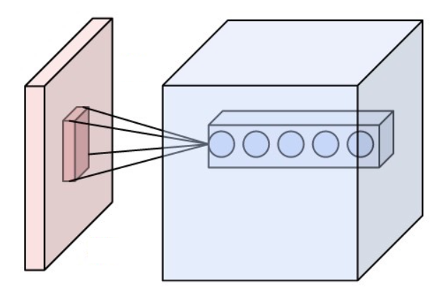
The receptive field, or sensory space, is a delimited medium where some physiological stimuli can evoke a sensory neuronal response in specific organisms. Complexity of the receptive field ranges from the unidimensional chemical structure of odorants to the multidimensional spacetime of human visual field, through the bidimensional skin surface, being a receptive field for touch perception. Receptive fields can positively or negatively alter the membrane potential with or without affecting the rate of action potentials. A sensory space can be dependent of an animal's location. For a particular sound wave traveling in an appropriate transmission medium, by means of sound localization, an auditory space would amount to a reference system that continuously shifts as the animal moves (taking into consideration the space inside the ears as well). Conversely, receptive fields can be largely independent of the animal's location, as in the case of place cells. A sensory space can also ... [...More Info...] [...Related Items...] OR: [Wikipedia] [Google] [Baidu] |
Complex Cell
Complex cells can be found in the primary visual cortex (V1), the secondary visual cortex (V2), and Brodmann area 19 ( V3). Like a simple cell, a complex cell will respond primarily to oriented edges and gratings, however it has a degree of spatial invariance. This means that its receptive field cannot be mapped into fixed excitatory and inhibitory zones. Rather, it will respond to patterns of light in a certain orientation within a large receptive field, regardless of the exact location. Some complex cells respond optimally only to movement in a certain direction. These cells were discovered by Torsten Wiesel and David Hubel David Hunter Hubel (February 27, 1926 – September 22, 2013) was an American Canadian neurophysiologist noted for his studies of the structure and function of the visual cortex. He was co-recipient with Torsten Wiesel of the 1981 Nobel Pr ... in the early 1960s. They refrained from reporting on the complex cells in (Hubel 1959) because they did ... [...More Info...] [...Related Items...] OR: [Wikipedia] [Google] [Baidu] |
Simple Cell
A simple cell in the visual cortex, primary visual cortex is a cell that responds primarily to oriented edges and gratings (bars of particular orientations). Torsten Wiesel and David Hubel discovered these cells in the late 1950s. Such cells are tuned to different frequencies and orientations, even with different phase relationships, possibly to extract disparity (depth) information and to attribute depth to detected lines and edges. This may result in a 3D 'wire-frame' representation as used in computer graphics. The fact that input from the left and right eyes is very close in the so-called cortical hypercolumns indicates that depth processing occurs very early, aiding the recognition of 3D objects. Later, many other cells with specific functions have been discovered: (a) end-stopped cells, which are thought to detect singularities like line and edge crossings, vertices, and line endings; (b) bar and grating cells. The latter are not linear operators because a bar cell does not ... [...More Info...] [...Related Items...] OR: [Wikipedia] [Google] [Baidu] |


