|
Neural Plate
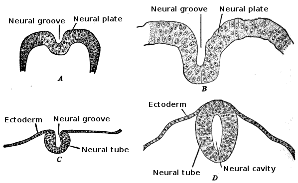
In embryology, the neural plate is a key Development of the human body, developmental structure that serves as the basis for the nervous system. Cranial to the primitive node of the embryonic primitive streak, Ectoderm, ectodermal tissue thickens and flattens to become the neural plate. The region anterior to the primitive node can be generally referred to as the neural plate. Cells take on a columnar appearance in the process as they continue to lengthen and narrow. The ends of the neural plate, known as the neural folds, push the ends of the plate up and together, folding into the neural tube, a structure critical to brain and spinal cord development. This process as a whole is termed primary neurulation. Signaling proteins are also important in neural plate development, and aid in Cellular differentiation, differentiating the tissue destined to become the neural plate. Examples of such proteins include bone morphogenetic proteins and cadherins. Expression of these proteins i ... [...More Info...] [...Related Items...] OR: [Wikipedia] [Google] [Baidu] |
Central Nervous System
The central nervous system (CNS) is the part of the nervous system consisting primarily of the brain, spinal cord and retina. The CNS is so named because the brain integrates the received information and coordinates and influences the activity of all parts of the bodies of bilateria, bilaterally symmetric and triploblastic animals—that is, all multicellular animals except sponges and Coelenterata, diploblasts. It is a structure composed of nervous tissue positioned along the Anatomical_terms_of_location#Rostral,_cranial,_and_caudal, rostral (nose end) to caudal (tail end) axis of the body and may have an enlarged section at the rostral end which is a brain. Only arthropods, cephalopods and vertebrates have a true brain, though precursor structures exist in onychophorans, gastropods and lancelets. The rest of this article exclusively discusses the vertebrate central nervous system, which is radically distinct from all other animals. Overview In vertebrates, the brain and spinal ... [...More Info...] [...Related Items...] OR: [Wikipedia] [Google] [Baidu] |
Notochord
The notochord is an elastic, rod-like structure found in chordates. In vertebrates the notochord is an embryonic structure that disintegrates, as the vertebrae develop, to become the nucleus pulposus in the intervertebral discs of the vertebral column. In non-vertebrate chordates a notochord persists. The notochord is derived from the embryonic mesoderm and consists of an inner core of vacuolated cells filled with glycoproteins, covered by two helical collagen-elastin sheaths. It lies longitudinally along the rostral-caudal (head to tail) axis of the body, dorsal to the gut tube, and ventral to the dorsal nerve cord. Some chordate invertebrates, such as tunicates, develop a notochord during the larval stage but lose it through subsequent stages into adulthood. The notochord is important for signaling the dorso-ventral patterning of cells coming from the mesodermal progenitors. This helps form the precursors needed for certain organs and the embryo to develop ... [...More Info...] [...Related Items...] OR: [Wikipedia] [Google] [Baidu] |
Neuroepithelial Cells
Neuroepithelial cells, or neuroectodermal cells, form the wall of the closed neural tube in early embryonic development. The neuroepithelial cells span the thickness of the tube's wall, connecting with the pia mater, pial surface and with the ventricular or lumen (anatomy), lumenal surface. They are joined at the lumen of the tube by adherens junction, junctional complexes, where they form a Pseudostratified columnar epithelium, pseudostratified layer of epithelium called neuroepithelium. Neuroepithelial cells are the stem cells of the central nervous system, known as neural stem cells, and generate the intermediate progenitor cells known as radial glial cells, that cellular differentiation, differentiate into neurons and glia in the process of neurogenesis. Embryonic neural development Brain development During the third week of human embryogenesis, embryonic growth, the brain begins to develop in the early fetus in a process called morphogenesis. Neuroepithelial cells of the ecto ... [...More Info...] [...Related Items...] OR: [Wikipedia] [Google] [Baidu] |
Endoderm
Endoderm is the innermost of the three primary germ layers in the very early embryo. The other two layers are the ectoderm (outside layer) and mesoderm (middle layer). Cells migrating inward along the archenteron form the inner layer of the gastrula, which develops into the endoderm. The endoderm consists at first of flattened cells, which subsequently become columnar. It forms the epithelial lining of multiple systems. In plant biology, endoderm corresponds to the innermost part of the cortex ( bark) in young shoots and young roots often consisting of a single cell layer. As the plant becomes older, more endoderm will lignify. Production The following chart shows the tissues produced by the endoderm. The embryonic endoderm develops into the interior linings of two tubes in the body, the digestive and respiratory tube. Liver and pancreas cells are believed to derive from a common precursor. In humans, the endoderm can differentiate into distinguishable organs after 5 w ... [...More Info...] [...Related Items...] OR: [Wikipedia] [Google] [Baidu] |
Mesoderm
The mesoderm is the middle layer of the three germ layers that develops during gastrulation in the very early development of the embryo of most animals. The outer layer is the ectoderm, and the inner layer is the endoderm.Langman's Medical Embryology, 11th edition. 2010. The mesoderm forms mesenchyme, mesothelium and coelomocytes. Mesothelium lines coeloms. Mesoderm forms the muscles in a process known as myogenesis, septa (cross-wise partitions) and mesenteries (length-wise partitions); and forms part of the gonads (the rest being the gametes). Myogenesis is specifically a function of mesenchyme. The mesoderm differentiates from the rest of the embryo through intercellular signaling, after which the mesoderm is polarized by an organizing center. The position of the organizing center is in turn determined by the regions in which beta-catenin is protected from degradation by GSK-3. Beta-catenin acts as a co-factor that alters the activity of the transcription facto ... [...More Info...] [...Related Items...] OR: [Wikipedia] [Google] [Baidu] |
Germ Layer
A germ layer is a primary layer of cell (biology), cells that forms during embryonic development. The three germ layers in vertebrates are particularly pronounced; however, all eumetazoans (animals that are sister taxa to the sponges) produce two or three primary germ layers. Some animals, like cnidarians, produce two germ layers (the ectoderm and endoderm) making them diploblastic. Other animals such as bilaterians produce a third layer (the mesoderm) between these two layers, making them triploblastic. Germ layers eventually give rise to all of an animal's Tissue (biology), tissues and organ (anatomy), organs through the process of organogenesis. History Caspar Friedrich Wolff observed organization of the early embryo in leaf-like layers. In 1817, Heinz Christian Pander discovered three primordial germ layers while studying chick embryos. Between 1850 and 1855, Robert Remak had further refined the germ cell layer (''Keimblatt'') concept, stating that the external, internal ... [...More Info...] [...Related Items...] OR: [Wikipedia] [Google] [Baidu] |
Glia
Glia, also called glial cells (gliocytes) or neuroglia, are non-neuronal cells in the central nervous system (the brain and the spinal cord) and in the peripheral nervous system that do not produce electrical impulses. The neuroglia make up more than one half the volume of neural tissue in the human body. They maintain homeostasis, form myelin, and provide support and protection for neurons. In the central nervous system, glial cells include oligodendrocytes (that produce myelin), astrocytes, ependymal cells and microglia, and in the peripheral nervous system they include Schwann cells (that produce myelin), and satellite cells. Function They have four main functions: * to surround neurons and hold them in place * to supply nutrients and oxygen to neurons * to insulate one neuron from another * to destroy pathogens and remove dead neurons. They also play a role in neurotransmission and synaptic connections, and in physiological processes such as breathing. While glia we ... [...More Info...] [...Related Items...] OR: [Wikipedia] [Google] [Baidu] |
Neurons
A neuron (American English), neurone (British English), or nerve cell, is an membrane potential#Cell excitability, excitable cell (biology), cell that fires electric signals called action potentials across a neural network (biology), neural network in the nervous system. They are located in the nervous system and help to receive and conduct impulses. Neurons communicate with other cells via synapses, which are specialized connections that commonly use minute amounts of chemical neurotransmitters to pass the electric signal from the presynaptic neuron to the target cell through the synaptic gap. Neurons are the main components of nervous tissue in all Animalia, animals except sponges and placozoans. Plants and fungi do not have nerve cells. Molecular evidence suggests that the ability to generate electric signals first appeared in evolution some 700 to 800 million years ago, during the Tonian period. Predecessors of neurons were the peptidergic secretory cells. They eventually ga ... [...More Info...] [...Related Items...] OR: [Wikipedia] [Google] [Baidu] |
Epidermis (skin)
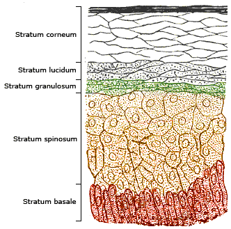
The epidermis is the outermost of the three layers that comprise the skin, the inner layers being the dermis and hypodermis. The epidermal layer provides a barrier to infection from environmental pathogens and regulates the amount of water released from the body into the atmosphere through transepidermal water loss. The epidermis is composed of multiple layers of flattened cells that overlie a base layer ( stratum basale) composed of columnar cells arranged perpendicularly. The layers of cells develop from stem cells in the basal layer. The thickness of the epidermis varies from 31.2μm for the penis to 596.6μm for the sole of the foot with most being roughly 90μm. Thickness does not vary between the sexes but becomes thinner with age. The human epidermis is an example of epithelium, particularly a stratified squamous epithelium. The word epidermis is derived through Latin , itself and . Something related to or part of the epidermis is termed epidermal. Structure ... [...More Info...] [...Related Items...] OR: [Wikipedia] [Google] [Baidu] |
Ectoderm
The ectoderm is one of the three primary germ layers formed in early embryonic development. It is the outermost layer, and is superficial to the mesoderm (the middle layer) and endoderm (the innermost layer). It emerges and originates from the outer layer of germ cells. The word ectoderm comes from the Greek language, Greek ''ektos'' meaning "outside", and ''derma'' meaning "skin".Gilbert, Scott F. Developmental Biology. 9th ed. Sunderland, MA: Sinauer Associates, 2010: 333-370. Print. Generally speaking, the ectoderm differentiates to form epithelial tissue, epithelial and nervous system, neural tissues (spinal cord, nerves and brain). This includes the Epidermis (skin), skin, linings of the mouth, anus, nostrils, sweat glands, hair and nails, and tooth enamel. Other types of epithelium are derived from the endoderm. In vertebrate embryos, the ectoderm can be divided into two parts: the dorsal surface ectoderm also known as the external ectoderm, and the neural plate, which inv ... [...More Info...] [...Related Items...] OR: [Wikipedia] [Google] [Baidu] |


