|
Midline Shift
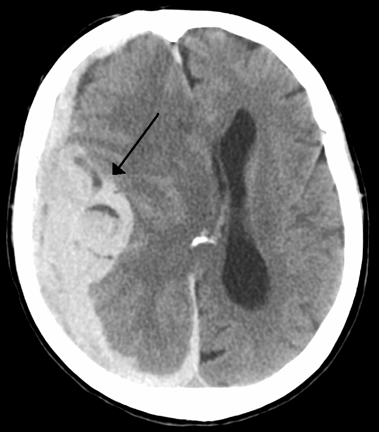
Midline shift is a shift of the brain past its center line. The sign may be evident on neuroimaging such as CT scanning. The sign is considered ominous because it is commonly associated with a distortion of the brain stem that can cause serious dysfunction evidenced by abnormal posturing and failure of the pupils to constrict in response to light. Midline shift is often associated with high intracranial pressure (ICP), which can be deadly. In fact, midline shift is a measure of ICP; presence of the former is an indication of the latter. Presence of midline shift is an indication for neurosurgeons to take measures to monitor and control ICP. Immediate surgery may be indicated when there is a midline shift of over 5 mm. The sign can be caused by conditions including traumatic brain injury, stroke, hematoma, or birth deformity that leads to a raised intracranial pressure. Methods of Detection Doctors detect midline shift using a variety of methods. The most prominent measurem ... [...More Info...] [...Related Items...] OR: [Wikipedia] [Google] [Baidu] |
Sonography
Medical ultrasound includes diagnostic techniques (mainly medical imaging, imaging techniques) using ultrasound, as well as therapeutic ultrasound, therapeutic applications of ultrasound. In diagnosis, it is used to create an image of internal body structures such as tendons, muscles, joints, blood vessels, and internal organs, to measure some characteristics (e.g. distances and velocities) or to generate an informative audible sound. Its aim is usually to find a source of disease or to exclude pathology. The usage of ultrasound to produce visual images for medicine is called medical ultrasonography or simply sonography. The practice of examining pregnant women using ultrasound is called obstetric ultrasonography, and was an early development of clinical ultrasonography. Ultrasound is composed of sound waves with frequency, frequencies which are significantly higher than the range of human hearing (>20,000 Hz). Ultrasonic images, also known as sonograms, are created by se ... [...More Info...] [...Related Items...] OR: [Wikipedia] [Google] [Baidu] |
Neurotrauma
Neurotrauma, brain damage or brain injury (BI) is the destruction or degeneration of brain cells. Brain injuries occur due to a wide range of internal and external factors. In general, brain damage refers to significant, undiscriminating trauma-induced damage. A common category with the greatest number of injuries is traumatic brain injury (TBI) following physical trauma or head injury from an outside source, and the term acquired brain injury (ABI) is used in appropriate circles to differentiate brain injuries occurring after birth from injury, from a genetic disorder (GBI), or from a congenital disorder (CBI). Primary and secondary brain injuries identify the processes involved, while focal and diffuse brain injury describe the severity and localization. Recent research has demonstrated that neuroplasticity, which allows the brain to reorganize itself by forming new neural connections throughout life, provides for rearrangement of its workings. This allows the brain to ... [...More Info...] [...Related Items...] OR: [Wikipedia] [Google] [Baidu] |
Mass Effect (medicine)
In medicine, a mass effect is the effect of a growing mass that results in secondary pathological effects by pushing on or displacing surrounding tissue. In oncology, the mass typically refers to a tumor. For example, cancer of the thyroid gland may cause symptoms due to compressions of certain structures of the head and neck; pressure on the laryngeal nerves may cause voice changes, narrowing of the windpipe may cause stridor, pressure on the gullet may cause dysphagia and so on. Surgical removal or debulking is sometimes used to palliate symptoms of the mass effect even if the underlying pathology is not curable. In neurology, a mass effect is the effect exerted by any mass, including, for example, hydrocephalus (cerebrospinal fluid buildup) or an evolving intracranial hemorrhage (bleeding within the skull) presenting with a clinically significant hematoma. The hematoma can exert a mass effect on the brain, increasing intracranial pressure and potentially causing midline sh ... [...More Info...] [...Related Items...] OR: [Wikipedia] [Google] [Baidu] |
Infarction
Infarction is tissue death (necrosis) due to inadequate blood supply to the affected area. It may be caused by artery blockages, rupture, mechanical compression, or vasoconstriction. The resulting lesion is referred to as an infarct (from the Latin ''infarctus'', "stuffed into"). Causes Infarction occurs as a result of prolonged ischemia, which is the insufficient supply of oxygen and nutrition to an area of tissue due to a disruption in blood supply. The blood vessel supplying the affected area of tissue may be blocked due to an obstruction in the vessel (e.g., an arterial embolus, thrombus, or atherosclerotic plaque), compressed by something outside of the vessel causing it to narrow (e.g., tumor, volvulus, or hernia), ruptured by trauma causing a loss of blood pressure downstream of the rupture, or vasoconstricted, which is the narrowing of the blood vessel by contraction of the muscle wall rather than an external force (e.g., cocaine vasoconstriction leading ... [...More Info...] [...Related Items...] OR: [Wikipedia] [Google] [Baidu] |
Subarachnoid Hemorrhage
Subarachnoid hemorrhage (SAH) is bleeding into the subarachnoid space—the area between the arachnoid membrane and the pia mater surrounding the brain. Symptoms may include a severe headache of rapid onset, vomiting, decreased level of consciousness, fever, and sometimes seizures. Neck stiffness or neck pain are also relatively common. In about a quarter of people a small bleed with resolving symptoms occurs within a month of a larger bleed. SAH may occur as a result of a head injury or spontaneously, usually from a ruptured cerebral aneurysm. Risk factors for spontaneous cases include high blood pressure, smoking, family history, alcoholism, and cocaine use. Generally, the diagnosis can be determined by a CT scan of the head if done within six hours of symptom onset. Occasionally, a lumbar puncture is also required. After confirmation further tests are usually performed to determine the underlying cause. Treatment is by prompt neurosurgery or endovascular coiling. Medicat ... [...More Info...] [...Related Items...] OR: [Wikipedia] [Google] [Baidu] |
Epidural Hematoma
Epidural hematoma is when bleeding occurs between the tough outer membrane covering the brain (dura mater) and the skull. Often there is loss of consciousness following a head injury, a brief regaining of consciousness, and then loss of consciousness again. Other symptoms may include headache, confusion, vomiting, and an inability to move parts of the body. Complications may include seizures. The cause is typically head injury that results in a break of the temporal bone and bleeding from the middle meningeal artery. Occasionally it can occur as a result of a bleeding disorder or blood vessel malformation. Diagnosis is typically by a CT scan or MRI. When this condition occurs in the spine it is known as a spinal epidural hematoma. Treatment is generally by urgent surgery in the form of a craniotomy or burr hole. Without treatment, death typically results. The condition occurs in one to four percent of head injuries. Typically it occurs in young adults. Males are more often ... [...More Info...] [...Related Items...] OR: [Wikipedia] [Google] [Baidu] |
Pathology
Pathology is the study of the causes and effects of disease or injury. The word ''pathology'' also refers to the study of disease in general, incorporating a wide range of biology research fields and medical practices. However, when used in the context of modern medical treatment, the term is often used in a narrower fashion to refer to processes and tests that fall within the contemporary medical field of "general pathology", an area which includes a number of distinct but inter-related medical specialties that diagnose disease, mostly through analysis of tissue, cell, and body fluid samples. Idiomatically, "a pathology" may also refer to the predicted or actual progression of particular diseases (as in the statement "the many different forms of cancer have diverse pathologies", in which case a more proper choice of word would be " pathophysiologies"), and the affix ''pathy'' is sometimes used to indicate a state of disease in cases of both physical ailment (as in cardiomy ... [...More Info...] [...Related Items...] OR: [Wikipedia] [Google] [Baidu] |
Pineal Gland
The pineal gland, conarium, or epiphysis cerebri, is a small endocrine gland in the brain of most vertebrates. The pineal gland produces melatonin, a serotonin-derived hormone which modulates sleep, sleep patterns in both circadian rhythm, circadian and Season, seasonal cycles. The shape of the gland resembles a pine cone, which gives it its name. The pineal gland is located in the epithalamus, near the center of the brain, between the two cerebral hemisphere, hemispheres, tucked in a groove where the two halves of the thalamus join. The pineal gland is one of the neuroendocrinology, neuroendocrine Circumventricular organs, secretory circumventricular organs in which capillaries are mostly Vascular permeability, permeable to solutes in the blood. Nearly all vertebrate species possess a pineal gland. The most important exception is a primitive vertebrate, the hagfish. Even in the hagfish, however, there may be a "pineal equivalent" structure in the dorsal diencephalon. The lanc ... [...More Info...] [...Related Items...] OR: [Wikipedia] [Google] [Baidu] |
Third Ventricle
The third ventricle is one of the four connected ventricles of the ventricular system within the mammalian brain. It is a slit-like cavity formed in the diencephalon between the two thalami, in the midline between the right and left lateral ventricles, and is filled with cerebrospinal fluid (CSF). Running through the third ventricle is the interthalamic adhesion, which contains thalamic neurons and fibers that may connect the two thalami. Structure The third ventricle is a narrow, laterally flattened, vaguely rectangular region, filled with cerebrospinal fluid, and lined by ependyma. It is connected at the superior anterior corner to the lateral ventricles, by the interventricular foramina, and becomes the cerebral aqueduct (''aqueduct of Sylvius'') at the posterior caudal corner. Since the interventricular foramina are on the lateral edge, the corner of the third ventricle itself forms a bulb, known as the ''anterior recess'' (it is also known as the ''bulb of the ventricl ... [...More Info...] [...Related Items...] OR: [Wikipedia] [Google] [Baidu] |
Septum Pellucidum
The septum pellucidum (Latin for "translucent wall") is a thin, triangular, vertical double membrane separating the anterior horns of the left and right lateral ventricles of the brain. It runs as a sheet from the corpus callosum down to the fornix. The septum is not present in the syndrome septo-optic dysplasia. Structure The septum pellucidum is located in the septal area in the midline of the brain between the two cerebral hemispheres. The septal area is also the location of the septal nuclei. It is attached to the lower part of the corpus callosum, the large collection of nerve fibers that connect the two cerebral hemispheres. It is attached to the front forward part of the fornix. The lateral ventricles sit on either side of the septum. The septum pellucidum consists of two layers or ''laminae'' of both white and gray matter. During fetal development, there is a space between the two laminae called the cave of septum pellucidum that, in ninety percent of cases, disappear ... [...More Info...] [...Related Items...] OR: [Wikipedia] [Google] [Baidu] |
Human Error
Human error refers to something having been done that was " not intended by the actor; not desired by a set of rules or an external observer; or that led the task or system outside its acceptable limits".Senders, J.W. and Moray, N.P. (1991) Human Error: Cause, Prediction, and Reduction'. Lawrence Erlbaum Associates, p.25. . Human error has been cited as a primary cause contributing factor in disasters and accidents in industries as diverse as nuclear power (e.g., the Three Mile Island accident), aviation (see pilot error), space exploration (e.g., the Space Shuttle Challenger disaster and Space Shuttle Columbia disaster), and medicine (see medical error). Prevention of human error is generally seen as a major contributor to reliability and safety of (complex) systems. Human error is one of the many contributing causes of risk events. Definition Human error refers to something having been done that was "not intended by the actor; not desired by a set of rules or an external observer ... [...More Info...] [...Related Items...] OR: [Wikipedia] [Google] [Baidu] |


.jpg)

