|
Membrane Nanotubule
A tunneling nanotube (TNT) or membrane nanotube is a term that has been applied to protrusions that extend from the plasma membrane which enable different animal cells to touch over long distances, sometimes over 100 Micrometre, μm between T cells. Two types of structures have been called nanotubes. The first type are less than 0.7 micrometers in diameter, contain actin and carry portions of plasma membrane between cells in both directions. The second type are larger (>0.7 μm), contain both actin and microtubules, and can carry components of the cytoplasm such as vesicle (biology), vesicles and organelles between cells, including Mitochondrion, whole mitochondria. The diameter of TNTs ranges from 50 to 200 nm and they can reach lengths of several cell diameters. These structures may be involved in cell-to-cell communication, transfer of nucleic acids such as Messenger RNA, mRNA and MicroRNA, miRNA between cells in culture or in a Tissue (biology), tissue, and the spread of ... [...More Info...] [...Related Items...] OR: [Wikipedia] [Google] [Baidu] |
Nanotubes
file:Chiraltube.png, A scanning tunneling microscopy image of a single-walled carbon nanotube file:Kohlenstoffnanoroehre Animation.gif, Rotating single-walled zigzag carbon nanotube A carbon nanotube (CNT) is a tube made of carbon with diameters typically measured in nanometers. ''Single-wall carbon nanotubes'' (''SWCNTs'') are one of the allotropes of carbon, intermediate between fullerene cages and flat graphene, with diameters in the range of a nanometre. Although not made this way, single-wall carbon nanotubes can be idealized as cutouts from a two-dimensional Hexagonal tiling, hexagonal lattice of carbon atoms rolled up along one of the Bravais lattice vectors of the hexagonal lattice to form a hollow cylinder. In this construction, periodic boundary conditions are imposed over the length of this roll-up vector to yield a helical lattice of seamlessly bonded carbon atoms on the cylinder surface. ''Multi-wall carbon nanotubes'' (''MWCNTs'') consisting of nested single-wall ... [...More Info...] [...Related Items...] OR: [Wikipedia] [Google] [Baidu] |
Imaginal Disc
An imaginal disc is one of the parts of a holometabolous insect larva that will become a portion of the outside of the adult insect during the pupal transformation. Contained within the body of the larva, there are pairs of discs that will form, for instance, the wings or legs or antennae or other structures in the adult. The role of the imaginal disc in insect development was first elucidated by Jan Swammerdam. During the pupal stage, many larval structures are broken down, and adult structures, including the discs, undergo rapid development. Each disc everts and elongates, with the central portion of the disc becoming the distal part of whichever appendage it is forming: the wing, leg, antenna, etc. During the larval stage, the cells in the growing disc appear undifferentiated, but their developmental fate in the adult is already determined. The experiment that demonstrates this developmental commitment is to take an imaginal disc from a third instar larva, about to undergo ... [...More Info...] [...Related Items...] OR: [Wikipedia] [Google] [Baidu] |
T Cell
A T cell is a type of lymphocyte. T cells are one of the important white blood cells of the immune system and play a central role in the adaptive immune response. T cells can be distinguished from other lymphocytes by the presence of a T-cell receptor (TCR) on their cell surface. T cells are born from hematopoietic stem cells, found in the bone marrow. Developing T cells then migrate to the thymus gland to develop (or mature). T cells derive their name from the thymus. After migration to the thymus, the precursor cells mature into several distinct types of T cells. T cell differentiation also continues after they have left the thymus. Groups of specific, differentiated T cell subtypes have a variety of important functions in controlling and shaping the immune response. One of these functions is immune-mediated cell death, and it is carried out by two major subtypes: CD8+ "killer" and CD4+ "helper" T cells. (These are named for the presence of the cell surface proteins CD8 or ... [...More Info...] [...Related Items...] OR: [Wikipedia] [Google] [Baidu] |
Caspase 3
Caspase-3 is a caspase protein that interacts with caspase-8 and caspase-9. It is encoded by the ''CASP3'' gene. ''CASP3'' orthologs have been identified in numerous mammals for which complete genome data are available. Unique orthologs are also present in birds, lizards, lissamphibians, and teleosts. The CASP3 protein is a member of the cysteine-aspartic acid protease (caspase) family. Sequential activation of caspases plays a central role in the execution-phase of cell apoptosis. Caspases exist as inactive proenzymes that undergo proteolytic processing at conserved aspartic residues to produce two subunits, large and small, that dimerize to form the active enzyme. This protein cleaves and activates caspases 6 and 7; and the protein itself is processed and activated by caspases 8, 9, and 10. It is the predominant caspase involved in the cleavage of amyloid-beta 4A precursor protein, which is associated with neuronal death in Alzheimer's disease. Alternative splicing of this ... [...More Info...] [...Related Items...] OR: [Wikipedia] [Google] [Baidu] |
S100A4
Protein S100-A4 (S100A4) is a protein that in humans is encoded by the ''S100A4'' gene. Function The protein encoded by this gene is a member of the S100 family of proteins containing 2 EF-hand calcium-binding motifs. S100 proteins are localized in the cytoplasm and/or nucleus of a wide range of cells, and involved in the regulation of a number of cellular processes such as cell cycle progression and differentiation. S100 genes include at least 13 members which are located as a cluster on chromosome 1q21. This protein may function in motility, invasion, and tubulin polymerization. Chromosomal rearrangements and altered expression of this gene have been implicated in tumor metastasis. Multiple alternatively spliced variants, encoding the same protein, have been identified. Interactions S100A4 has been shown to interact with S100 calcium binding protein A1. Therapeutic targeting for cancer S100A4, a member of the S100 calcium-binding protein family secreted by tumor and stro ... [...More Info...] [...Related Items...] OR: [Wikipedia] [Google] [Baidu] |
Mesenchymal Stem Cell
Mesenchymal stem cells (MSCs) also known as mesenchymal stromal cells or medicinal signaling cells are multipotent stromal cells that can differentiate into a variety of cell types, including osteoblasts (bone cells), chondrocytes (cartilage cells), myocytes (muscle cells) and adipocytes (fat cells which give rise to marrow adipose tissue). Structure Definition While the terms ''mesenchymal stem cell'' (MSC) and ''marrow stromal cell'' have been used interchangeably for many years, neither term is sufficiently descriptive: * Mesenchyme is embryonic connective tissue that is derived from the mesoderm and that differentiates into hematopoietic and connective tissue, whereas MSCs do not differentiate into hematopoietic cells. * Stromal cells are connective tissue cells that form the supportive structure in which the functional cells of the tissue reside. While this is an accurate description for one function of MSCs, the term fails to convey the relatively recently discove ... [...More Info...] [...Related Items...] OR: [Wikipedia] [Google] [Baidu] |
Phosphatidylserine
Phosphatidylserine (abbreviated Ptd-L-Ser or PS) is a phospholipid and is a component of the cell membrane. It plays a key role in cell cycle signaling, specifically in relation to apoptosis. It is a key pathway for viruses to enter cells via apoptotic mimicry. Its exposure on the outer surface of a membrane marks the cell for destruction via apoptosis. Structure Phosphatidylserine is a phospholipid—more specifically a glycerophospholipid—which consists of two fatty acids attached in ester linkage to the first and second carbon of glycerol and serine attached through a phosphodiester linkage to the third carbon of the glycerol. Phosphatidylserine sourced from plants differs in fatty acid composition from that sourced from animals. It is commonly found in the inner (cytoplasmic) leaflet of biological membranes. It is almost entirely found in the inner monolayer of the membrane with only less than 10% of it in the outer monolayer. Introduction Phosphatidylserine (PS) is ... [...More Info...] [...Related Items...] OR: [Wikipedia] [Google] [Baidu] |
Chemotaxis
Chemotaxis (from '' chemo-'' + ''taxis'') is the movement of an organism or entity in response to a chemical stimulus. Somatic cells, bacteria, and other single-cell or multicellular organisms direct their movements according to certain chemicals in their environment. This is important for bacteria to find food (e.g., glucose) by swimming toward the highest concentration of food molecules, or to flee from poisons (e.g., phenol). In multicellular organisms, chemotaxis is critical to early development (e.g., movement of sperm towards the egg during fertilization) and development (e.g., migration of neurons or lymphocytes) as well as in normal function and health (e.g., migration of leukocytes during injury or infection). In addition, it has been recognized that mechanisms that allow chemotaxis in animals can be subverted during cancer metastasis. The aberrant chemotaxis of leukocytes and lymphocytes also contribute to inflammatory diseases such as atherosclerosis, asthma, and arthr ... [...More Info...] [...Related Items...] OR: [Wikipedia] [Google] [Baidu] |
Cytoneme
Cytonemes are thin, cellular projections that are specialized for exchange of signaling proteins between cells. Cytonemes emanate from cells that make signaling proteins, extending directly to cells that receive signaling proteins. Cytonemes also extend directly from cells that receive signaling proteins to cells that make them. A cytoneme is a type of filopodium - a thin, tubular extension of a cell’s plasma membrane that has a core composed of tightly bundled, parallel actin filaments. Filopodia can extend more than 100 μm and have been measured as thin as 0.1 μm and as thick as 0.5 μm. Cytonemes with a diameter of approximately 0.2 μm and as long as 80 μm have been observed in the Drosophila wing imaginal disc. Many cell types have filopodia. The functions of filopodia have been attributed to pathfinding of neurons, early stages of synapse formation, antigen presentation by dendritic cells of the immune system, force generation by macrophages and virus transmission. The ... [...More Info...] [...Related Items...] OR: [Wikipedia] [Google] [Baidu] |
Lamellipodium
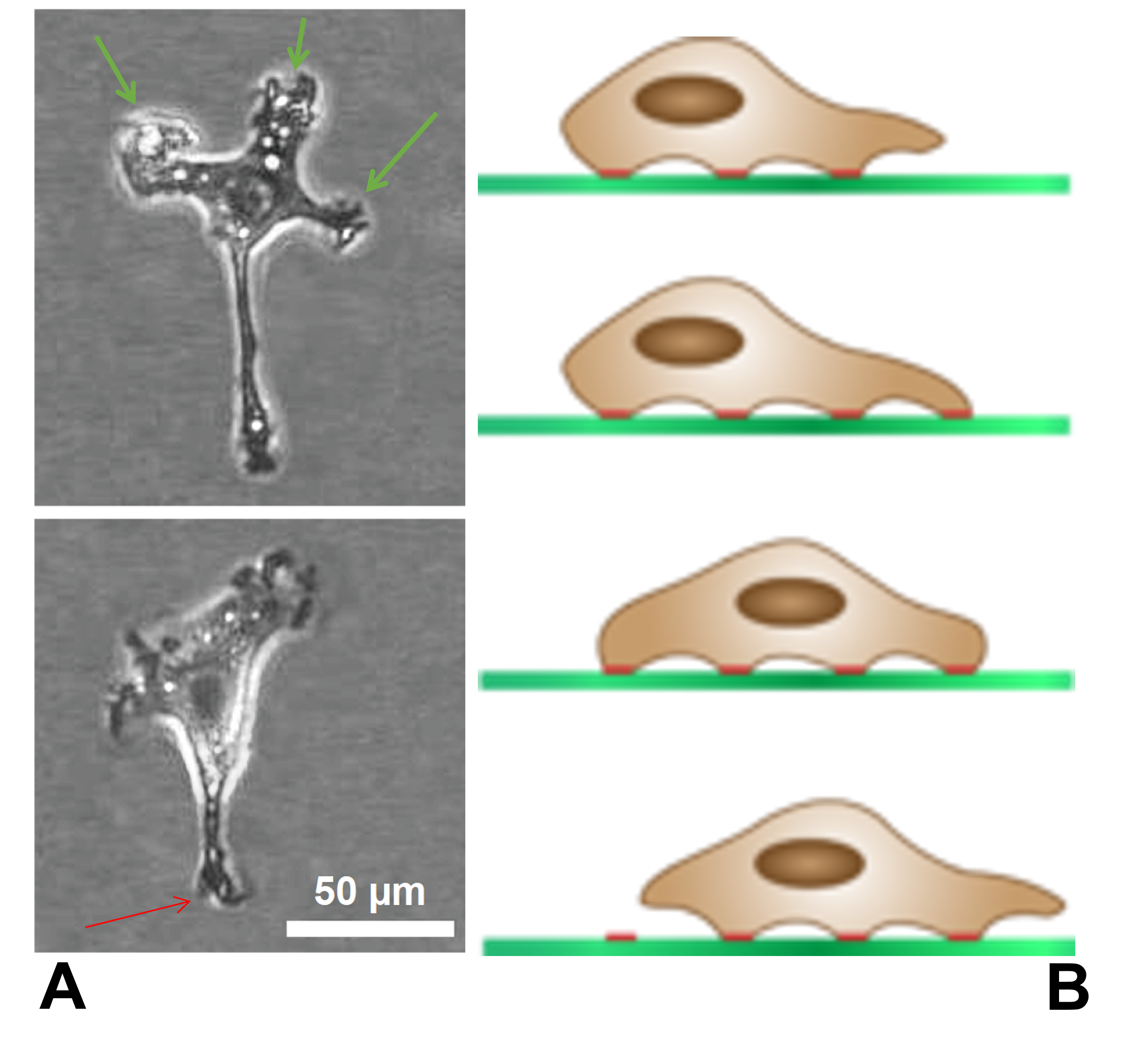
The lamellipodium (plural lamellipodia) (from Latin ''lamella'', related to ', "thin sheet", and the Greek radical ''pod-'', "foot") is a cytoskeletal protein actin projection on the leading edge of the cell. It contains a quasi-two-dimensional actin mesh; the whole structure propels the cell across a substrate. Within the lamellipodia are ribs of actin called microspikes, which, when they spread beyond the lamellipodium frontier, are called filopodia. The lamellipodium is born of actin nucleation in the plasma membrane of the cell and is the primary area of actin incorporation or microfilament formation of the cell. Description Lamellipodia are found primarily in all mobile cells, such as the keratinocytes of fish and frogs, which are involved in the quick repair of wounds. The lamellipodia of these keratinocytes allow them to move at speeds of 10–20 μm / min over epithelial surfaces. When separated from the main part of a cell, a lamellipodium can still cra ... [...More Info...] [...Related Items...] OR: [Wikipedia] [Google] [Baidu] |
Monocyte
Monocytes are a type of leukocyte or white blood cell. They are the largest type of leukocyte in blood and can differentiate into macrophages and conventional dendritic cells. As a part of the vertebrate innate immune system monocytes also influence adaptive immune responses and exert tissue repair functions. There are at least three subclasses of monocytes in human blood based on their phenotypic receptors. Structure Monocytes are amoeboid in appearance, and have nongranulated cytoplasm. Thus they are classified as agranulocytes, although they might occasionally display some azurophil granules and/or vacuoles. With a diameter of 15–22 μm, monocytes are the largest cell type in peripheral blood. Monocytes are mononuclear cells and the ellipsoidal nucleus is often lobulated/indented, causing a bean-shaped or kidney-shaped appearance. Monocytes compose 2% to 10% of all leukocytes in the human body. Development Monocytes are produced by the bone marrow from precursors ca ... [...More Info...] [...Related Items...] OR: [Wikipedia] [Google] [Baidu] |


.png)