|
Magnetic Particle Imaging
Magnetic particle imaging (MPI) is an emerging non-invasive tomographic technique that directly detects superparamagnetic nanoparticle tracers. The technology has potential applications in diagnostic imaging and material science. Currently, it is used in medical research to measure the 3-D location and concentration of nanoparticles. Imaging does not use ionizing radiation and can produce a signal at any depth within the body. MPI was first conceived in 2001 by scientists working at the Royal Philips Research lab in Hamburg. The first system was established and reported in 2005. Since then, the technology has been advanced by academic researchers at several universities around the world. The first commercial MPI scanners have recently become available froMagnetic Insightan The hardware used for MPI is very different from MRI. MPI systems use changing magnetic fields to generate a signal from superparamagnetic iron oxide (SPIO) nanoparticles. These fields are specifically design ... [...More Info...] [...Related Items...] OR: [Wikipedia] [Google] [Baidu] |
Tomography
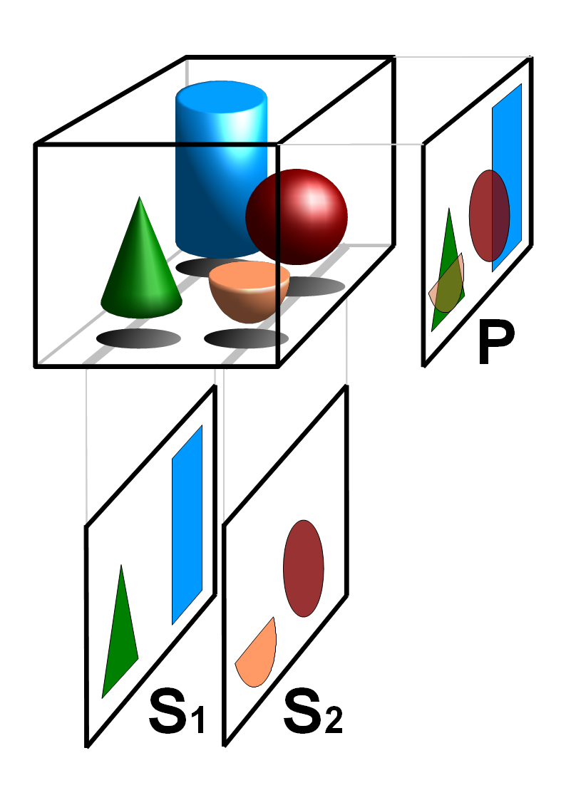
Tomography is imaging by sections or sectioning that uses any kind of penetrating wave. The method is used in radiology, archaeology, biology, atmospheric science, geophysics, oceanography, plasma physics, materials science, astrophysics, quantum information, and other areas of science. The word ''tomography'' is derived from Ancient Greek τόμος ''tomos'', "slice, section" and γράφω ''graphō'', "to write" or, in this context as well, "to describe." A device used in tomography is called a tomograph, while the image produced is a tomogram. In many cases, the production of these images is based on the mathematical procedure tomographic reconstruction, such as X-ray computed tomography technically being produced from multiple projectional radiographs. Many different reconstruction algorithms exist. Most algorithms fall into one of two categories: filtered back projection (FBP) and iterative reconstruction (IR). These procedures give inexact results: they represent a compr ... [...More Info...] [...Related Items...] OR: [Wikipedia] [Google] [Baidu] |
Mononuclear Phagocyte System
In immunology, the mononuclear phagocyte system or mononuclear phagocytic system (MPS) also known as the reticuloendothelial system or macrophage system is a part of the immune system that consists of the phagocytic cells located in reticular connective tissue. The cells are primarily monocytes and macrophages, and they accumulate in lymph nodes and the spleen. The Kupffer cells of the liver and tissue histiocytes are also part of the MPS. The mononuclear phagocyte system and the monocyte macrophage system refer to two different entities, often mistakenly understood as one. "Reticuloendothelial system" is an older term for the mononuclear phagocyte system, but it is used less commonly now, as it is understood that most endothelial cells are not macrophages. The mononuclear phagocyte system is also a somewhat dated concept trying to combine a broad range of cells, and should be used with caution. Cell types and locations The spleen is the second largest unit of the mononuclear ph ... [...More Info...] [...Related Items...] OR: [Wikipedia] [Google] [Baidu] |
Regenerative Medicine
Regenerative medicine deals with the "process of replacing, engineering or regenerating human or animal cells, tissues or organs to restore or establish normal function". This field holds the promise of engineering damaged tissues and organs by stimulating the body's own repair mechanisms to functionally heal previously irreparable tissues or organs. Regenerative medicine also includes the possibility of growing tissues and organs in the laboratory and implanting them when the body cannot heal itself. When the cell source for a regenerated organ is derived from the patient's own tissue or cells, the challenge of organ transplant rejection via immunological mismatch is circumvented. This approach could alleviate the problem of the shortage of organs available for donation. Some of the biomedical approaches within the field of regenerative medicine may involve the use of stem cells. Examples include the injection of stem cells or progenitor cells obtained through directed differenti ... [...More Info...] [...Related Items...] OR: [Wikipedia] [Google] [Baidu] |
Cell Therapy
Cell therapy (also called cellular therapy, cell transplantation, or cytotherapy) is a therapy in which viable cells are injected, grafted or implanted into a patient in order to effectuate a medicinal effect, for example, by transplanting T-cells capable of fighting cancer cells via cell-mediated immunity in the course of immunotherapy, or grafting stem cells to regenerate diseased tissues. Cell therapy originated in the nineteenth century when scientists experimented by injecting animal material in an attempt to prevent and treat illness. Although such attempts produced no positive benefit, further research found in the mid twentieth century that human cells could be used to help prevent the human body rejecting transplanted organs, leading in time to successful bone marrow transplantation as has become common practice in treatment for patients that have compromised bone marrow after disease, infection, radiation or chemotherapy. In recent decades, however, stem cell and cel ... [...More Info...] [...Related Items...] OR: [Wikipedia] [Google] [Baidu] |
Radiation Exposure
Radiation is a moving form of energy, classified into ionizing and non-ionizing type. Ionizing radiation is further categorized into electromagnetic radiation (without matter) and particulate radiation (with matter). Electromagnetic radiation consists of photons, which can be thought of as energy packets, traveling in the form of a wave. Examples of electromagnetic radiation includes X-rays and gamma rays (see photo "Types of Electromagnetic Radiation"). These types of radiation can easily penetrate the human body because of high energy. Radiation exposure is a measure of the ionization of air due to ionizing radiation from photons. It is defined as the electric charge freed by such radiation in a specified volume of air divided by the mass of that air. Medical exposure is defined by the International Commission on Radiological Protection as exposure incurred by patients as part of their own medical or dental diagnosis or treatment; by persons, other than those occupationally expos ... [...More Info...] [...Related Items...] OR: [Wikipedia] [Google] [Baidu] |
Nuclear Medicine
Nuclear medicine or nucleology is a medical specialty involving the application of radioactive substances in the diagnosis and treatment of disease. Nuclear imaging, in a sense, is "radiology done inside out" because it records radiation emitting from within the body rather than radiation that is generated by external sources like X-rays. In addition, nuclear medicine scans differ from radiology, as the emphasis is not on imaging anatomy, but on the function. For such reason, it is called a physiological imaging modality. Single photon emission computed tomography (SPECT) and positron emission tomography (PET) scans are the two most common imaging modalities in nuclear medicine. Diagnostic medical imaging Diagnostic In nuclear medicine imaging, radiopharmaceuticals are taken internally, for example, through inhalation, intravenously or orally. Then, external detectors (gamma cameras) capture and form images from the radiation emitted by the radiopharmaceuticals. This process ... [...More Info...] [...Related Items...] OR: [Wikipedia] [Google] [Baidu] |
Screening (medicine)
Screening, in medicine, is a strategy used to look for as-yet-unrecognised conditions or risk markers. This testing can be applied to individuals or to a whole population. The people tested may not exhibit any signs or symptoms of a disease, or they might exhibit only one or two symptoms, which by themselves do not indicate a definitive diagnosis. Screening interventions are designed to identify conditions which could at some future point turn into disease, thus enabling earlier intervention and management in the hope to reduce mortality and suffering from a disease. Although screening may lead to an earlier diagnosis, not all screening tests have been shown to benefit the person being screened; overdiagnosis, misdiagnosis, and creating a false sense of security are some potential adverse effects of screening. Additionally, some screening tests can be inappropriately overused. For these reasons, a test used in a screening program, especially for a disease with low incidence, must ... [...More Info...] [...Related Items...] OR: [Wikipedia] [Google] [Baidu] |
Cancer
Cancer is a group of diseases involving abnormal cell growth with the potential to invade or spread to other parts of the body. These contrast with benign tumors, which do not spread. Possible signs and symptoms include a lump, abnormal bleeding, prolonged cough, unexplained weight loss, and a change in bowel movements. While these symptoms may indicate cancer, they can also have other causes. Over 100 types of cancers affect humans. Tobacco use is the cause of about 22% of cancer deaths. Another 10% are due to obesity, poor diet, lack of physical activity or excessive drinking of alcohol. Other factors include certain infections, exposure to ionizing radiation, and environmental pollutants. In the developing world, 15% of cancers are due to infections such as ''Helicobacter pylori'', hepatitis B, hepatitis C, human papillomavirus infection, Epstein–Barr virus and human immunodeficiency virus (HIV). These factors act, at least partly, by changing the genes of ... [...More Info...] [...Related Items...] OR: [Wikipedia] [Google] [Baidu] |
Micrometastasis
A micrometastasis is a small collection of cancer cells that has been shed from the original tumor and spread to another part of the body through the lymphovascular system. Micrometastases are too few, in size and quantity, to be picked up in a screening or diagnostic test, and therefore cannot be seen with imaging tests such as a mammogram, MRI, ultrasound, PET, or CT scans. These migrant cancer cells may group together to form a second tumor, which is so small that it can only be seen under a microscope. Approximately ninety percent of people who die from cancer die from metastatic disease, since these cells are so challenging to detect. It is important for these cancer cells to be treated immediately after discovery, in order to prevent the relapse (regrowth of the cancer) and the likely death of the patient. Detection of micrometastatic cells The major concern with micrometastases is that the only way to determine if they are present in distant tissue is to remove cells from wh ... [...More Info...] [...Related Items...] OR: [Wikipedia] [Google] [Baidu] |
Enhanced Permeability And Retention Effect
The enhanced permeability and retention (EPR) effect is a controversial concept by which molecules of certain sizes (typically liposomes, nanoparticles, and macromolecular drugs) tend to accumulate in tumor tissue much more than they do in normal tissues. The general explanation that is given for this phenomenon is that, in order for tumor cells to grow quickly, they must stimulate the production of blood vessels. VEGF and other growth factors are involved in cancer angiogenesis. Tumor cell aggregates as small as 150–200 μm, start to become dependent on blood supply carried out by neovasculature for their nutritional and oxygen supply. These newly formed tumor vessels are usually abnormal in form and architecture. They are poorly aligned defective endothelial cells with wide fenestrations, lacking a smooth muscle layer, or innervation with a wider lumen, and impaired functional receptors for angiotensin II. Furthermore, tumor tissues usually lack effective lymphatic drainage. Al ... [...More Info...] [...Related Items...] OR: [Wikipedia] [Google] [Baidu] |
Neoplasm
A neoplasm () is a type of abnormal and excessive growth of tissue. The process that occurs to form or produce a neoplasm is called neoplasia. The growth of a neoplasm is uncoordinated with that of the normal surrounding tissue, and persists in growing abnormally, even if the original trigger is removed. This abnormal growth usually forms a mass, when it may be called a tumor. ICD-10 classifies neoplasms into four main groups: benign neoplasms, in situ neoplasms, malignant neoplasms, and neoplasms of uncertain or unknown behavior. Malignant neoplasms are also simply known as cancers and are the focus of oncology. Prior to the abnormal growth of tissue, as neoplasia, cells often undergo an abnormal pattern of growth, such as metaplasia or dysplasia. However, metaplasia or dysplasia does not always progress to neoplasia and can occur in other conditions as well. The word is from Ancient Greek 'new' and 'formation, creation'. Types A neoplasm can be benign, potentially ma ... [...More Info...] [...Related Items...] OR: [Wikipedia] [Google] [Baidu] |
Cardiac Imaging
Cardiac imaging refers to non-invasive imaging of the heart using ultrasound, magnetic resonance imaging (MRI), computed tomography (CT), or nuclear medicine (NM) imaging with PET or SPECT. These cardiac techniques are otherwise referred to as echocardiography, Cardiac MRI, Cardiac CT, Cardiac PET and Cardiac SPECT including myocardial perfusion imaging. Indications A physician may recommend cardiac imaging to support a diagnosis of a heart condition. Medical specialty professional organizations discourage the use of routine cardiac imaging during pre-operative assessment for patients about to undergo low or mid-risk non-cardiac surgery because the procedure carries risks and is unlikely to result in the change of a patient's management., citing * * Stress cardiac imaging is discouraged in the evaluation of patients without cardiac symptoms or in routine follow-ups. Echocardiography ''Transthoracic echocardiography'' uses ultrasonic waves for continuous heart chamber and bl ... [...More Info...] [...Related Items...] OR: [Wikipedia] [Google] [Baidu] |


.jpg)
