|
Monckeberg's Arteriosclerosis
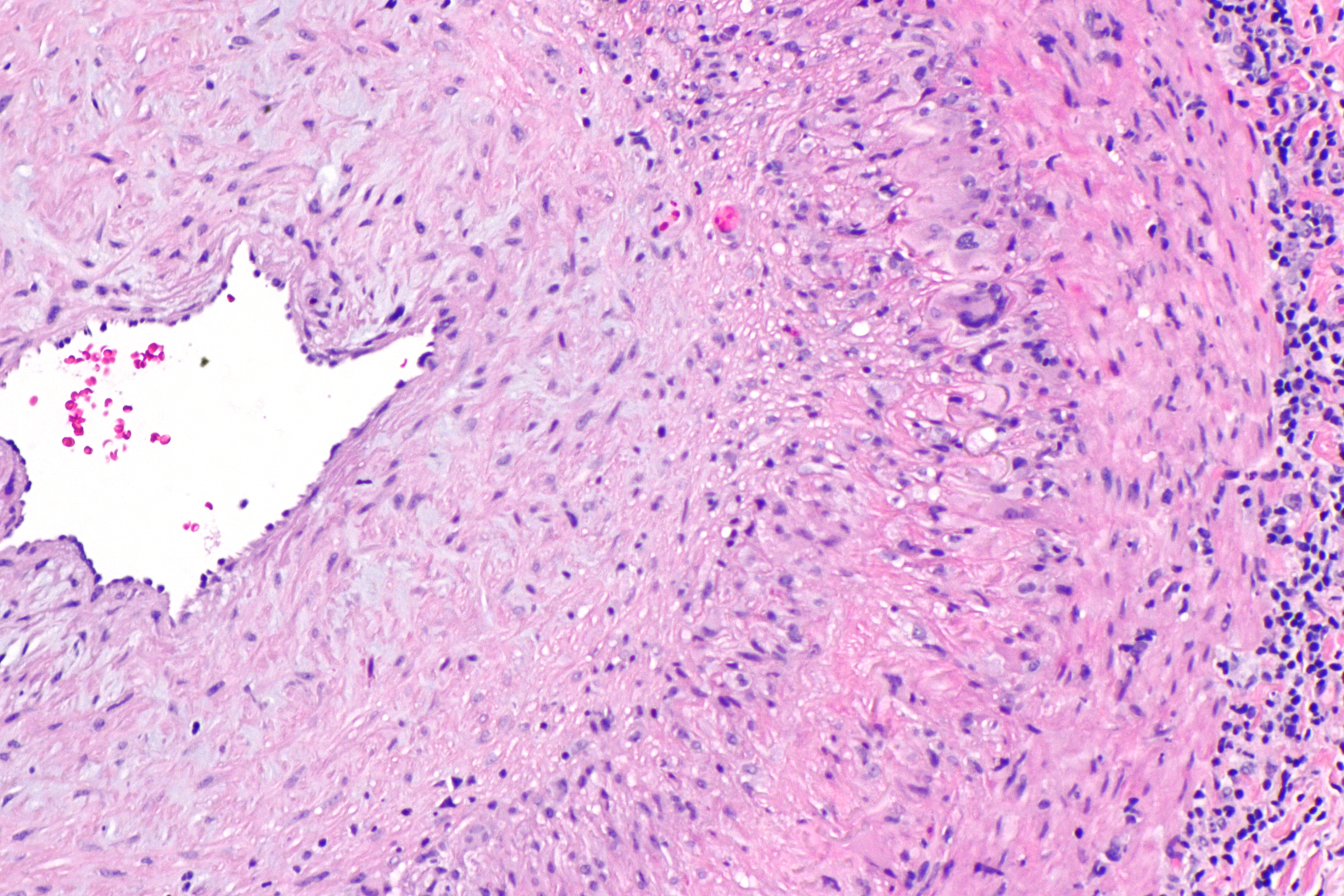
Mönckeberg's arteriosclerosis, or Mönckeberg's sclerosis, is a non-inflammatory form of arteriosclerosis (artery hardening), which differs from atherosclerosis traditionally. Calcium deposits are found in the muscular middle layer of the walls of arteries (the tunica media) with no obstruction of the lumen. It is an example of dystrophic calcification. This condition occurs as an age-related degenerative process. However, it can occur in pseudoxanthoma elasticum and idiopathic arterial calcification of infancy as a pathological condition, as well. Its clinical significance and cause are not well understood and its relationship to atherosclerosis and other forms of vascular calcification are the subject of disagreement. Mönckeberg's arteriosclerosis is named after Johann Georg Mönckeberg, who first described it in 1903. The severity of calcium deposits formed by Mönckeberg's arteriosclerosis can be categorized into stages based on the histological appearance. Understandi ... [...More Info...] [...Related Items...] OR: [Wikipedia] [Google] [Baidu] |
Haematoxylin
Haematoxylin American and British English spelling differences#ae and oe, or hematoxylin (), also called natural black 1 or Colour Index International, C.I. 75290, is a chemical compound, compound extracted from wood#Heartwood and sapwood, heartwood of the logwood tree (''Haematoxylum campechianum'') with a chemical formula of . This Natural dye, naturally derived dye has been used as a staining, histologic stain, as an ink and as a dye in the textile and leather industry. As a dye, haematoxylin has been called palo de Campeche, logwood extract, bluewood and blackwood. In histology, haematoxylin staining is commonly followed by counterstain, counterstaining with eosin. When paired, this staining procedure is known as H&E staining and is one of the most commonly used combinations in histology. In addition to its use in the H&E stain, haematoxylin is also a component of the Papanicolaou stain (or Pap stain) which is widely used in the study of cytology specimens. Although the stain ... [...More Info...] [...Related Items...] OR: [Wikipedia] [Google] [Baidu] |
Arterial Calcification Due To CD73 Deficiency
Arterial calcification due to CD73 deficiency or Calcification of joints and arteries is a rare genetic disorder affecting adults. Signs and symptoms This condition is characterised by calcification of the peripheral arteries. Causes This condition is caused by mutations in the 5'-Nucleotidase Ecto ( NT5E) gene. Diagnosis Medical evaluation and genetic test are used to ascertain Arterial calcification due to CD73 deficiency Epidemiology This a rare disorder, up to 2020 less than 20 individuals have been reported to have the condition History This condition was first described in 2011. References {{DEFAULTSORT:Calcification of joints and arteries Rare genetic syndromes Congenital disorders Rare diseases Autosomal recessive disorders ... [...More Info...] [...Related Items...] OR: [Wikipedia] [Google] [Baidu] |
Diabetes Mellitus
Diabetes mellitus, commonly known as diabetes, is a group of common endocrine diseases characterized by sustained hyperglycemia, high blood sugar levels. Diabetes is due to either the pancreas not producing enough of the hormone insulin, or the cells of the body becoming unresponsive to insulin's effects. Classic symptoms include polydipsia (excessive thirst), polyuria (excessive urination), polyphagia (excessive hunger), Weight loss#Unintentional, weight loss, and blurred vision. If left untreated, the disease can lead to various health complications, including disorders of the Cardiovascular disease, cardiovascular system, Diabetic retinopathy, eye, Diabetic nephropathy, kidney, and Diabetic neuropathy, nerves. Diabetes accounts for approximately 4.2 million deaths every year, with an estimated 1.5 million caused by either untreated or poorly treated diabetes. The major types of diabetes are Type 1 diabetes, type 1 and Type 2 diabetes, type 2. The most common treatment for ty ... [...More Info...] [...Related Items...] OR: [Wikipedia] [Google] [Baidu] |
Autopsy
An autopsy (also referred to as post-mortem examination, obduction, necropsy, or autopsia cadaverum) is a surgical procedure that consists of a thorough examination of a corpse by dissection to determine the cause, mode, and manner of death; or the exam may be performed to evaluate any disease or injury that may be present for research or educational purposes. The term ''necropsy'' is generally used for non-human animals. Autopsies are usually performed by a specialized medical doctor called a pathologist. Only a small portion of deaths require an autopsy to be performed, under certain circumstances. In most cases, a medical examiner or coroner can determine the cause of death. Purposes of performance Autopsies are performed for either legal or medical purposes. Autopsies can be performed when any of the following information is desired: * Manner of death must be determined ** Determine if death was natural or unnatural ** Injury source and extent on the corpse * Pos ... [...More Info...] [...Related Items...] OR: [Wikipedia] [Google] [Baidu] |
Infarction
Infarction is tissue death (necrosis) due to Ischemia, inadequate blood supply to the affected area. It may be caused by Thrombosis, artery blockages, rupture, mechanical compression, or vasoconstriction. The resulting lesion is referred to as an infarct (from the Latin ''infarctus'', "stuffed into"). Causes Infarction occurs as a result of prolonged ischemia, which is the insufficient supply of oxygen and nutrition to an area of tissue due to a disruption in blood supply. The blood vessel supplying the affected area of tissue may be blocked due to an obstruction in the vessel (e.g., an arterial embolus, thrombus, or atherosclerotic plaque), compressed by something outside of the vessel causing it to narrow (e.g., tumor, volvulus, or hernia), ruptured by trauma causing a loss of blood pressure downstream of the rupture, or vasoconstricted, which is the narrowing of the blood vessel by contraction of the muscle wall rather than an external force (e.g., cocaine vaso ... [...More Info...] [...Related Items...] OR: [Wikipedia] [Google] [Baidu] |
Ischemia
Ischemia or ischaemia is a restriction in blood supply to any tissue, muscle group, or organ of the body, causing a shortage of oxygen that is needed for cellular metabolism (to keep tissue alive). Ischemia is generally caused by problems with blood vessels, with resultant damage to or dysfunction of tissue, i.e., hypoxia and microvascular dysfunction. It also implies local hypoxia in a part of a body resulting from constriction (such as vasoconstriction, thrombosis, or embolism). Ischemia causes not only insufficiency of oxygen but also reduced availability of nutrients and inadequate removal of metabolic wastes. Ischemia can be partial (poor perfusion) or total blockage. The inadequate delivery of oxygenated blood to the organs must be resolved either by treating the cause of the inadequate delivery or reducing the oxygen demand of the system that needs it. For example, patients with myocardial ischemia have a decreased blood flow to the heart and are prescribe ... [...More Info...] [...Related Items...] OR: [Wikipedia] [Google] [Baidu] |
Giant Cell Arteritis
Giant cell arteritis (GCA), also called temporal arteritis, is an inflammatory autoimmune disease of large blood vessels. Symptoms may include headache, pain over the temples, flu-like symptoms, double vision, and difficulty opening the mouth. Complications can include blockage of the artery to the eye with resulting blindness, as well as aortic dissection, and aortic aneurysm. GCA is frequently associated with polymyalgia rheumatica. The cause is unknown. The underlying mechanism involves inflammation of the small blood vessels that supply the walls of larger arteries. This mainly affects arteries around the head and neck, though some in the chest may also be affected. Diagnosis is suspected based on symptoms, blood tests, and medical imaging, and confirmed by biopsy of the temporal artery. However, in about 10% of people the temporal artery is normal. Treatment is typical with high doses of steroids such as prednisone or prednisolone. Once symptoms have resolved, ... [...More Info...] [...Related Items...] OR: [Wikipedia] [Google] [Baidu] |
Kidneys
In humans, the kidneys are two reddish-brown bean-shaped blood-filtering organs that are a multilobar, multipapillary form of mammalian kidneys, usually without signs of external lobulation. They are located on the left and right in the retroperitoneal space, and in adult humans are about in length. They receive blood from the paired renal arteries; blood exits into the paired renal veins. Each kidney is attached to a ureter, a tube that carries excreted urine to the bladder. The kidney participates in the control of the volume of various body fluids, fluid osmolality, acid-base balance, various electrolyte concentrations, and removal of toxins. Filtration occurs in the glomerulus: one-fifth of the blood volume that enters the kidneys is filtered. Examples of substances reabsorbed are solute-free water, sodium, bicarbonate, glucose, and amino acids. Examples of substances secreted are hydrogen, ammonium, potassium and uric acid. The nephron is the structural and functi ... [...More Info...] [...Related Items...] OR: [Wikipedia] [Google] [Baidu] |
Heart
The heart is a muscular Organ (biology), organ found in humans and other animals. This organ pumps blood through the blood vessels. The heart and blood vessels together make the circulatory system. The pumped blood carries oxygen and nutrients to the tissue, while carrying metabolic waste such as carbon dioxide to the lungs. In humans, the heart is approximately the size of a closed fist and is located between the lungs, in the middle compartment of the thorax, chest, called the mediastinum. In humans, the heart is divided into four chambers: upper left and right Atrium (heart), atria and lower left and right Ventricle (heart), ventricles. Commonly, the right atrium and ventricle are referred together as the right heart and their left counterparts as the left heart. In a healthy heart, blood flows one way through the heart due to heart valves, which prevent cardiac regurgitation, backflow. The heart is enclosed in a protective sac, the pericardium, which also contains a sma ... [...More Info...] [...Related Items...] OR: [Wikipedia] [Google] [Baidu] |
Pulse Pressure
Pulse pressure is the difference between systolic and diastolic blood pressure. It is measured in millimeters of mercury (mmHg). It represents the force that the heart generates each time it contracts. Healthy pulse pressure is around 40 mmHg. A pulse pressure that is consistently 60 mmHg or greater is likely to be associated with disease, and a pulse pressure of 50 mmHg or more increases the risk of cardiovascular disease. Pulse pressure is considered low if it is less than 25% of the systolic. (For example, if the systolic pressure is 120 mmHg, then the pulse pressure would be considered low if it were less than 30 mmHg, since 30 is 25% of 120.) A very low pulse pressure can be a symptom of disorders such as congestive heart failure. Calculation Pulse pressure is calculated as the difference between the systolic blood pressure and the diastolic blood pressure. The systemic pulse pressure is approximately proportional to stroke volume, or the amoun ... [...More Info...] [...Related Items...] OR: [Wikipedia] [Google] [Baidu] |
Arterial Stiffness
Arterial stiffness occurs as a consequence of biological aging, arteriosclerosis and genetic disorders, such as Marfan, Williams, and Ehlers-Danlos syndromes. Inflammation plays a major role in arteriosclerosis and arterial stiffness. Increased arterial stiffness is associated with an increased risk of cardiovascular events such as myocardial infarction, hypertension, heart failure, and stroke. The World Health Organization identified cardiovascular disease as the leading cause of death globally in 2019. Degenerative changes that occur with age in the walls of large elastic arteries are thought to contribute to increased stiffening over time, including the disruption of lamellar elastin structures within the wall, possibly due to repeated cycles of mechanical stress; inflammation; changes in arterial collagen proteins, partially as a compensatory mechanism against the loss of arterial elastin and partially due to fibrosis; and crosslinking of adjacent collagen fibers by ... [...More Info...] [...Related Items...] OR: [Wikipedia] [Google] [Baidu] |
Calciphylaxis
Calciphylaxis, also known as calcific uremic arteriolopathy (CUA) or “Grey Scale”, is a rare syndrome characterized by painful skin lesions. The pathogenesis of calciphylaxis is unclear but believed to involve calcification of the small blood vessels located within the fatty tissue and deeper layers of the skin, blood clots, and eventual death of skin cells due to lack of blood flow. It is seen mostly in people with end-stage kidney disease but can occur in the earlier stages of chronic kidney disease and rarely in people with normally functioning kidneys. Calciphylaxis is a rare but serious disease, believed to affect 1-4% of all dialysis patients. It results in chronic non-healing wounds and indicates poor prognosis, with typical life expectancy of less than one year. Calciphylaxis is one type of extraskeletal calcification. Similar extraskeletal calcifications are observed in some people with high levels of calcium in the blood, including people with milk-alkali sy ... [...More Info...] [...Related Items...] OR: [Wikipedia] [Google] [Baidu] |


