|
McBurney's Point
McBurney's point is the point over the right side of the abdomen that is one-third of the distance from the anterior superior iliac spine to the Navel, umbilicus (navel). This is near the most common location of the Vermiform appendix, appendix. Location McBurney's point is located one third of the distance from the right anterior superior iliac spine to the Navel, umbilicus (navel). This point roughly corresponds to the most common location of the base of the Vermiform appendix, appendix, where it is attached to the cecum. Appendicitis Deep Pain, tenderness at McBurney's point, known as McBurney's sign, is a sign of acute appendicitis. The clinical sign of referred pain in the epigastrium when pressure is applied is also known as Aaron's sign. Specific localization of tenderness to McBurney's point indicates that inflammation is no longer limited to the lumen of the bowel (which localizes pain poorly), and is irritating the lining of the peritoneum at the place where the pe ... [...More Info...] [...Related Items...] OR: [Wikipedia] [Google] [Baidu] |
Navel
The navel (clinically known as the umbilicus; : umbilici or umbilicuses; also known as the belly button or tummy button) is a protruding, flat, or hollowed area on the abdomen at the attachment site of the umbilical cord. Structure The umbilicus is used to visually separate the abdomen into quadrants. The umbilicus is a prominent Scar#Umbilical, scar on the abdomen, with its position being relatively consistent among humans. The skin around the waist at the level of the umbilicus is supplied by the tenth thoracic spinal nerve (T10 dermatome (anatomy), dermatome). The umbilicus itself typically lies at a vertical level corresponding to the junction between the L3 and L4 vertebrae, with a normal variation among people between the L3 and L5 vertebrae. Parts of the adult navel include the "umbilical cord remnant" or "umbilical tip", which is the often protruding scar left by the detachment of the umbilical cord. This is located in the center of the navel, sometimes described ... [...More Info...] [...Related Items...] OR: [Wikipedia] [Google] [Baidu] |
Laparoscopy
Laparoscopy () is an operation performed in the abdomen or pelvis using small incisions (usually 0.5–1.5 cm) with the aid of a camera. The laparoscope aids diagnosis or therapeutic interventions with a few small cuts in the abdomen.MedlinePlus > Laparoscopy Update Date: 21 August 2009. Updated by: James Lee, MD // No longer valid Laparoscopic surgery, also called minimally invasive procedure, bandaid surgery, or keyhole surgery, is a modern surgical technique. There are a number of advantages to the patient with laparoscopic surgery versus an exploratory laparotomy. These include reduced pain due to smaller incisions, reduced hemorrhaging, and shorter recovery time. The key element is the use of a laparoscope, a long fiber optic cable system that allows viewing of the affected area by snaking the cable from a more distant, but more easily accessible location. Laparoscopic surgery includes operations within the abdominal or pelvic cavities, whereas keyhole surgery per ... [...More Info...] [...Related Items...] OR: [Wikipedia] [Google] [Baidu] |
New York Medical Journal (1865)
The ''New York Medical Journal'' is an American medical journal A medical journal is a peer-reviewed scientific journal that communicates medical information to physicians, other health professionals. Journals that cover many medical specialties are sometimes called general medical journals. History The first .... It was first published in 1865. References General medical journals {{general-medical-journal-stub ... [...More Info...] [...Related Items...] OR: [Wikipedia] [Google] [Baidu] |
Charles McBurney (surgeon)
Charles Heber McBurney (17 February 1845 in Roxbury, Massachusetts – 7 November 1913 in Brookline, Massachusetts) was an American surgeon, well known for describing McBurney's point in appendicitis. Life and career Charles McBurney was born in 1845. He graduated in the arts from Harvard College in 1866, and qualified in medicine at the College of Physicians and Surgeons of Columbia University in New York City with an M.D. in 1870. He trained further in Europe for 2 years, and started practice in New York in 1873. He became assistant surgeon to the Bellevue Hospital in 1880, and surgeon-in-chief of the Roosevelt Hospital (now Mount Sinai West) in 1888. Here he did his most famous work on appendicitis, presenting his report on operative management to the New York Surgical Society in 1889. He described the point of greatest tenderness in appendicitis, which is now known as McBurney's point. He was professor of surgery from 1889 to 1907, and thereafter became emeritus professor ... [...More Info...] [...Related Items...] OR: [Wikipedia] [Google] [Baidu] |
Surgeon
In medicine, a surgeon is a medical doctor who performs surgery. Even though there are different traditions in different times and places, a modern surgeon is a licensed physician and received the same medical training as physicians before specializing in surgery. In some countries and jurisdictions, the title of 'surgeon' is restricted to maintain the integrity of the craft group in the medical profession. A specialist regarded as a legally recognized surgeon includes podiatry, dentistry, and veterinary medicine. It is estimated that surgeons perform over 300 million surgical procedures globally each year. History The first person to document a surgery was the 6th century BC Indian physician-surgeon, Sushruta. He specialized in cosmetic plastic surgery and even documented an open rhinoplasty procedure.Papel, Ira D. and Frodel, John (2008) ''Facial Plastic and Reconstructive Surgery''. Thieme Medical Pub. His Masterpiece, magnum opus ''Suśruta-saṃhitā'' is one of the m ... [...More Info...] [...Related Items...] OR: [Wikipedia] [Google] [Baidu] |
Americans
Americans are the Citizenship of the United States, citizens and United States nationality law, nationals of the United States, United States of America.; ; Law of the United States, U.S. federal law does not equate nationality with Race (human categorization), race or ethnicity but rather with citizenship.* * * * * * * The U.S. has 37 American ancestries, ancestry groups with more than one million individuals. White Americans form the largest race (human classification), racial and ethnic group at 61.6% of the U.S. population, with Non-Hispanic whites, non-Hispanic Whites making up 57.8% of the population. Hispanic and Latino Americans form the second-largest group and are 18.7% of the American population. African Americans, Black Americans constitute the country's third-largest ancestry group and are 12.4% of the total U.S. population. Asian Americans are the country's fourth-largest group, composing 6% of the American population. The country's 3.7 million Native Americans i ... [...More Info...] [...Related Items...] OR: [Wikipedia] [Google] [Baidu] |
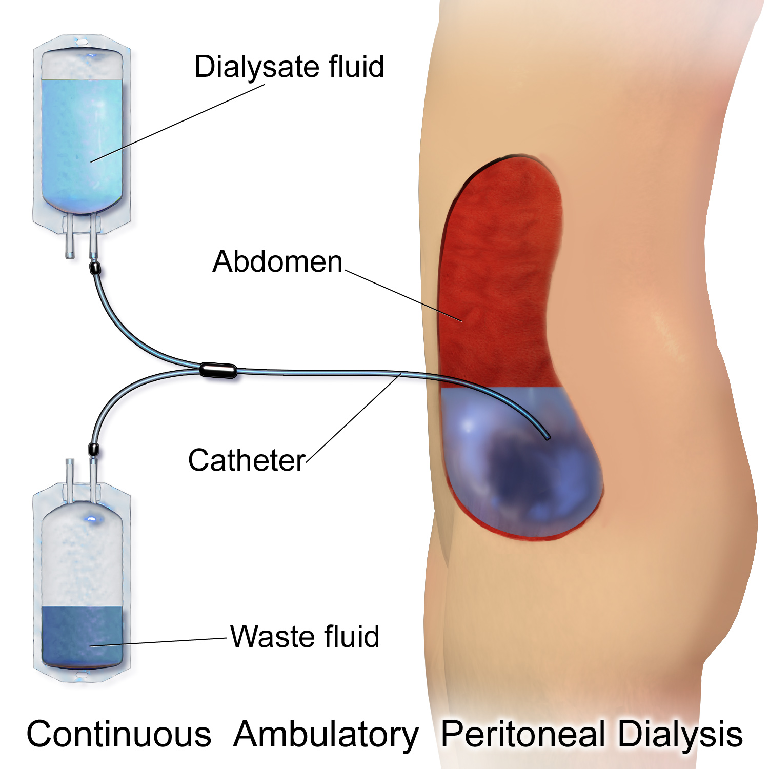
Catheter
In medicine, a catheter ( ) is a thin tubing (material), tube made from medical grade materials serving a broad range of functions. Catheters are medical devices that can be inserted in the body to treat diseases or perform a surgical procedure. Catheters are manufactured for specific applications, such as cardiovascular, urological, gastrointestinal, neurovascular and ophthalmic procedures. The process of inserting a catheter is called ''catheterization''. In most uses, a catheter is a thin, flexible tube (''soft'' catheter) though catheters are available in varying levels of stiffness depending on the application. A catheter left inside the body, either temporarily or permanently, may be referred to as an "indwelling catheter" (for example, a peripherally inserted central catheter). A permanently inserted catheter may be referred to as a "permcath" (originally a trademark). Catheters can be inserted into a body cavity, duct, or vessel, brain, skin or adipose tissue. Functional ... [...More Info...] [...Related Items...] OR: [Wikipedia] [Google] [Baidu] |
Peritoneal Dialysis
Peritoneal dialysis (PD) is a type of kidney dialysis, dialysis that uses the peritoneum in a person's abdomen as the membrane through which fluid and dissolved substances are exchanged with the blood. It is used to remove excess fluid, correct electrolyte problems, and remove toxins in those with kidney failure. Peritoneal dialysis has better outcomes than hemodialysis during the first two years. Other benefits include greater flexibility and better tolerability in those with significant heart disease. Side effects Complications may include peritonitis, infections within the abdomen, hernias, high blood sugar, bleeding in the abdomen, and blockage of the catheter. Peritoneal dialysis is not possible in those with significant prior abdominal surgery or inflammatory bowel disease. It requires some degree of technical skill to be done properly. Mechanism In peritoneal dialysis, a specific solution is introduced and then removed through a permanent tube in the lower abdomen. Th ... [...More Info...] [...Related Items...] OR: [Wikipedia] [Google] [Baidu] |
Intercostal Space
The intercostal space (ICS) is the anatomic space between two ribs (Lat. costa). Since there are 12 ribs on each side, there are 11 intercostal spaces, each numbered for the rib superior to it. Structures in intercostal space * several kinds of intercostal muscle * intercostal arteries and intercostal veins * intercostal lymph nodes * intercostal nerves Order of components Muscles There are 3 muscular layers in each intercostal space, consisting of the external intercostal muscle, the internal intercostal muscle, and the thinner innermost intercostal muscle. These muscles help to move the ribs during breathing. Neurovascular bundles Neurovascular bundles are located between the internal intercostal muscle and the innermost intercostal muscle. The neurovascular bundle has a strict order of vein-artery-nerve (VAN), from top to bottom. This neurovascular bundle runs high in the intercostal space, and the smaller collateral neurovascular bundle runs just superior ... [...More Info...] [...Related Items...] OR: [Wikipedia] [Google] [Baidu] |
Vascular Surgery
Vascular surgery is a surgical subspecialty in which vascular diseases involving the arteries, veins, or lymphatic vessels, are managed by medical therapy, minimally-invasive catheter procedures and surgical reconstruction. The specialty evolved from general surgery, general and cardiac surgery, cardiovascular surgery where it refined the management of just the vessels, no longer treating the heart or other organs. Modern vascular surgery includes open surgery techniques, endovascular (minimally invasive) techniques and medical management of vascular diseases - unlike the parent specialities. The vascular surgeon is trained in the diagnosis and management of diseases affecting all parts of the vascular system excluding the coronaries and intracranial vasculature. Vascular surgeons also are called to assist other physicians to carry out surgery near vessels, or to salvage vascular injuries that include hemorrhage control, dissection, occlusion or simply for safe exposure of vascul ... [...More Info...] [...Related Items...] OR: [Wikipedia] [Google] [Baidu] |
Aorta
The aorta ( ; : aortas or aortae) is the main and largest artery in the human body, originating from the Ventricle (heart), left ventricle of the heart, branching upwards immediately after, and extending down to the abdomen, where it splits at the aortic bifurcation into two smaller arteries (the common iliac artery, common iliac arteries). The aorta distributes Oxygen saturation (medicine), oxygenated blood to all parts of the body through the systemic circulation. Structure Sections In anatomical sources, the aorta is usually divided into sections. One way of classifying a part of the aorta is by anatomical compartment, where the thoracic aorta (or thoracic portion of the aorta) runs from the heart to the thoracic diaphragm, diaphragm. The aorta then continues downward as the abdominal aorta (or abdominal portion of the aorta) from the diaphragm to the aortic bifurcation. Another system divides the aorta with respect to its course and the direction of blood flow. In this s ... [...More Info...] [...Related Items...] OR: [Wikipedia] [Google] [Baidu] |
Pseudoaneurysm
A pseudoaneurysm, also known as a false aneurysm, is a locally contained hematoma outside an artery or the heart due to damage to the vessel wall. The injury passes through all three layers of the arterial wall, causing a leak, which is contained by a new, weak "wall" formed by the products of the clotting cascade. A pseudoaneurysm (PSA) does not contain any layer of the vessel wall. This differentiates it from a true aneurysm, which is contained by all three layers of the vessel wall, and a dissecting aneurysm, which has a breach in the innermost layer of an artery and subsequent dissection/separation of the tunica intima from the tunica media. A pseudoaneurysm, being associated with a vessel, can be pulsatile; it may be confused with a true aneurysm or dissecting aneurysm. The most common presentation of pseudoaneurysm is femoral artery pseudoaneurysm following access for an endovascular procedure, and this event may complicate up to 8% of vascular interventional proc ... [...More Info...] [...Related Items...] OR: [Wikipedia] [Google] [Baidu] |


