|
MAChR
Muscarinic acetylcholine receptors (mAChRs) are acetylcholine receptors that form G protein-coupled receptor complexes in the cell membranes of certain neurons and other cells. They play several roles, including acting as the main end-receptor stimulated by acetylcholine released from postganglionic fibers. They are mainly found in the parasympathetic nervous system, but also have a role in the sympathetic nervous system in the control of sweat glands. Muscarinic receptors are so named because they are more sensitive to muscarine than to nicotine. Their counterparts are nicotinic acetylcholine receptors (nAChRs), receptor ion channels that are also important in the autonomic nervous system. Many drugs and other substances (for example pilocarpine and scopolamine) manipulate these two distinct receptors by acting as selective agonists or antagonists. Function Acetylcholine (ACh) is a neurotransmitter found in the brain, neuromuscular junctions and the autonomic ganglia. Mu ... [...More Info...] [...Related Items...] OR: [Wikipedia] [Google] [Baidu] |
Acetylcholine Receptor
An acetylcholine receptor (abbreviated AChR) or a cholinergic receptor is an integral membrane protein that responds to the binding of acetylcholine, a neurotransmitter. Classification Like other transmembrane receptors, acetylcholine receptors are classified according to their "pharmacology," or according to their relative affinities and sensitivities to different molecules. Although all acetylcholine receptors, by definition, respond to acetylcholine, they respond to other molecules as well. * Nicotinic acetylcholine receptors (''nAChR'', also known as " ionotropic" acetylcholine receptors) are particularly responsive to nicotine. The nicotine ACh receptor is also a Na+, K+ and Ca2+ ion channel. * Muscarinic acetylcholine receptors (''mAChR'', also known as " metabotropic" acetylcholine receptors) are particularly responsive to muscarine. Nicotinic and muscarinic are two main kinds of "cholinergic" receptors. Receptor types Molecular biology has shown that the nicotinic ... [...More Info...] [...Related Items...] OR: [Wikipedia] [Google] [Baidu] |
Muscarinic Acetylcholine Receptor M2-3UON
A muscarinic acetylcholine receptor agonist, also simply known as a muscarinic agonist or as a muscarinic agent, is an agent that activates the activity of the muscarinic acetylcholine receptor. The muscarinic receptor has different subtypes, labelled M1-M5, allowing for further differentiation. Clinical significance M1 M1-type muscarinic acetylcholine receptors play a role in cognitive processing. In Alzheimer disease (AD), amyloid formation may decrease the ability of these receptors to transmit signals, leading to decreased cholinergic activity. As these receptors themselves appear relatively unchanged in the disease process, they have become a potential therapeutic target when trying to improve cognitive function in patients with AD. A number of muscarinic agonists have been developed and are under investigation to treat AD. These agents show promise as they are neurotrophic, decrease amyloid depositions, and improve damage due to oxidative stress. Tau-phosphorylation ... [...More Info...] [...Related Items...] OR: [Wikipedia] [Google] [Baidu] |
Acetylcholine
Acetylcholine (ACh) is an organic compound that functions in the brain and body of many types of animals (including humans) as a neurotransmitter. Its name is derived from its chemical structure: it is an ester of acetic acid and choline. Parts in the body that use or are affected by acetylcholine are referred to as cholinergic. Acetylcholine is the neurotransmitter used at the neuromuscular junction. In other words, it is the chemical that motor neurons of the nervous system release in order to activate muscles. This property means that drugs that affect cholinergic systems can have very dangerous effects ranging from paralysis to convulsions. Acetylcholine is also a neurotransmitter in the autonomic nervous system, both as an internal transmitter for both the sympathetic nervous system, sympathetic and the parasympathetic nervous system, and as the final product released by the parasympathetic nervous system. Acetylcholine is the primary neurotransmitter of the parasympathet ... [...More Info...] [...Related Items...] OR: [Wikipedia] [Google] [Baidu] |
Pilocarpine
Pilocarpine, sold under the brand name Pilopine HS among others, is a lactone alkaloid originally extracted from plants of the Pilocarpus genus. It is used as a medication to reduce pressure inside the eye and treat dry mouth. As an eye drop it is used to manage angle closure glaucoma until surgery can be performed, ocular hypertension, primary open angle glaucoma, and to constrict the pupil after dilation. However, due to its side effects, it is no longer typically used for long-term management. Onset of effects with the drops is typically within an hour and lasts for up to a day. By mouth it is used for dry mouth as a result of Sjögren syndrome or radiation therapy. Common side effects of the eye drops include irritation of the eye, increased tearing, headache, and blurry vision. Other side effects include allergic reactions and retinal detachment. Use is generally not recommended during pregnancy. Pilocarpine is in the miotics family of medication. It works by activ ... [...More Info...] [...Related Items...] OR: [Wikipedia] [Google] [Baidu] |
Adrenal Medulla
The adrenal medulla () is the inner part of the adrenal gland. It is located at the center of the gland, being surrounded by the adrenal cortex. It is the innermost part of the adrenal gland, consisting of chromaffin cells that secrete catecholamines, including epinephrine (adrenaline), norepinephrine (noradrenaline), and a small amount of dopamine, in response to stimulation by sympathetic preganglionic neurons. Structure The adrenal medulla consists of irregularly shaped cells grouped around blood vessels. These cells are intimately connected with the sympathetic division of the autonomic nervous system (ANS). These adrenal medullary cells are modified postganglionic neurons, and preganglionic autonomic nerve fibers lead to them directly from the central nervous system. The adrenal medulla affects energy availability, heart rate, and basal metabolic rate. Recent research indicates that the adrenal medulla may receive input from higher-order cognitive centers in the prefron ... [...More Info...] [...Related Items...] OR: [Wikipedia] [Google] [Baidu] |
Postganglionic Fiber
In the autonomic nervous system, nerve fibers from the ganglion to the effector organ are called postganglionic nerve fibers. Neurotransmitters The neurotransmitters of postganglionic fibers differ: * In the parasympathetic division, neurons are ''cholinergic''. That is to say acetylcholine is the primary neurotransmitter responsible for the communication between neurons on the parasympathetic pathway. * In the sympathetic division, neurons are mostly ''adrenergic'' (that is, epinephrine and norepinephrine function as the primary neurotransmitters). Notable exceptions to this rule include the sympathetic innervation of sweat glands and arrectores pilorum muscles where the neurotransmitter at both pre and post ganglionic synapses is acetylcholine. Another notable structure is the medulla of the adrenal gland, where chromaffin cells function as modified post-ganglionic nerves. Instead of releasing epinephrine and norepinephrine into a synaptic cleft, these cells of the adrenal ... [...More Info...] [...Related Items...] OR: [Wikipedia] [Google] [Baidu] |
IPSP
An inhibitory postsynaptic potential (IPSP) is a kind of synaptic potential that makes a postsynaptic neuron less likely to generate an action potential.Purves et al. Neuroscience. 4th ed. Sunderland (MA): Sinauer Associates, Incorporated; 2008. The opposite of an inhibitory postsynaptic potential is an excitatory postsynaptic potential (EPSP), which is a synaptic potential that makes a postsynaptic neuron ''more'' likely to generate an action potential. IPSPs can take place at all chemical synapses, which use the secretion of neurotransmitters to create cell-to-cell signalling. EPSPs and IPSPs compete with each other at numerous synapses of a neuron. This determines whether an action potential occurring at the presynaptic terminal produces an action potential at the postsynaptic membrane. Some common neurotransmitters involved in IPSPs are GABA and glycine. Inhibitory presynaptic neurons release neurotransmitters that then bind to the postsynaptic receptors; this induces a ch ... [...More Info...] [...Related Items...] OR: [Wikipedia] [Google] [Baidu] |
Excitatory Postsynaptic Potential
In neuroscience, an excitatory postsynaptic potential (EPSP) is a postsynaptic potential that makes the postsynaptic neuron more likely to fire an action potential. This temporary depolarization of postsynaptic membrane potential, caused by the flow of positively charged ions into the postsynaptic cell, is a result of opening ligand-gated ion channels. These are the opposite of inhibitory postsynaptic potentials (IPSPs), which usually result from the flow of ''negative'' ions into the cell or positive ions ''out'' of the cell. EPSPs can also result from a decrease in outgoing positive charges, while IPSPs are sometimes caused by an increase in positive charge outflow. The flow of ions that causes an EPSP is an excitatory postsynaptic current (EPSC). EPSPs, like IPSPs, are graded (i.e. they have an additive effect). When multiple EPSPs occur on a single patch of postsynaptic membrane, their combined effect is the sum of the individual EPSPs. Larger EPSPs result in greater membra ... [...More Info...] [...Related Items...] OR: [Wikipedia] [Google] [Baidu] |
Autonomic Ganglion
An autonomic ganglion is a cluster of nerve cell bodies (a ganglion) in the autonomic nervous system The autonomic nervous system (ANS), sometimes called the visceral nervous system and formerly the vegetative nervous system, is a division of the nervous system that operates viscera, internal organs, smooth muscle and glands. The autonomic nervo .... The two types are the sympathetic ganglion and the parasympathetic ganglion. References Autonomic nervous system {{neuroanatomy-stub ... [...More Info...] [...Related Items...] OR: [Wikipedia] [Google] [Baidu] |
Autonomic Ganglia
An autonomic ganglion is a cluster of neuron, nerve cell Cell body, bodies (a ganglion) in the autonomic nervous system. The two types are the sympathetic ganglion and the parasympathetic ganglion. References Autonomic ganglia, Autonomic nervous system {{neuroanatomy-stub ... [...More Info...] [...Related Items...] OR: [Wikipedia] [Google] [Baidu] |
Neuromuscular Junctions
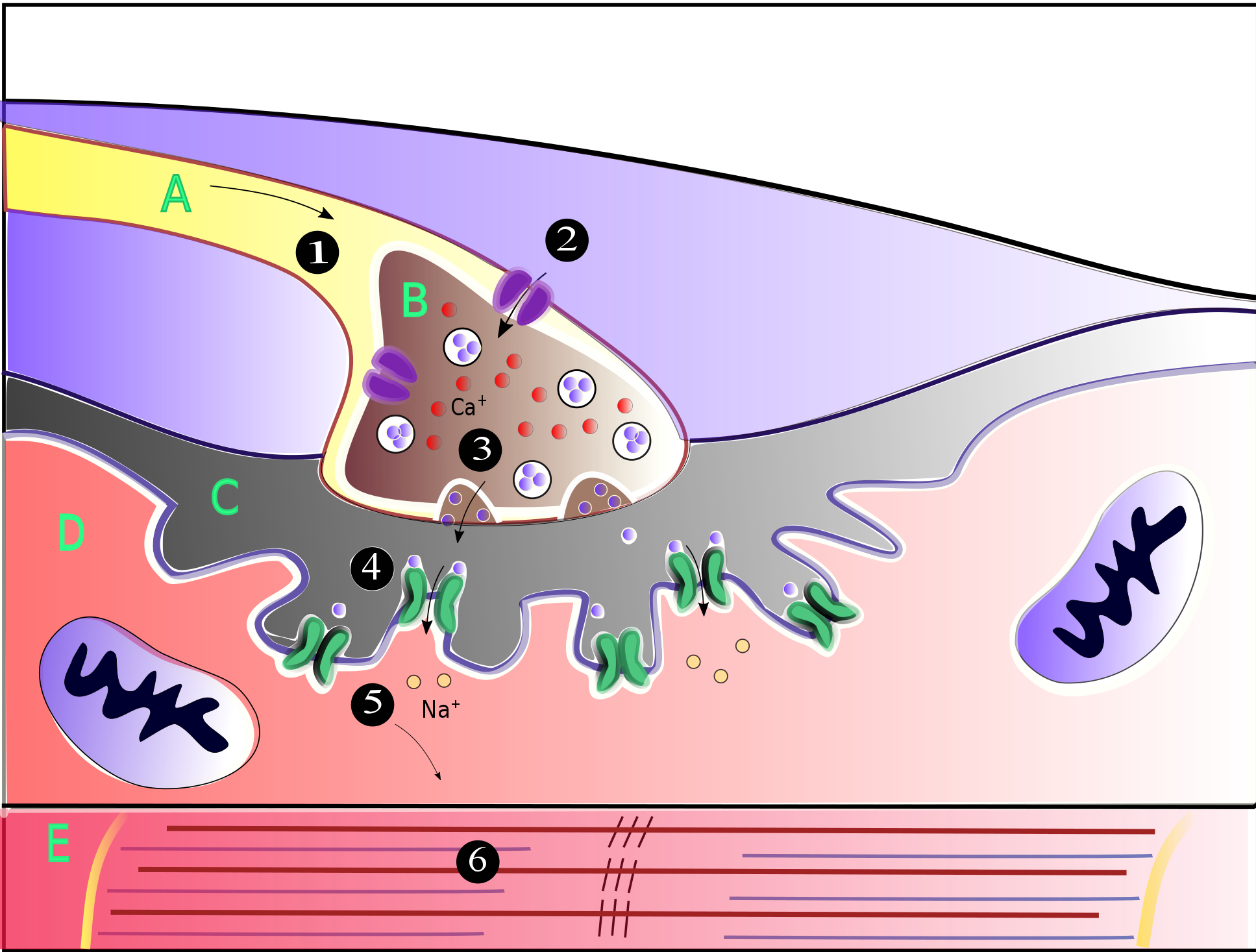
A neuromuscular junction (or myoneural junction) is a chemical synapse between a motor neuron and a muscle fiber. It allows the motor neuron to transmit a signal to the muscle fiber, causing muscle contraction. Muscles require innervation to function—and even just to maintain muscle tone, avoiding atrophy. In the neuromuscular system, nerves from the central nervous system and the peripheral nervous system are linked and work together with muscles. Synaptic transmission at the neuromuscular junction begins when an action potential reaches the presynaptic terminal of a motor neuron, which activates voltage-gated calcium channels to allow calcium ions to enter the neuron. Calcium ions bind to sensor proteins (synaptotagmins) on synaptic vesicles, triggering vesicle fusion with the cell membrane and subsequent neurotransmitter release from the motor neuron into the synaptic cleft. In vertebrates, motor neurons release acetylcholine (ACh), a small molecule neurotransmitter, which ... [...More Info...] [...Related Items...] OR: [Wikipedia] [Google] [Baidu] |
Brain
The brain is an organ (biology), organ that serves as the center of the nervous system in all vertebrate and most invertebrate animals. It consists of nervous tissue and is typically located in the head (cephalization), usually near organs for special senses such as visual perception, vision, hearing, and olfaction. Being the most specialized organ, it is responsible for receiving information from the sensory nervous system, processing that information (thought, cognition, and intelligence) and the coordination of motor control (muscle activity and endocrine system). While invertebrate brains arise from paired segmental ganglia (each of which is only responsible for the respective segmentation (biology), body segment) of the ventral nerve cord, vertebrate brains develop axially from the midline dorsal nerve cord as a brain vesicle, vesicular enlargement at the rostral (anatomical term), rostral end of the neural tube, with centralized control over all body segments. All vertebr ... [...More Info...] [...Related Items...] OR: [Wikipedia] [Google] [Baidu] |


