|
List Of Homologues Of The Human Reproductive System
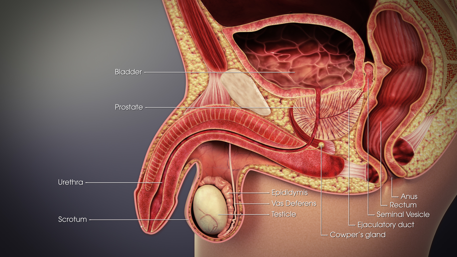
This list of related male and female reproductive organs shows how the male and female reproductive organs and the development of the reproductive system are related, sharing a common developmental path. This makes them biological homologues. These organs differentiate into the respective sex organs in males and females. List Internal organs External organs The external genitalia of both males and females have similar origins. They arise from the genital tubercle that forms anterior to the cloacal folds (proliferating mesenchymal cells around the cloacal membrane). The caudal aspect of the cloacal folds further subdivides into the posterior anal folds and the anterior urethral folds. Bilateral to the urethral fold, genital swellings (tubercles) become prominent. These structures are the future scrotal swellings and labia majora in males and females, respectively. The genital tubercles of an eight-week-old embryo of either sex are identical. They both have a glans area, whi ... [...More Info...] [...Related Items...] OR: [Wikipedia] [Google] [Baidu] |
Cervix
The cervix or cervix uteri (Latin, 'neck of the uterus') is the lower part of the uterus (womb) in the human female reproductive system. The cervix is usually 2 to 3 cm long (~1 inch) and roughly cylindrical in shape, which changes during pregnancy. The narrow, central cervical canal runs along its entire length, connecting the uterine cavity and the lumen of the vagina. The opening into the uterus is called the internal os, and the opening into the vagina is called the external os. The lower part of the cervix, known as the vaginal portion of the cervix (or ectocervix), bulges into the top of the vagina. The cervix has been documented anatomically since at least the time of Hippocrates, over 2,000 years ago. The cervical canal is a passage through which sperm must travel to fertilize an egg cell after sexual intercourse. Several methods of contraception, including cervical caps and cervical diaphragms, aim to block or prevent the passage of sperm through the cervic ... [...More Info...] [...Related Items...] OR: [Wikipedia] [Google] [Baidu] |
Urogenital Sinus
The urogenital sinus is a part of the human body only present in the development of the urinary and reproductive organs. It is the ventral part of the cloaca, formed after the cloaca separates from the anal canal during the fourth to seventh weeks of development. In males, the UG sinus is divided into three regions: upper, pelvic, and phallic. The upper part gives rise to the urinary bladder and the pelvic part gives rise to the prostatic and membranous parts of the urethra, the prostate and the bulbourethral gland (Cowper's). The phallic portion gives rise to the spongy (bulbar) part of the urethra and the urethral glands (Littre's). Note that the penile part of the urethra originates from urogenital fold. In females, the pelvic part of the UG sinus gives rise to the sinovaginal bulbs, structures that will eventually form the inferior two thirds of the vagina. This process begins when the lower tip of the paramesonephric ducts, the structures that will eventually form the ... [...More Info...] [...Related Items...] OR: [Wikipedia] [Google] [Baidu] |
Seminal Vesicle
The seminal vesicles (also called vesicular glands, or seminal glands) are a pair of two convoluted tubular glands that lie behind the urinary bladder of some male mammals. They secrete fluid that partly composes the semen. The vesicles are 5–10 cm in size, 3–5 cm in diameter, and are located between the bladder and the rectum. They have multiple outpouchings which contain secretory glands, which join together with the vas deferens at the ejaculatory duct. They receive blood from the vesiculodeferential artery, and drain into the vesiculodeferential veins. The glands are lined with column-shaped and cuboidal cells. The vesicles are present in many groups of mammals, but not marsupials, monotremes or carnivores. Inflammation of the seminal vesicles is called seminal vesiculitis, most often is due to bacterial infection as a result of a sexually transmitted disease or following a surgical procedure. Seminal vesiculitis can cause pain in the lower abdomen, scrot ... [...More Info...] [...Related Items...] OR: [Wikipedia] [Google] [Baidu] |
Vas Deferens
The vas deferens or ductus deferens is part of the male reproductive system of many vertebrates. The ducts transport sperm from the epididymis to the ejaculatory ducts in anticipation of ejaculation. The vas deferens is a partially coiled tube which exits the abdominal cavity through the inguinal canal. Etymology ''Vas deferens'' is Latin, meaning "carrying-away vessel"; the plural version is ''vasa deferentia''. ''Ductus deferens'' is also Latin, meaning "carrying-away duct"; the plural version is ''ducti deferentes''. Structure There are two vasa deferentia, connecting the left and right epididymis with the seminal vesicles to form the ejaculatory duct in order to move sperm. The (human) vas deferens measures 30–35 cm in length, and 2–3 mm in diameter. The vas deferens is continuous proximally with the tail of the epididymis. The vas deferens exhibits a tortuous, convoluted initial/proximal section (which measures 2–3 cm in length). Distally, it form ... [...More Info...] [...Related Items...] OR: [Wikipedia] [Google] [Baidu] |
Epididymis
The epididymis (; plural: epididymides or ) is a tube that connects a testicle to a vas deferens in the male reproductive system. It is a single, narrow, tightly-coiled tube in adult humans, in length. It serves as an interconnection between the multiple efferent ducts at the rear of a testicle (proximally), and the vas deferens (distally). Anatomy The epididymis is situated posterior and somewhat lateral to the testis. The epididymis is invested completely by the tunica vaginalis (which is continuous with the tunica vaginalis covering the testis). The epididymis can be divided into three main regions: * The head ( la, caput). The head of the epididymis receives spermatozoa via the efferent ducts of the mediastinum testis, mediastinium of the testis at the superior pole of the testis. The head is characterized histologically by a thick epithelium with long stereocilia (described below) and a little smooth muscle. It is involved in absorbing fluid to make the sperm more concentra ... [...More Info...] [...Related Items...] OR: [Wikipedia] [Google] [Baidu] |
Gartner's Duct
Gartner's duct, also known as Gartner's canal or the ductus longitudinalis epoophori, is a potential embryological remnant in human female development of the mesonephric duct in the development of the urinary and reproductive organs. It was discovered and described in 1822 by Hermann Treschow Gartner. Gartner's duct is located in the uterus' broad ligament. Its position is parallel with the lateral uterine tube and lateral walls of vagina and cervix. The paired mesonephric ducts in the male, in contrast, go on to form the paired epididymis, ductus deferens, ejaculatory duct and seminal vesicle. In females, they may persist between the layer of the broad ligament of the uterus and in the wall of the vagina. Clinical significance These may give rise to Gartner's duct cysts. See also *List of homologues of the human reproductive system This list of related male and female reproductive organs shows how the male and female reproductive organs and the development of the re ... [...More Info...] [...Related Items...] OR: [Wikipedia] [Google] [Baidu] |
Mesonephric Duct
The mesonephric duct (also known as the Wolffian duct, archinephric duct, Leydig's duct or nephric duct) is a paired organ that forms during the embryonic development of humans and other mammals and gives rise to male reproductive organs. Structure The mesonephric duct connects the primitive kidney, the ''mesonephros'', to the cloaca. It also serves as the primordium for male urogenital structures including the epididymis, vas deferens, and seminal vesicles. Development In both male and female the mesonephric duct develops into the trigone of urinary bladder, a part of the bladder wall, but the sexes differentiate in other ways during development of the urinary and reproductive organs. Male In a male, it develops into a system of connected organs between the efferent ducts of the testis and the prostate, namely the epididymis, the vas deferens, and the seminal vesicle. The prostate forms from the urogenital sinus and the efferent ducts form from the mesonephric tu ... [...More Info...] [...Related Items...] OR: [Wikipedia] [Google] [Baidu] |
Paradidymis
The term paradidymis (organ of Giraldés) is applied to a small collection of convoluted tubules, situated in front of the lower part of the spermatic cord, above the head of the epididymis. These tubes are lined with columnar ciliated epithelium, and probably represent the remains of a part of the Wolffian body, like the epididymis, but are functionless and vestigial. The Wolffian body operates as a kidney (mesonephros The mesonephros ( el, middle kidney) is one of three excretory organs that develop in vertebrates. It serves as the main excretory organ of aquatic vertebrates and as a temporary kidney in reptiles, birds, and mammals. The mesonephros is included ...) in fishes and amphibians, but the corresponding tissue is co-opted to form parts of the male reproductive system in other classes of vertebrate. The paradidymis represents a remnant of an unused, atrophied part of the Wolffian body. References {{Authority control Scrotum ... [...More Info...] [...Related Items...] OR: [Wikipedia] [Google] [Baidu] |
Efferent Ducts
The efferent ducts (or efferent ductules or ductuli efferentes or ductus efferentes or vasa efferentia) connect the rete testis with the initial section of the epididymis.Hess 2018 There are two basic designs for efferent ductule structure: * a) multiple entries into the epididymis, as seen in most large mammals. In humans and other large mammals, there are approximately 15 to 20 efferent ducts, which also occupy nearly one third of the head of the epididymis. * b) single entry, as seen in most small animals such as rodents, where by the 3–6 ductules merge into a single small ductule prior to entering the epididymis. The ductuli are unilaminar and composed of columnar ciliated and non-ciliated (absorptive) cells. The ciliated cells serve to stir the luminal fluids, possibly to help ensure homogeneous absorption of water from the fluid produced by the testis, which results in an increase in the concentration of luminal sperm. The epithelium is surrounded by a band of smooth m ... [...More Info...] [...Related Items...] OR: [Wikipedia] [Google] [Baidu] |
Paroophoron
The paroophoron (of Johnson) consists of a few scattered rudimentary tubules, best seen in the child, situated in the broad ligament between the epoöphoron and the uterus. Named for the Welsh anatomist David Johnson who originally described the structure at the University of Wales, Aberystwyth. It is a remnant of the mesonephric tubules Mesonephric tubules are genital ridges that are next to the mesonephros. In males, some of the mesonephric kidney tubules, instead of being used to filter blood like the rest, "grow" over to the developing testes, penetrate them, and become conn .... See also * Epoophoron References External links * * Mammal female reproductive system {{genitourinary-stub ... [...More Info...] [...Related Items...] OR: [Wikipedia] [Google] [Baidu] |
Epoophoron
The epoophoron or epoöphoron (also called organ of RosenmüllerJ. C. Rosenmüller. De ovariis embryonum et foetuum humanorum. 1802. or the parovarium) is a remnant of the mesonephric tubules that can be found next to the ovary and fallopian tube. Anatomy It may contain 10–15 transverse small ducts or tubules that lead to the Gartner’s duct (also longitudinal duct of epoophoron) that represents the caudal remnant of the mesonephric duct and passes through the broad ligament and the lateral wall of the cervix and vagina. The epoophoron is a homologue to the epididymis in the male. While the epoophoron is located in the lateral portion of the mesosalpinx and mesovarium, the paroophoron (residual remnant of that part of the mesonephric duct that forms the paradidymis in the male) lies more medially in the mesosalpinx. Histology It has a unique histological profile. Clinical significance Clinically the organ may give rise to a local paraovarian cyst or adenoma. See a ... [...More Info...] [...Related Items...] OR: [Wikipedia] [Google] [Baidu] |

