|
Leopold's Maneuvers
In obstetrics, Leopold maneuvers are a common and systematic way to determine the position of a fetus inside the woman's uterus. They are named after the gynecologist Christian Gerhard Leopold. They are also used to estimate term fetal weight. The maneuvers consist of four distinct actions, each helping to determine the position of the fetus. The maneuvers are important because they help determine the position and lie of the fetus, which in conjunction with correct Health assessment, assessment of the shape of the mother, maternal human pelvis, pelvis can indicate whether the childbirth, delivery is going to be complicated, or whether a caesarean section is necessary. The Physical examination, examiner's skill and practice in performing the maneuvers are the primary factor in whether the fetal lie is correctly ascertained. Alternately, position can be determined by ultrasound performed by a sonographer or physician. Performing the maneuvers Leopold maneuvers are difficult to p ... [...More Info...] [...Related Items...] OR: [Wikipedia] [Google] [Baidu] |
Palpation
Palpation is the process of using one's hands to check the body, especially while perceiving/diagnosing a disease or illness. Usually performed by a health care practitioner, it is the process of feeling an object in or on the body to determine its size, shape, firmness, or location (for example, a veterinarian can feel the stomach of a pregnant animal to ensure good health and successful delivery). Palpation is an important part of the physical examination; the sense of touch is just as important in this examination as the sense of sight is. Physicians develop great skill in palpating problems below the surface of the body, becoming able to detect things that untrained persons would not. Mastery of anatomy and much practice are required to achieve a high level of skill. The concept of being able to detect or notice subtle tactile signs and to recognize their significance or implications is called appreciating them (just as in general vocabulary one can speak of appreciating ... [...More Info...] [...Related Items...] OR: [Wikipedia] [Google] [Baidu] |
Human Head
In human anatomy, the head is at the top of the human body. It supports the face and is maintained by the skull, which itself encloses the brain. Structure The human head consists of a fleshy outer portion, which surrounds the bony skull. The brain is enclosed within the skull. There are 22 bones in the human head. The head rests on the neck, and the seven cervical vertebrae support it. The human head typically weighs between Over 98% of humans fit into this range. There have been odd incidences where human beings have abnormally small or large heads. The Zika virus was responsible for underdeveloped heads in the early 2000s. The face is the anterior part of the head, containing the eyes, nose, and mouth. On either side of the mouth, the cheeks provide a fleshy border to the oral cavity. The ears sit to either side of the head. Blood supply The head receives blood supply through the internal and external carotid arteries. These supply the area outside of the skull (exte ... [...More Info...] [...Related Items...] OR: [Wikipedia] [Google] [Baidu] |
Bone
A bone is a rigid organ that constitutes part of the skeleton in most vertebrate animals. Bones protect the various other organs of the body, produce red and white blood cells, store minerals, provide structure and support for the body, and enable mobility. Bones come in a variety of shapes and sizes and have complex internal and external structures. They are lightweight yet strong and hard and serve multiple functions. Bone tissue (osseous tissue), which is also called bone in the uncountable sense of that word, is hard tissue, a type of specialized connective tissue. It has a honeycomb-like matrix internally, which helps to give the bone rigidity. Bone tissue is made up of different types of bone cells. Osteoblasts and osteocytes are involved in the formation and mineralization of bone; osteoclasts are involved in the resorption of bone tissue. Modified (flattened) osteoblasts become the lining cells that form a protective layer on the bone surface. The minera ... [...More Info...] [...Related Items...] OR: [Wikipedia] [Google] [Baidu] |
Buttocks
The buttocks (singular: buttock) are two rounded portions of the exterior anatomy of most mammals, located on the posterior of the pelvic region. In humans, the buttocks are located between the lower back and the perineum. They are composed of a layer of exterior skin and underlying subcutaneous fat superimposed on a left and right gluteus maximus and gluteus medius muscles. The two gluteus maximus muscles are the largest muscles in the human body. They are responsible for movements such as straightening the body into the upright (standing) posture when it is bent at the waist; maintaining the body in the upright posture by keeping the hip joints extended; and propelling the body forward via further leg (hip) extension when walking or running. In the seated position, the buttocks bear the weight of the upper body and take that weight off the feet. In many cultures, the buttocks play a role in sexual attraction. Many cultures have also used the buttocks as a primary target f ... [...More Info...] [...Related Items...] OR: [Wikipedia] [Google] [Baidu] |
Torso
The torso or trunk is an anatomical term for the central part, or the core, of the body of many animals (including humans), from which the head, neck, limbs, tail and other appendages extend. The tetrapod torso — including that of a human — is usually divided into the '' thoracic'' segment (also known as the upper torso, where the forelimbs extend), the ''abdominal'' segment (also known as the "mid-section" or " midriff"), and the '' pelvic'' and '' perineal'' segments (sometimes known together with the abdomen as the lower torso, where the hindlimbs extend). Anatomy Major organs In humans, most critical organs, with the notable exception of the brain, are housed within the torso. In the upper chest, the heart and lungs are protected by the rib cage, and the abdomen contains most of the organs responsible for digestion: the stomach, which breaks down partially digested food via gastric acid; the liver, which respectively produces bile necessary for digestion; ... [...More Info...] [...Related Items...] OR: [Wikipedia] [Google] [Baidu] |
Fetal
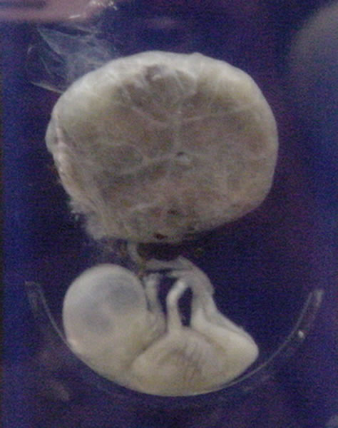
A fetus or foetus (; plural fetuses, feti, foetuses, or foeti) is the unborn offspring that develops from an animal embryo. Following embryonic development the fetal stage of development takes place. In human prenatal development, fetal development begins from the ninth week after fertilization (or eleventh week gestational age) and continues until birth. Prenatal development is a continuum, with no clear defining feature distinguishing an embryo from a fetus. However, a fetus is characterized by the presence of all the major body organs, though they will not yet be fully developed and functional and some not yet situated in their final anatomical location. Etymology The word ''fetus'' (plural '' fetuses'' or '' feti'') is related to the Latin '' fētus'' ("offspring", "bringing forth", "hatching of young") and the Greek "φυτώ" to plant. The word "fetus" was used by Ovid in Metamorphoses, book 1, line 104. The predominant British, Irish, and Commonwealth spelling is '' ... [...More Info...] [...Related Items...] OR: [Wikipedia] [Google] [Baidu] |
Abdomen
The abdomen (colloquially called the belly, tummy, midriff, tucky or stomach) is the part of the body between the thorax (chest) and pelvis, in humans and in other vertebrates. The abdomen is the front part of the abdominal segment of the torso. The area occupied by the abdomen is called the abdominal cavity. In arthropods it is the posterior tagma of the body; it follows the thorax or cephalothorax. In humans, the abdomen stretches from the thorax at the thoracic diaphragm to the pelvis at the pelvic brim. The pelvic brim stretches from the lumbosacral joint (the intervertebral disc between L5 and S1) to the pubic symphysis and is the edge of the pelvic inlet. The space above this inlet and under the thoracic diaphragm is termed the abdominal cavity. The boundary of the abdominal cavity is the abdominal wall in the front and the peritoneal surface at the rear. In vertebrates, the abdomen is a large body cavity enclosed by the abdominal muscles, at front and to ... [...More Info...] [...Related Items...] OR: [Wikipedia] [Google] [Baidu] |
Knee
In humans and other primates, the knee joins the thigh with the leg and consists of two joints: one between the femur and tibia (tibiofemoral joint), and one between the femur and patella (patellofemoral joint). It is the largest joint in the human body. The knee is a modified hinge joint, which permits flexion and extension as well as slight internal and external rotation. The knee is vulnerable to injury and to the development of osteoarthritis. It is often termed a ''compound joint'' having tibiofemoral and patellofemoral components. (The fibular collateral ligament is often considered with tibiofemoral components.) Structure The knee is a modified hinge joint, a type of synovial joint, which is composed of three functional compartments: the patellofemoral articulation, consisting of the patella, or "kneecap", and the patellar groove on the front of the femur through which it slides; and the medial and lateral tibiofemoral articulations linking the femur, or thigh ... [...More Info...] [...Related Items...] OR: [Wikipedia] [Google] [Baidu] |
Shoulder
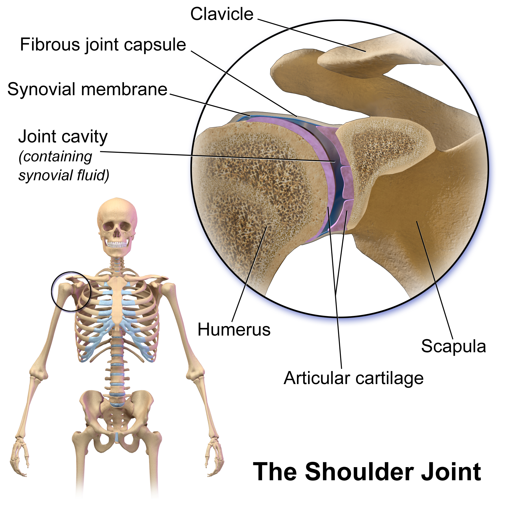
The human shoulder is made up of three bones: the clavicle (collarbone), the scapula (shoulder blade), and the humerus (upper arm bone) as well as associated muscles, ligaments and tendons. The articulations between the bones of the shoulder make up the shoulder joints. The shoulder joint, also known as the glenohumeral joint, is the major joint of the shoulder, but can more broadly include the acromioclavicular joint. In human anatomy, the shoulder joint comprises the part of the body where the humerus attaches to the scapula, and the head sits in the glenoid cavity. The shoulder is the group of structures in the region of the joint. The shoulder joint is the main joint of the shoulder. It is a ball and socket joint that allows the arm to rotate in a circular fashion or to hinge out and up away from the body. The joint capsule is a soft tissue envelope that encircles the glenohumeral joint and attaches to the scapula, humerus, and head of the biceps. It is lined by a thin, ... [...More Info...] [...Related Items...] OR: [Wikipedia] [Google] [Baidu] |
Micturate
Urination, also known as micturition, is the release of urine from the urinary bladder through the urethra to the outside of the body. It is the urinary system's form of excretion. It is also known medically as micturition, voiding, uresis, or, rarely, emiction, and known colloquially by various names including peeing, weeing, and pissing. In healthy humans (and many other animals), the process of urination is under voluntary control. In infants, some elderly individuals, and those with neurological injury, urination may occur as a reflex. It is normal for adult humans to urinate up to seven times during the day. In some animals, in addition to expelling waste material, urination can mark territory or express submissiveness. Physiologically, urination involves coordination between the central, autonomic, and somatic nervous systems. Brain centres that regulate urination include the pontine micturition center, periaqueductal gray, and the cerebral cortex. In placental mam ... [...More Info...] [...Related Items...] OR: [Wikipedia] [Google] [Baidu] |
Catheter
In medicine, a catheter (/ˈkæθətər/) is a thin tubing (material), tube made from medical grade materials serving a broad range of functions. Catheters are medical devices that can be inserted in the body to treat diseases or perform a surgical procedure. Catheters are manufactured for specific applications, such as cardiovascular, urological, gastrointestinal, neurovascular and ophthalmic procedures. The process of inserting a catheter is ''catheterization''. In most uses, a catheter is a thin, flexible tube (''soft'' catheter) though catheters are available in varying levels of stiffness depending on the application. A catheter left inside the body, either temporarily or permanently, may be referred to as an "indwelling catheter" (for example, a peripherally inserted central catheter). A permanently inserted catheter may be referred to as a "permcath" (originally a trademark). Catheters can be inserted into a body cavity, duct, or vessel, brain, skin or adipose tissue. Fun ... [...More Info...] [...Related Items...] OR: [Wikipedia] [Google] [Baidu] |


