|
List Of Skeletal Muscles Of The Human Body
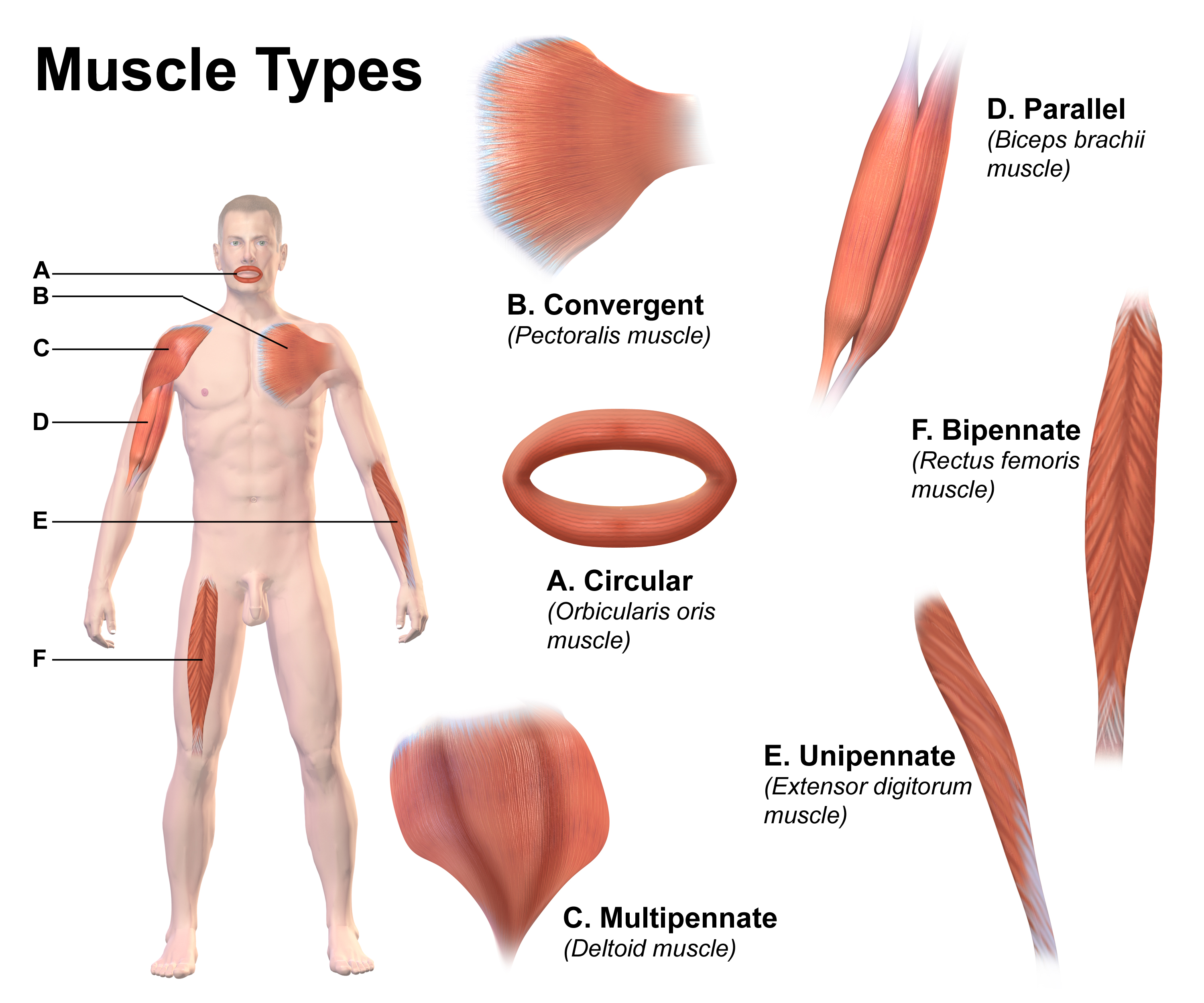
This is a table of skeletal muscles of the human anatomy, with muscle counts and other information. Table Table explanation and summary The muscles are described using anatomical terminology. The columns are as follows: For Origin, Insertion and Action please name a specific Rib, Thoracic vertebrae or Cervical vertebrae, by using C1-7, T1-12 or R1-12. Summary in numbers There does not appear to be a definitive source counting all skeletal muscles. Different sources group muscles differently, regarding physical features as different parts of a single muscle or as several muscles. There are also vestigial muscles that are present in some people but absent in others, such as the palmaris longus muscle. There are between 600 and 840 muscles within the typical human body, depending on how they are counted. In the present table, using statistical counts of the instances of each muscle, and ignoring gender-specific muscles, there are 753 skeletal muscles. Sometimes mal ... [...More Info...] [...Related Items...] OR: [Wikipedia] [Google] [Baidu] |
Skeletal Muscle
Skeletal muscle (commonly referred to as muscle) is one of the three types of vertebrate muscle tissue, the others being cardiac muscle and smooth muscle. They are part of the somatic nervous system, voluntary muscular system and typically are attached by tendons to bones of a skeleton. The skeletal muscle cells are much longer than in the other types of muscle tissue, and are also known as ''muscle fibers''. The tissue of a skeletal muscle is striated muscle tissue, striated – having a striped appearance due to the arrangement of the sarcomeres. A skeletal muscle contains multiple muscle fascicle, fascicles – bundles of muscle fibers. Each individual fiber and each muscle is surrounded by a type of connective tissue layer of fascia. Muscle fibers are formed from the cell fusion, fusion of developmental myoblasts in a process known as myogenesis resulting in long multinucleated cells. In these cells, the cell nucleus, nuclei, termed ''myonuclei'', are located along the inside ... [...More Info...] [...Related Items...] OR: [Wikipedia] [Google] [Baidu] |
Temporal Branches Of The Facial Nerve
The temporal branches of the facial nerve (frontal branch of the facial nerve) crosses the zygomatic arch to the temporal region, supplying the auriculares anterior and superior, and joining with the zygomaticotemporal branch of the maxillary nerve, and with the auriculotemporal branch of the mandibular nerve. The more anterior branches supply the frontalis, the orbicularis oculi, and corrugator supercilii, and join the supraorbital and lacrimal branches of the ophthalmic. The temporal branch acts as the efferent limb of the corneal reflex. Anatomic location The temporal branch of the facial nerve is typically found between the temporoparietal fascia (i.e., superficial temporal fascia) and temporal fascia (i.e., deep temporal fascia). This layer is also known as the innominate fascia. There are several methods using anatomic landmarks that may be used to find the temporal branch of the facial nerve The facial nerve, also known as the seventh cranial nerve, cranial n ... [...More Info...] [...Related Items...] OR: [Wikipedia] [Google] [Baidu] |
Corrugator Supercilii Muscle
The corrugator supercilii muscle is a small, narrow, pyramidal muscle of the face. It arises from the medial end of the superciliary arch; it inserts into the deep surface of the skin of the eyebrow. It draws the eyebrow downward and medially, producing the vertical "frowning" wrinkles of the forehead. It may be thought as the principal muscle in the facial expression of suffering. It also shields the eyes from strong sunlight. Structure The corrugator supercilii muscle is located at the medial end of the eyebrow. Its fibers pass laterally and somewhat superiorly from its origin to its insertion. Origin It arises from bone at the medial extremity of the superciliary arch. Insertion It inserts between the palpebral and orbital portions of the orbicularis oculi muscle. It inserts into the deep surface of the skin of the eyebrow, above the middle of the orbital arch. Innervation Motor innervation is provided by the temporal branches of facial nerve (CN VII). Vasculatu ... [...More Info...] [...Related Items...] OR: [Wikipedia] [Google] [Baidu] |
Lacrimal Bone
The lacrimal bones are two small and fragile bones of the facial skeleton; they are roughly the size of the little fingernail and situated at the front part of the medial wall of the orbit. They each have two surfaces and four borders. Several bony landmarks of the lacrimal bones function in the process of lacrimation. Specifically, the lacrimal bones help form the nasolacrimal canal necessary for tear translocation. A depression on the anterior inferior portion of one bone, the lacrimal fossa, houses the membranous lacrimal sac. Tears, from the lacrimal glands, collect in this sac during excessive lacrimation. The fluid then flows through the nasolacrimal duct and into the nasopharynx. This drainage results in what is commonly referred to a runny nose during excessive crying or tear production. Injury or fracture of the lacrimal bone can result in posttraumatic obstruction of the lacrimal pathways. Structure Lateral or orbital surface The lateral or orbital surface is divided b ... [...More Info...] [...Related Items...] OR: [Wikipedia] [Google] [Baidu] |
Medial Palpebral Ligament
The medial palpebral ligament (medial canthal tendon) is a ligament of the face. It attaches to the frontal process of the maxilla, the lacrimal groove, and the tarsus of each eyelid. It has a superficial (anterior) and a deep (posterior) layer, with many surrounding attachments. It connects the medial canthus of each eyelid to the medial part of the orbit. It is a useful point of fixation during eyelid reconstructive surgery. Structure The anterior attachment of the medial palpebral ligament is to the frontal process of the maxilla in front of the lacrimal groove (near the nasal bone and the frontal bone), and its posterior attachment is the lacrimal bone. Crossing the lacrimal sac, it divides into two parts, upper and lower, each attached to the medial end of the corresponding tarsus of each eyelid. As the ligament crosses the lacrimal sac, a strong aponeurotic lamina is given off from its posterior surface; this expands over the sac, and is attached to the poste ... [...More Info...] [...Related Items...] OR: [Wikipedia] [Google] [Baidu] |
Levator Palpebrae Superioris Muscle
The levator palpebrae superioris () is the muscle in the orbit that elevates the upper eyelid. Structure The levator palpebrae superioris originates from inferior surface of the lesser wing of the sphenoid bone, just above the optic foramen. It broadens and decreases in thickness (becomes thinner) and becomes the levator aponeurosis. This portion inserts on the skin of the upper eyelid, as well as the superior tarsal plate. It is a skeletal muscle. The superior tarsal muscle, a smooth muscle, is attached to the levator palpebrae superioris, and inserts on the superior tarsal plate as well. Blood supply The levator palebrae superioris receives its blood supply from branches of the ophthalmic artery, specifically, muscular branches and the supraorbital artery. Blood is drained into the superior ophthalmic vein. Nerve supply The levator palpebrae superioris receives motor innervation from the superior division of the oculomotor nerve. The smooth muscle that originates from ... [...More Info...] [...Related Items...] OR: [Wikipedia] [Google] [Baidu] |
Eyelids
An eyelid ( ) is a thin fold of skin that covers and protects an eye. The levator palpebrae superioris muscle retracts the eyelid, exposing the cornea to the outside, giving vision. This can be either voluntarily or involuntarily. "Palpebral" (and "blepharal") means relating to the eyelids. Its key function is to regularly spread the tears and other secretions on the eye surface to keep it moist, since the cornea must be continuously moist. They keep the eyes from drying out when asleep. Moreover, the blink reflex protects the eye from foreign bodies. A set of specialized hairs known as lashes grow from the upper and lower eyelid margins to further protect the eye from dust and debris. The appearance of the human upper eyelid often varies between different populations. The prevalence of an epicanthic fold covering the inner corner of the eye account for the majority of East Asian and Southeast Asian populations, and is also found in varying degrees among other populatio ... [...More Info...] [...Related Items...] OR: [Wikipedia] [Google] [Baidu] |
Zygomatic Branch
The zygomatic branches of the facial nerve (malar branches) are nerves of the face. They run across the zygomatic bone to the lateral angle of the orbit. Here, they supply the orbicularis oculi muscle, and join with filaments from the lacrimal nerve and the zygomaticofacial branch of the maxillary nerve (CN V2). Structure The zygomatic branches of the facial nerve are branches of the facial nerve (CN VII). They run across the zygomatic bone to the lateral angle of the orbit. This is deep to zygomaticus major muscle. They send fibres to orbicularis oculi muscle. Connections The zygomatic branches of the facial nerve have many nerve connections. Along their course, there may be connections with the buccal branches of the facial nerve. They join with filaments from the lacrimal nerve and the zygomaticofacial nerve from the maxillary nerve (CN V2). They also join with the inferior palpebral nerve and the superior labial nerve, both from the infraorbital nerve. Function The ... [...More Info...] [...Related Items...] OR: [Wikipedia] [Google] [Baidu] |
Angular Artery
The angular artery is an artery of the face. It is the terminal part of the facial artery. It ascends to the medial angle of the eye's orbit. It is accompanied by the angular vein. It ends by anastomosing with the dorsal nasal branch of the ophthalmic artery. It supplies the lacrimal sac, the orbicularis oculi muscle, and the outer side of the nose. Structure The angular artery is the terminal part of the facial artery. It ascends to the medial angle of the eye's orbit (the medial canthus). It is embedded in the fibers of the angular head of the levator labii superioris muscle. It is accompanied by the angular vein. On the cheek, it distributes branches which anastomose with the infraorbital artery. It ends by anastomosing with the dorsal nasal branch of the ophthalmic artery. Function The angular artery supplies the lacrimal sac, most of the outer side of the nose, part of the lower eyelid, and the orbicularis oculi muscle. Clinical significance The angular art ... [...More Info...] [...Related Items...] OR: [Wikipedia] [Google] [Baidu] |
Zygomatico-orbital Artery
The middle temporal artery occasionally gives off a zygomatico-orbital branch, which runs along the upper border of the zygomatic arch, between the two layers of the temporal fascia, to the lateral angle of the orbit. This branch, which may arise directly from the superficial temporal artery, supplies the orbicularis oculi, and anastomoses with the lacrimal and palpebral branches of the ophthalmic artery The ophthalmic artery (OA) is an artery of the head. It is the first branch of the internal carotid artery distal to the cavernous sinus. Branches of the ophthalmic artery supply all the structures in the orbit around the eye, as well as some .... References External links Arteries of the head and neck {{circulatory-stub ... [...More Info...] [...Related Items...] OR: [Wikipedia] [Google] [Baidu] |
Lateral Palpebral Raphe
The lateral palpebral raphe is a ligamentous band near the eye. Its existence is contentious, and many sources describe it as the continuation of nearby muscles. It is formed from the lateral ends of the orbicularis oculi muscle. It connects the orbicularis oculi muscle, the frontosphenoidal process of the zygomatic bone, and the tarsi of the eyelids. Structure The lateral palpebral raphe is formed from the lateral ends of the orbicularis oculi muscle. It may also be formed from the pretarsal muscles of the eyelids. It is attached to the margin of the frontosphenoidal process of the zygomatic bone. It passes towards the midline to the lateral commissure of the eyelids. Here, it divides into two slips, which are attached to the margins of the respective tarsi of the eyelids. The lateral palpebral ligament has a tensile strength of around 12 newtons. Relations The lateral palpebral raphe is a much weaker structure than the medial palpebral ligament on the other side of th ... [...More Info...] [...Related Items...] OR: [Wikipedia] [Google] [Baidu] |

