|
Lacunar Stroke
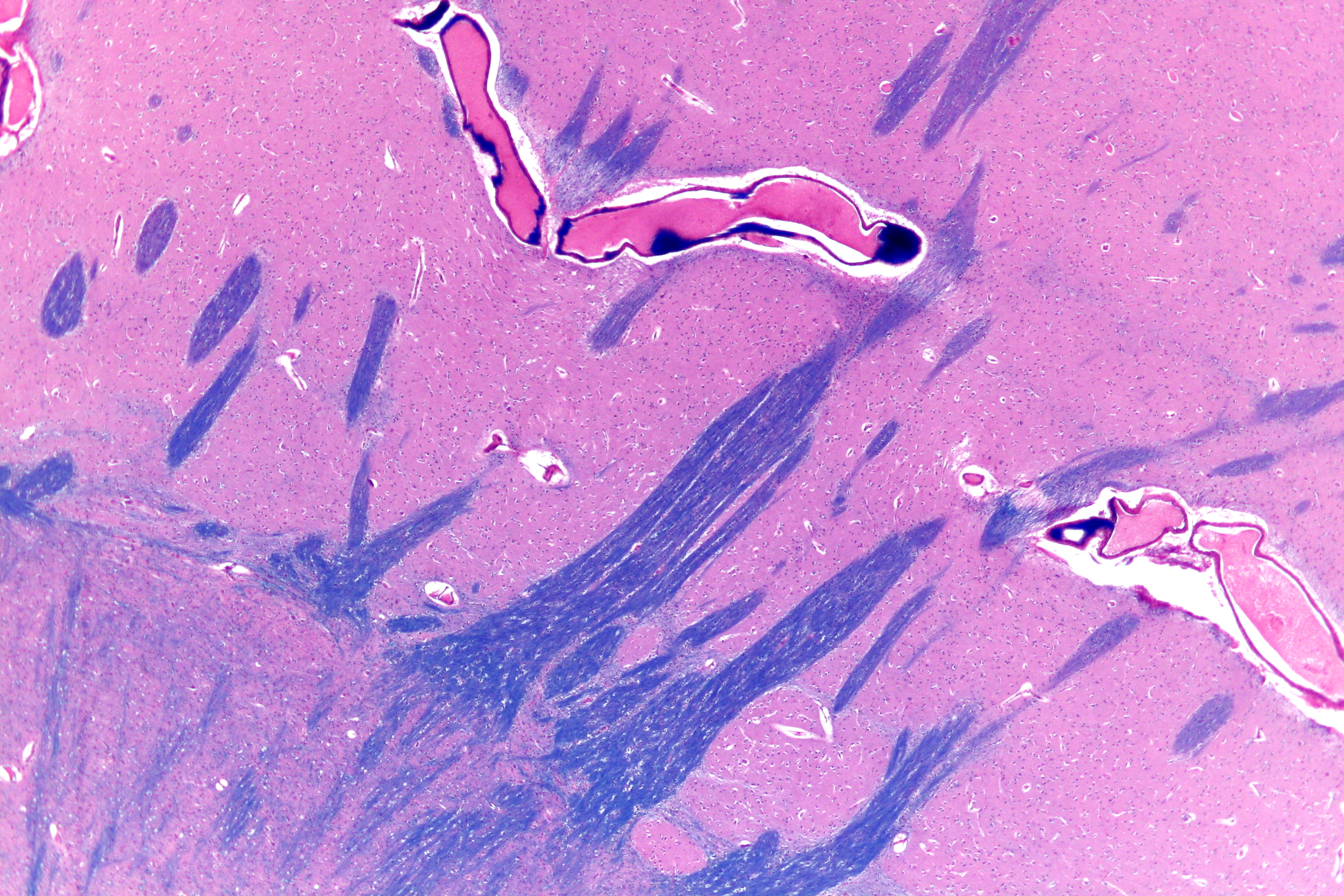
Lacunar stroke or lacunar cerebral infarct (LACI) is the most common type of ischemic stroke, resulting from the Vascular occlusion, occlusion of small penetrating artery, arteries that provide blood to the brain's deep structures. Patients who present with symptoms of a lacunar stroke, but who have not yet had diagnostic imaging performed, may be described as having lacunar stroke syndrome (LACS). Much of the current knowledge of lacunar strokes comes from C. Miller Fisher's cadaver dissections of post-mortem stroke patients. He observed "lacunae" (empty spaces) in the deep brain structures after occlusion of 200–800 μm penetrating arteries and connected them with five classic syndromes. These syndromes are still noted today, though lacunar infarcts are diagnosed based on clinical judgment and radiology, radiologic imaging. Signs and symptoms Each of the five classical lacunar syndromes has a relatively distinct symptom complex. Symptoms may occur suddenly, progressiv ... [...More Info...] [...Related Items...] OR: [Wikipedia] [Google] [Baidu] |
CT Scan
A computed tomography scan (CT scan), formerly called computed axial tomography scan (CAT scan), is a medical imaging technique used to obtain detailed internal images of the body. The personnel that perform CT scans are called radiographers or radiology technologists. CT scanners use a rotating X-ray tube and a row of detectors placed in a gantry (medical), gantry to measure X-ray Attenuation#Radiography, attenuations by different tissues inside the body. The multiple X-ray measurements taken from different angles are then processed on a computer using tomographic reconstruction algorithms to produce Tomography, tomographic (cross-sectional) images (virtual "slices") of a body. CT scans can be used in patients with metallic implants or pacemakers, for whom magnetic resonance imaging (MRI) is Contraindication, contraindicated. Since its development in the 1970s, CT scanning has proven to be a versatile imaging technique. While CT is most prominently used in medical diagnosis, i ... [...More Info...] [...Related Items...] OR: [Wikipedia] [Google] [Baidu] |
Superior Cerebellar Artery
The superior cerebellar artery (SCA) is an artery of the head. It arises near the end of the basilar artery. It is a branch of the basilar artery. It supplies parts of the cerebellum, the midbrain, and other nearby structures. It is the cause of trigeminal neuralgia in some patients. Structure The superior cerebellar artery arises near the end of the basilar artery. It passes laterally around the brainstem. This is immediately below the oculomotor nerve, which separates it from the posterior cerebral artery. It then winds around the cerebral peduncle, close to the trochlear nerve. It also lies close to the cerebellar tentorium. When it arrives at the upper surface of the cerebellum, it divides into branches which ramify in the pia mater and anastomose with those of the anterior inferior cerebellar artery, anterior inferior cerebellar arteries and the Posterior inferior cerebellar artery, posterior inferior cerebellar arteries. Several branches are given to the pineal body, the a ... [...More Info...] [...Related Items...] OR: [Wikipedia] [Google] [Baidu] |
Putamen
The putamen (; from Latin, meaning "nutshell") is a subcortical nucleus (neuroanatomy), nucleus with a rounded structure, in the basal ganglia nuclear group. It is located at the base of the forebrain and above the midbrain. The putamen and caudate nucleus together form the dorsal striatum. Through various pathways, the putamen is connected to the substantia nigra, the globus pallidus, the claustrum, and the thalamus, in addition to many regions of the cerebral cortex. A primary function of the putamen is to regulate movements at various stages such as in preparation and execution; and to influence various types of learning. It employs GABA, acetylcholine, and enkephalin to perform its functions. The putamen also plays a role in neurodegenerative diseases, such as Parkinson's disease. History The word "putamen" is from Latin, referring to that which "falls off in pruning", from "putare", meaning "to prune, to think, or to consider". Most MRI research was focused broadly on th ... [...More Info...] [...Related Items...] OR: [Wikipedia] [Google] [Baidu] |
Basilar Artery
The basilar artery (U.K.: ; U.S.: ) is one of the arteries that supplies the brain with oxygen-rich blood. The two vertebral arteries and the basilar artery are known as the vertebral basilar system, which supplies blood to the posterior part of the circle of Willis and joins with blood supplied to the anterior part of the circle of Willis from the internal carotid arteries. Structure The diameter of the basilar artery range from 1.5 to 6.6 mm. Origin The basilar artery arises from the union of the two vertebral arteries at the junction between the medulla oblongata and the pons between the abducens nerves (CN VI). Course It ascends along the basilar sulcus of the ventral pons. It divides at the junction of the midbrain and pons into the posterior cerebral arteries. Branches Its branches from caudal to rostral include: *anterior inferior cerebellar artery *labyrinthine artery (<15% of people, usually branches from the anterior inferior cerebellar artery) * [...More Info...] [...Related Items...] OR: [Wikipedia] [Google] [Baidu] |
Cerebellar Arteries
A cerebellar artery is an artery that provides blood to the cerebellum. Types include: * Superior cerebellar artery * Anterior inferior cerebellar artery * Posterior inferior cerebellar artery The posterior inferior cerebellar artery (PICA) is the largest branch of the vertebral artery. It is one of the three main arteries that supply blood to the cerebellum, a part of the brain. Blockage of the posterior inferior cerebellar artery can ... Arteries of the head and neck {{circulatory-stub ... [...More Info...] [...Related Items...] OR: [Wikipedia] [Google] [Baidu] |
Circle Of Willis
The circle of Willis (also called Willis' circle, loop of Willis, cerebral arterial circle, and Willis polygon) is a circulatory anastomosis that supplies blood to the brain and surrounding structures in reptiles, birds and mammals, including humans. It is named after Thomas Willis (1621–1675), an English physician. Structure The circle of Willis is a part of the cerebral circulation and is composed of the following arteries: * Anterior cerebral artery (left and right) at their A1 segments * Anterior communicating artery * Internal carotid artery (left and right) at its distal tip (carotid terminus) * Posterior cerebral artery (left and right) at their P1 segments * Posterior communicating artery (left and right) The middle cerebral arteries, supplying the brain, are also considered part of the Circle of Willis Origin of arteries The left and right internal carotid arteries arise from the left and right common carotid arteries. The posterior communicating artery is given ... [...More Info...] [...Related Items...] OR: [Wikipedia] [Google] [Baidu] |
Arteries Beneath Brain Gray Closer
An artery () is a blood vessel in humans and most other animals that takes oxygenated blood away from the heart in the systemic circulation to one or more parts of the body. Exceptions that carry deoxygenated blood are the pulmonary arteries in the pulmonary circulation that carry blood to the lungs for oxygenation, and the umbilical arteries in the fetal circulation that carry deoxygenated blood to the placenta. It consists of a multi-layered artery wall wrapped into a tube-shaped channel. Arteries contrast with veins, which carry deoxygenated blood back towards the heart; or in the pulmonary and fetal circulations carry oxygenated blood to the lungs and fetus respectively. Structure The anatomy of arteries can be separated into gross anatomy, at the macroscopic level, and microanatomy, which must be studied with a microscope. The arterial system of the human body is divided into systemic arteries, carrying blood from the heart to the whole body, and pulmonary arteries, c ... [...More Info...] [...Related Items...] OR: [Wikipedia] [Google] [Baidu] |
Cognitive Functions
Cognitive skills are skills of the mind, as opposed to other types of skills such as motor skills, social skills or life skills. Some examples of cognitive skills are literacy, self-reflection, logical reasoning, abstract thinking, critical thinking, introspection and mental arithmetic. Cognitive skills vary in processing complexity, and can range from more fundamental processes such as perception and various memory functions, to more sophisticated processes such as decision making, problem solving and metacognition. Specialisation of functions Cognitive science has provided theories of how the brain works, and these have been of great interest to researchers who work in the empirical fields of brain science. A fundamental question is whether cognitive functions, for example visual processing and language, are autonomous modules, or to what extent the functions depend on each other. Research evidence points towards a middle position, and it is now generally accepted that there ... [...More Info...] [...Related Items...] OR: [Wikipedia] [Google] [Baidu] |
Computed Axial Tomography
A computed tomography scan (CT scan), formerly called computed axial tomography scan (CAT scan), is a medical imaging technique used to obtain detailed internal images of the body. The personnel that perform CT scans are called radiographers or radiology technologists. CT scanners use a rotating X-ray tube and a row of detectors placed in a gantry to measure X-ray attenuations by different tissues inside the body. The multiple X-ray measurements taken from different angles are then processed on a computer using tomographic reconstruction algorithms to produce tomographic (cross-sectional) images (virtual "slices") of a body. CT scans can be used in patients with metallic implants or pacemakers, for whom magnetic resonance imaging (MRI) is contraindicated. Since its development in the 1970s, CT scanning has proven to be a versatile imaging technique. While CT is most prominently used in medical diagnosis, it can also be used to form images of non-living objects. The 1979 Nobel ... [...More Info...] [...Related Items...] OR: [Wikipedia] [Google] [Baidu] |
Silent Stroke
A silent stroke (or asymptomatic cerebral infarction) is a stroke that does not have any outward symptoms associated with stroke, and the patient is typically unaware they have suffered a stroke. Despite not causing identifiable symptoms, a silent stroke still causes damage to the brain and places the patient at increased risk for both transient ischemic attack and major stroke in the future. In a broad study in 1998, more than 11 million people were estimated to have experienced a stroke in the United States. Approximately 770,000 of these strokes were symptomatic and 11 million were first-ever silent MRI infarcts or hemorrhages. Silent strokes typically cause lesions which are detected via the use of neuroimaging such as MRI. The risk of silent stroke increases with age but may also affect younger adults. Women appear to be at increased risk for silent stroke, with hypertension and current cigarette smoking being amongst the predisposing factors. These types of strokes includ ... [...More Info...] [...Related Items...] OR: [Wikipedia] [Google] [Baidu] |
Histopathology Of A Lacunar Cerebral Infarct
Histopathology (compound of three Greek words: 'tissue', 'suffering', and ''-logia'' 'study of') is the microscopic examination of tissue in order to study the manifestations of disease. Specifically, in clinical medicine, histopathology refers to the examination of a biopsy or surgical specimen by a pathologist, after the specimen has been processed and histological sections have been placed onto glass slides. In contrast, cytopathology examines free cells or tissue micro-fragments (as "cell blocks "). Collection of tissues Histopathological examination of tissues starts with surgery, biopsy, or autopsy. The tissue is removed from the Human body, body or plant, and then, often following expert dissection in the fresh state, placed in a fixation (histology), fixative which stabilizes the tissues to prevent Decomposition, decay. The most common fixative is 10% neutral buffered formalin (corresponding to 3.7% w/v formaldehyde in neutral buffered water, such as phosphate buf ... [...More Info...] [...Related Items...] OR: [Wikipedia] [Google] [Baidu] |
Ventral Posterolateral Nucleus
The ventral posterolateral nucleus (VPL) is one of the subdivisions of the ventral posterior nucleus in the ventral nuclear group of the thalamus. It relays sensory information from the second-order neurons of the neospinothalamic tract and medial lemniscus (of the dorsal column-medial lemniscus pathway) which synapse with the third-order neurons in the nucleus. These then project to the primary somatosensory cortex in the postcentral gyrus. There is uncertainty regarding the location of VMpo (posterior part of ventral medial nucleus), as determined by spinothalamic tract (STT) terminations and staining for calcium-binding proteins, and several authorities do not consider its existence as being proved. The term "ventral posterolateral nucleus" was introduced by Le Gros Clark in 1930. Anatomy Subdivisions The oral part of the ventral posterolateral nucleus (nucleus ventrointermedius) in the human, (VPLO) is a subdivision of the VPL with projections to the motor cortex ... [...More Info...] [...Related Items...] OR: [Wikipedia] [Google] [Baidu] |


