|
Internal Rectal Prolapse
Internal rectal prolapse (IRP) is medical condition involving a telescopic, funnel-shaped infolding of the wall of the rectum that occurs during defecation. The term IRP is used when the prolapsed section of rectal wall remains inside the body and is not visible outside the body. IRP is a type of rectal prolapse. The other main types of rectal prolapse are external rectal prolapse (where the prolapsed segment of rectum protrudes through the anus and is visible externally) and rectal mucosal prolapse (where only the mucosal layer of the wall of the rectum prolapses). IRP may not cause any symptoms, or may cause obstructed defecation syndrome (difficulty during defecation) and/or fecal incontinence. The causes are not clear. IRP may represent the first stage of a progressive condition that eventually may result in external rectal prolapse. However, it is uncommon for IRP to progress to external rectal prolapse. It is possible that chronic straining during defecation (dyssynergic def ... [...More Info...] [...Related Items...] OR: [Wikipedia] [Google] [Baidu] |
Colorectal Surgery
Colorectal surgery is a field in medicine dealing with disorders of the rectum, anus, and colon. The field is also known as proctology, but this term is now used infrequently within medicine and is most often employed to identify practices relating to the anus and rectum in particular. The word ''proctology'' is derived from the Greek words , meaning "anus" or "hindparts", and , meaning "science" or "study". Physicians specializing in this field of medicine are called colorectal surgeons or proctologists. In the United States, to become colorectal surgeons, surgical doctors have to complete a general surgery residency as well as a colorectal surgery fellowship, upon which they are eligible to be certified in their field of expertise by the American Board of Colon and Rectal Surgery or the American Osteopathic Board of Proctology. In other countries, certification to practice proctology is given to surgeons at the end of a 2–3 year subspecialty residency by the country's ... [...More Info...] [...Related Items...] OR: [Wikipedia] [Google] [Baidu] |
Rectocele
In gynecology, a rectocele ( ) or posterior vaginal wall prolapse results when the rectum bulges ( herniates) into the vagina. Two common causes of this defect are childbirth and hysterectomy. Rectocele also tends to occur with other forms of pelvic organ prolapse, such as enterocele, sigmoidocele and cystocele. Although the term applies most often to this condition in females, males can also develop it. Rectoceles in men are uncommon, and associated with prostatectomy. Signs and symptoms Mild cases may simply produce a sense of pressure or protrusion within the vagina, and the occasional feeling that the rectum has not been completely emptied after a bowel movement. Moderate cases may involve difficulty passing stool (because the attempt to evacuate pushes the stool into the rectocele instead of out through the anus), discomfort or pain during evacuation or intercourse, constipation, and a general sensation that something is "falling down" or "falling out" within the p ... [...More Info...] [...Related Items...] OR: [Wikipedia] [Google] [Baidu] |
Puborectalis Muscle
The levator ani is a broad, thin muscle group, situated on either side of the pelvis. It is formed from three muscle components: the pubococcygeus, the iliococcygeus, and the puborectalis. It is attached to the inner surface of each side of the lesser pelvis, and these unite to form the greater part of the pelvic floor. The coccygeus muscle completes the pelvic floor, which is also called the ''pelvic diaphragm''. It supports the viscera in the pelvic cavity, and surrounds the various structures that pass through it. The levator ani is the main pelvic floor muscle and contracts rhythmically during female orgasm, and painfully during vaginismus. Structure The levator ani is made up of 3 parts: * Iliococcygeus muscle * Pubococcygeus muscle * Puborectalis muscle The iliococcygeus arises from the inner side of the ischium (the lower and back part of the hip bone) and from the posterior part of the tendinous arch of the obturator fascia, and is attached to the coccyx and ... [...More Info...] [...Related Items...] OR: [Wikipedia] [Google] [Baidu] |
Anal Canal
The anal canal is the part that connects the rectum to the anus, located below the level of the pelvic diaphragm. It is located within the anal triangle of the perineum, between the right and left ischioanal fossa. As the final functional segment of the bowel, it functions to regulate release of excrement by two muscular sphincter complexes. The anus is the aperture at the terminal portion of the anal canal. Structure In humans, the anal canal is approximately long, from the anorectal junction to the anus. It is directed downwards and backwards. It is surrounded by inner involuntary and outer voluntary sphincters which keep the lumen closed in the form of an anteroposterior slit. The canal is differentiated from the rectum by a transition along the internal surface from endodermal to skin-like ectodermal tissue. The anal canal is traditionally divided into two segments, upper and lower, separated by the pectinate line (also known as the dentate line): * upper zo ... [...More Info...] [...Related Items...] OR: [Wikipedia] [Google] [Baidu] |
Levator Ani
The levator ani is a broad, thin muscle group, situated on either side of the pelvis. It is formed from three muscle components: the pubococcygeus, the iliococcygeus, and the puborectalis. It is attached to the inner surface of each side of the lesser pelvis, and these unite to form the greater part of the pelvic floor. The coccygeus muscle completes the pelvic floor, which is also called the ''pelvic diaphragm''. It supports the viscera in the pelvic cavity, and surrounds the various structures that pass through it. The levator ani is the main pelvic floor muscle and contracts rhythmically during female orgasm, and painfully during vaginismus. Structure The levator ani is made up of 3 parts: * Iliococcygeus muscle * Pubococcygeus muscle * Puborectalis muscle The iliococcygeus arises from the inner side of the ischium (the lower and back part of the hip bone) and from the posterior part of the tendinous arch of the obturator fascia, and is attached to the coccyx and ... [...More Info...] [...Related Items...] OR: [Wikipedia] [Google] [Baidu] |
Transverse Folds Of Rectum
The transverse folds of rectum (or Houston's valves or the valves of Houston) are semi-lunar transverse folds of the rectal wall that protrude into the rectum, not the anal canal as that lies below the rectum. Their use seems to be to support the weight of fecal matter, and prevent its urging toward the anus, which would produce a strong urge to defecate. Although the term rectum means straight, these transverse folds overlap each other during the empty state of the intestine to such an extent that, as Houston remarked, they require considerable maneuvering to conduct an instrument along the canal, as often occurs in sigmoidoscopy and colonoscopy. These folds are about 12 mm. in width and are composed of the circular muscle coat of the rectum. They are usually three in number; sometimes a fourth is found, and occasionally only two are present. * One is situated near the commencement of the rectum, on the right side. * A second extends inward from the left side of the tu ... [...More Info...] [...Related Items...] OR: [Wikipedia] [Google] [Baidu] |
Coccyx
The coccyx (: coccyges or coccyxes), commonly referred to as the tailbone, is the final segment of the vertebral column in all apes, and analogous structures in certain other mammals such as horse anatomy, horses. In tailless primates (e.g. humans and other great apes) since ''Nacholapithecus'' (a Miocene hominoid),Nakatsukasa 2004, ''Acquisition of bipedalism'' (SeFig. 5entitled ''First coccygeal/caudal vertebra in short-tailed or tailless primates.''.) the coccyx is the remnant of a Human vestigiality#Coccyx, vestigial tail. In animals with bony tails, it is known as Rump (animal), ''tailhead'' or ''dock'', in bird anatomy as ''tailfan''. It comprises three to five separate or fused coccygeal vertebrae below the sacrum, attached to the sacrum by a fibrocartilaginous joint, the sacrococcygeal symphysis, which permits limited movement between the sacrum and the coccyx. Structure The coccyx is formed of three, four or five rudimentary vertebrae. It articulates superiorly with ... [...More Info...] [...Related Items...] OR: [Wikipedia] [Google] [Baidu] |
Sacral Spinal Nerve 3
The sacral spinal nerve 3 (S3) is a spinal nerve of the sacral segment. Nervous System -- Groups of Nerves It originates from the from below the 3rd body of the . 
Muscles S3 supplies many muscles, either directly or through nerves originating from S3. They are not innervated with S3 as single origin, but partly by S3 and partly by other spinal nerves. The mus ...[...More Info...] [...Related Items...] OR: [Wikipedia] [Google] [Baidu] |
Sigmoid Mesocolon
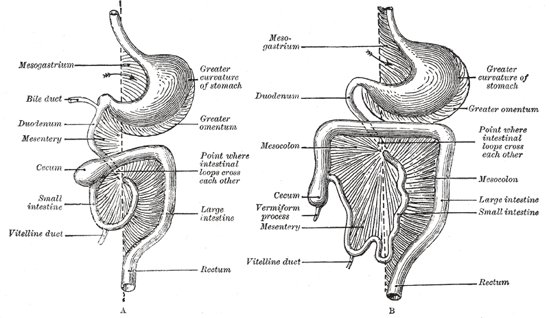
In human anatomy, the mesentery is an organ that attaches the intestines to the posterior abdominal wall, consisting of a double fold of the peritoneum. It helps (among other functions) in storing fat and allowing blood vessels, lymphatics, and nerves to supply the intestines. The (the part of the mesentery that attaches the colon to the abdominal wall) was formerly thought to be a fragmented structure, with all named parts—the ascending, transverse, descending, and sigmoid mesocolons, the mesoappendix, and the mesorectum—separately terminating their insertion into the posterior abdominal wall. However, in 2012, new microscopic and electron microscopic examinations showed the mesocolon to be a single structure derived from the duodenojejunal flexure and extending to the distal mesorectal layer. Thus the mesentery is an internal organ. Structure The mesentery of the small intestine arises from the root of the mesentery (or mesenteric root) and is the part connected ... [...More Info...] [...Related Items...] OR: [Wikipedia] [Google] [Baidu] |
Abdominal Wall
In anatomy, the abdominal wall represents the boundaries of the abdominal cavity. The abdominal wall is split into the anterolateral and posterior walls. There is a common set of layers covering and forming all the walls: the deepest being the visceral peritoneum, which covers many of the abdominal organs (most of the large and small intestines, for example), and the parietal peritoneum—which covers the visceral peritoneum below it, the extraperitoneal fat, the transversalis fascia, the internal and external oblique and transversus abdominis aponeurosis, and a layer of fascia, which has different names according to what it covers (e.g., transversalis, psoas fascia). In medical vernacular, the term 'abdominal wall' most commonly refers to the layers composing the anterior abdominal wall which, in addition to the layers mentioned above, includes the three layers of muscle: the transversus abdominis (transverse abdominal muscle), the internal oblique, internal (obliquus internus) a ... [...More Info...] [...Related Items...] OR: [Wikipedia] [Google] [Baidu] |
Sigmoid Colon
The sigmoid colon (or pelvic colon) is the part of the large intestine that is closest to the rectum and anus. It forms a loop that averages about in length. The loop is typically shaped like a Greek letter sigma (ς) or Latin letter S (thus ''sigma'' + '' -oid''). This part of the colon normally lies within the pelvis, but due to its freedom of movement it is liable to be displaced into the abdominal cavity. Structure The sigmoid colon begins at the superior aperture of the lesser pelvis, where it is continuous with the iliac colon, and passes transversely across the front of the sacrum to the right side of the pelvis. It then curves on itself and turns toward the left to reach the middle line at the level of the third piece of the sacrum, where it bends downward and ends in the rectum. Its function is to expel solid and gaseous waste from the gastrointestinal tract. The curving path it takes toward the anus allows it to store gas in the superior arched portion, enabling ... [...More Info...] [...Related Items...] OR: [Wikipedia] [Google] [Baidu] |
Small Intestine
The small intestine or small bowel is an organ (anatomy), organ in the human gastrointestinal tract, gastrointestinal tract where most of the #Absorption, absorption of nutrients from food takes place. It lies between the stomach and large intestine, and receives bile and pancreatic juice through the pancreatic duct to aid in digestion. The small intestine is about long and folds many times to fit in the abdomen. Although it is longer than the large intestine, it is called the small intestine because it is narrower in diameter. The small intestine has three distinct regions – the duodenum, jejunum, and ileum. The duodenum, the shortest, is where preparation for absorption through small finger-like protrusions called intestinal villus, intestinal villi begins. The jejunum is specialized for the absorption through its lining by enterocytes: small nutrient particles which have been previously digested by enzymes in the duodenum. The main function of the ileum is to absorb vitami ... [...More Info...] [...Related Items...] OR: [Wikipedia] [Google] [Baidu] |


