|
Infratemporal Fossa
The infratemporal fossa is an irregularly shaped cavity that is a part of the skull. It is situated below and medial to the zygomatic arch. It is not fully enclosed by bone in all directions. It contains superficial muscles, including the lower part of the temporalis muscle, the lateral pterygoid muscle, and the medial pterygoid muscle. It also contains important blood vessels such as the middle meningeal artery, the pterygoid plexus, and the retromandibular vein, and nerves such as the mandibular nerve (CN V3) and its branches. Structure Boundaries The boundaries of the infratemporal fossa occur: * ''anteriorly'', by the infratemporal surface of the maxilla, and the ridge which descends from its zygomatic process. This contains the alveolar canal. * ''posteriorly'', by the tympanic part of the temporal bone, and the spina angularis of the sphenoid. * ''superiorly'', by the greater wing of the sphenoid below the infratemporal crest, and by the under surface of the ... [...More Info...] [...Related Items...] OR: [Wikipedia] [Google] [Baidu] |
Skull
The skull, or cranium, is typically a bony enclosure around the brain of a vertebrate. In some fish, and amphibians, the skull is of cartilage. The skull is at the head end of the vertebrate. In the human, the skull comprises two prominent parts: the neurocranium and the facial skeleton, which evolved from the first pharyngeal arch. The skull forms the frontmost portion of the axial skeleton and is a product of cephalization and vesicular enlargement of the brain, with several special senses structures such as the eyes, ears, nose, tongue and, in fish, specialized tactile organs such as barbels near the mouth. The skull is composed of three types of bone: cranial bones, facial bones and ossicles, which is made up of a number of fused flat and irregular bones. The cranial bones are joined at firm fibrous junctions called sutures and contains many foramina, fossae, processes, and sinuses. In zoology, the openings in the skull are called fenestrae, the most ... [...More Info...] [...Related Items...] OR: [Wikipedia] [Google] [Baidu] |
Spina Angularis
The sphenoidal spine (Latin: "''spina angularis''") is a downwardly directed process at the apex of the great wings of the sphenoid bone that serves as the origin of the sphenomandibular ligament. Additional images File:Spine of sphenoid bone.png, Base of skull. Inferior surface. Spine of sphenoid bone marked with black circle References External links * - "Schematic view of key landmarks of the infratemporal fossa The infratemporal fossa is an irregularly shaped cavity that is a part of the skull. It is situated below and medial to the zygomatic arch. It is not fully enclosed by bone in all directions. It contains superficial muscles, including the lower ...." * Bones of the head and neck {{musculoskeletal-stub ... [...More Info...] [...Related Items...] OR: [Wikipedia] [Google] [Baidu] |
Lingula Of Mandible
The lingula of the mandible is a prominent bony ridge on the medial side of the mandible. It is next to the mandibular foramen. It gives attachment to the sphenomandibular ligament. Structure The lingula of the mandible is a prominent bony ridge on the medial side of the mandible In jawed vertebrates, the mandible (from the Latin ''mandibula'', 'for chewing'), lower jaw, or jawbone is a bone that makes up the lowerand typically more mobilecomponent of the mouth (the upper jaw being known as the maxilla). The jawbone i .... It is next to the mandibular foramen. It has a notch from which the mylohyoid groove originates. It gives attachment to the sphenomandibular ligament. Variation The lingula of the mandible can take many shapes, including triangular, truncated, and nodular. In a majority of people, this shape is symmetrical. See also * Lingula References External links * http://ect.downstate.edu/courseware/haonline/labs/l22/os2009.htm * * Bones o ... [...More Info...] [...Related Items...] OR: [Wikipedia] [Google] [Baidu] |
Inferior Alveolar Nerve
The inferior alveolar nerve (IAN) (also the inferior dental nerve) is a sensory branch of the mandibular nerve (CN V3) (which is itself the third branch of the trigeminal nerve (CN V)). The nerve provides sensory innervation to the lower/mandibular teeth and their corresponding gingiva as well as a small area of the face (via its mental nerve). Structure Origin The inferior alveolar nerve arises from the mandibular nerve. Course After branching from the mandibular nerve, the inferior alveolar nerve passes posterior to the lateral pterygoid muscle. It issues a branch (the mylohyoid nerve) before entering the mandibular foramen to come to pass in the mandibular canal within the mandible. Passing through the canal, it issues sensory branches for the molar and second premolar teeth; the branches first form the inferior dental plexus which then gives off small gingival and dental nerves to these teeth themselves. The nerve terminates distally/anteriorly (near the second ... [...More Info...] [...Related Items...] OR: [Wikipedia] [Google] [Baidu] |
Mandibular Canal
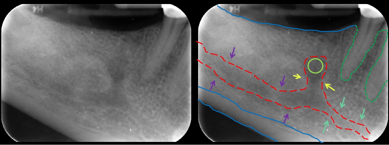
In human anatomy, the mandibular canal is a canal within the mandible that contains the inferior alveolar nerve, inferior alveolar artery, and inferior alveolar vein. It runs obliquely downward and forward in the ramus, and then horizontally forward in the body, where it is placed under the alveoli and communicates with them by small openings. On arriving at the incisor teeth, it turns back to communicate with the mental foramen, giving off a small canal known as the mandibular incisive canal, which run to the cavities containing the incisor teeth. It carries branches of the inferior alveolar nerve and artery. The mandibular canal is continuous with two foramina: the mental foramen which opens in the mental region of the mandible and carried the distal fibres of the inferior alveolar nerve as the mental nerve; and the mandibular foramen on medial aspect of ramus, into which the mandibular nerve enters to become the inferior alveolar nerve. The mandibular canal often runs cl ... [...More Info...] [...Related Items...] OR: [Wikipedia] [Google] [Baidu] |
Mandibular Foramen
The mandibular foramen is an opening on the internal surface of the ramus of the mandible. It allows for divisions of the mandibular nerve and blood vessels to pass through. Structure The mandibular foramen is an opening on the internal surface of the ramus of the mandible. It allows for divisions of the mandibular nerve and blood vessels to pass through. Variation There are two distinct anatomies to its rim. * In the common form the rim is V-shaped, with a groove separating the anterior and posterior parts. * In the horizontal-oval form there is no groove, and the rim is horizontally oriented and oval in shape, the anterior and posterior parts connected. Rarely, a bifid inferior alveolar nerve may be present, in which case a second mandibular foramen, more inferiorly placed, exists and can be detected by noting a doubled mandibular canal on a radiograph. Function The mandibular nerve is one of three branches of the trigeminal nerve, and the only one having motor innervat ... [...More Info...] [...Related Items...] OR: [Wikipedia] [Google] [Baidu] |
Ramus Of Mandible
In jawed vertebrates, the mandible (from the Latin ''mandibula'', 'for chewing'), lower jaw, or jawbone is a bone that makes up the lowerand typically more mobilecomponent of the mouth (the upper jaw being known as the maxilla). The jawbone is the skull's only movable, posable bone, sharing joints with the cranium's temporal bones. The mandible hosts the lower teeth (their depth delineated by the alveolar process). Many muscles attach to the bone, which also hosts nerves (some connecting to the teeth) and blood vessels. Amongst other functions, the jawbone is essential for chewing food. Owing to the Neolithic advent of agriculture (), human jaws evolved to be smaller. Although it is the strongest bone of the facial skeleton, the mandible tends to deform in old age; it is also subject to fracturing. Surgery allows for the removal of jawbone fragments (or its entirety) as well as regenerative methods. Additionally, the bone is of great forensic significance. Structure ... [...More Info...] [...Related Items...] OR: [Wikipedia] [Google] [Baidu] |
Lateral Pterygoid Plate
The pterygoid processes of the sphenoid (from Greek ''pteryx'', ''pterygos'', "wing"), one on either side, descend perpendicularly from the regions where the body and the greater wings of the sphenoid bone unite. Each process consists of a medial pterygoid plate and a lateral pterygoid plate, the latter of which serve as the origins of the medial and lateral pterygoid muscles. The medial pterygoid, along with the masseter allows the jaw to move in a vertical direction as it contracts and relaxes. The lateral pterygoid allows the jaw to move in a horizontal direction during mastication (chewing). Fracture of either plate are used in clinical medicine to distinguish the Le Fort fracture classification for high impact injuries to the sphenoid and maxillary bones. The superior portion of the pterygoid processes are fused anteriorly; a vertical groove, the pterygopalatine fossa, descends on the front of the line of fusion. The plates are separated below by an angular cleft, th ... [...More Info...] [...Related Items...] OR: [Wikipedia] [Google] [Baidu] |
Foramen Spinosum
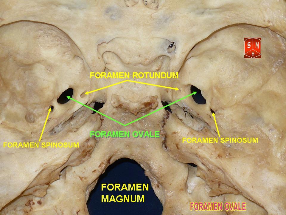
The foramen spinosum is a Foramen, small open hole in the greater wing of the sphenoid bone that gives passage to the middle meningeal artery and vein, and the meningeal branch of the mandibular nerve (sometimes it passes through the Foramen ovale (skull), foramen ovale instead). The foramen spinosum is often used as a landmark in neurosurgery due to its close relations with other cranial foramina. It was first described by Jakob Benignus Winslow in the 18th century. Structure The foramen spinosum is a small foramen in the Greater wing of sphenoid bone, greater wing of the sphenoid bone of the skull. It connects the middle cranial fossa (superiorly), and infratemporal fossa (inferiorly). Contents The foramen transmits the middle meningeal artery and vein, and sometimes the meningeal branch of the mandibular nerve (it may pass through the foramen ovale instead). Relations The foramen is situated just anterior to the sphenopetrosal suture. It is located posterolateral t ... [...More Info...] [...Related Items...] OR: [Wikipedia] [Google] [Baidu] |
Trigeminal Nerve
In neuroanatomy, the trigeminal nerve (literal translation, lit. ''triplet'' nerve), also known as the fifth cranial nerve, cranial nerve V, or simply CN V, is a cranial nerve responsible for Sense, sensation in the face and motor functions such as biting and chewing; it is the most complex of the cranial nerves. Its name (''trigeminal'', ) derives from each of the two nerves (one on each side of the pons) having three major branches: the ophthalmic nerve (V), the maxillary nerve (V), and the mandibular nerve (V). The ophthalmic and maxillary nerves are purely sensory, whereas the mandibular nerve supplies motor as well as sensory (or "cutaneous") functions. Adding to the complexity of this nerve is that Autonomic nervous system, autonomic nerve fibers as well as special sensory fibers (taste) are contained within it. The motor division of the trigeminal nerve derives from the Basal plate (neural tube), basal plate of the embryonic pons, and the sensory division originates in ... [...More Info...] [...Related Items...] OR: [Wikipedia] [Google] [Baidu] |
Foramen Ovale (skull)
The foramen ovale (En: oval window) is a hole in the posterior part of the sphenoid bone, posterolateral to the foramen rotundum. It is one of the larger of the several holes (the foramina) in the skull. It transmits the mandibular nerve, a branch of the trigeminal nerve. Structure The foramen ovale is an opening in the greater wing of the sphenoid bone. The foramen ovale is one of two cranial foramina in the greater wing, the other being the foramen spinosum. The foramen ovale is posterolateral to the foramen rotundum and anteromedial to the foramen spinosum. Posterior and medial to the foramen is the opening for the carotid canal. Contents The following structures pass through foramen ovale: * mandibular nerve (CN V) (a branch of the trigeminal nerve (CN V)) *accessory meningeal artery * lesser petrosal nerve (a branch of the glossopharyngeal nerve) * an emissary vein connecting the cavernous sinus with the pterygoid plexus * (occasionally) meningeal branch o ... [...More Info...] [...Related Items...] OR: [Wikipedia] [Google] [Baidu] |
Temporal Squama
The squamous part of temporal bone, or temporal squama, forms the front and upper part of the temporal bone, and is scale-like, thin, and translucent. Surfaces Its outer surface is smooth and convex; it affords attachment to the temporal muscle, and forms part of the temporal fossa; on its hinder part is a vertical groove for the middle temporal artery. A curved line, the ''temporal line'', or ''supramastoid crest'', runs backward and upward across its posterior part; it serves for the attachment of the temporal fascia, and limits the origin of the temporalis muscle. The boundary between the squamous part and the mastoid portion of the bone, as indicated by traces of the original suture, lies about 1 cm. below this line. Projecting from the lower part of the squamous part is a long, arched process, the ''zygomatic process''. This process is at first directed lateralward, its two surfaces looking upward and downward; it then appears as if twisted inward upon itself, and runs ... [...More Info...] [...Related Items...] OR: [Wikipedia] [Google] [Baidu] |


