|
Horner's Syndrome
Horner's syndrome, also known as oculosympathetic paresis, is a combination of symptoms that arises when a group of nerves known as the sympathetic trunk is damaged. The signs and symptoms occur on the same side (ipsilateral) as it is a lesion of the sympathetic trunk. It is characterized by miosis (a constricted pupil), partial ptosis (a weak, droopy eyelid), apparent anhidrosis (decreased sweating), with apparent enophthalmos (inset eyeball). The nerves of the sympathetic trunk arise from the spinal cord in the chest, and from there ascend to the neck and face. The nerves are part of the sympathetic nervous system, a division of the autonomic (or involuntary) nervous system. Once the syndrome has been recognized, medical imaging and response to particular eye drops may be required to identify the location of the problem and the underlying cause. Signs and symptoms Signs that are found in people with Horner's syndrome on the affected side of the face include the following ... [...More Info...] [...Related Items...] OR: [Wikipedia] [Google] [Baidu] |
Nerve
A nerve is an enclosed, cable-like bundle of nerve fibers (called axons). Nerves have historically been considered the basic units of the peripheral nervous system. A nerve provides a common pathway for the Electrochemistry, electrochemical nerve impulses called action potentials that are transmitted along each of the axons to peripheral organs or, in the case of sensory nerves, from the periphery back to the central nervous system. Each axon is an extension of an individual neuron, along with other supportive cells such as some Schwann cells that coat the axons in myelin. Each axon is surrounded by a layer of connective tissue called the endoneurium. The axons are bundled together into groups called Nerve fascicle, fascicles, and each fascicle is wrapped in a layer of connective tissue called the perineurium. The entire nerve is wrapped in a layer of connective tissue called the epineurium. Nerve cells (often called neurons) are further classified as either Sensory neuron, sens ... [...More Info...] [...Related Items...] OR: [Wikipedia] [Google] [Baidu] |
Red Eye (medicine)
A red eye is an eye that appears red due to illness or injury. It is usually hyperemia, injection and prominence of the superficial blood vessels of the conjunctiva, which may be caused by disorders of these or adjacent structures. Conjunctivitis and subconjunctival hemorrhage are two of the less serious but more common causes. Management includes assessing whether emergency action (including referral) is needed, or whether treatment can be accomplished without additional resources. slit lamp, Slit lamp examination is invaluable in diagnosis but initial assessment can be performed using a careful history, testing vision (visual acuity), and carrying out a swinging-flashlight test, penlight examination. Diagnosis Particular sign (medicine), signs and symptoms may indicate that the cause is serious and requires immediate attention. Seven such signs are: * #Reduced visual acuity, Reduced visual acuity * #Ciliary flush, Ciliary flush (circumcorneal injection) * #Corneal abnormal ... [...More Info...] [...Related Items...] OR: [Wikipedia] [Google] [Baidu] |
Nictitating Membrane
The nictitating membrane (from Latin '' nictare'', to blink) is a transparent or translucent third eyelid present in some animals that can be drawn across the eye from the medial canthus to protect and moisten it while maintaining vision. Most Anura (tailless amphibians), some reptiles, birds, and sharks, and some mammals (such as cats, beavers, polar bears, seals and aardvarks) have full nictitating membranes; in many other mammals, a small, vestigial portion of the nictitating membrane remains in the corner of the eye. It is often informally called a third eyelid or haw; the scientific terms for it are the ''plica semilunaris'', ''membrana nictitans'', or ''palpebra tertia''. Description The nictitating membrane is a transparent or translucent third eyelid present in some animals that can be drawn across the eye for protection and to moisten it while maintaining vision. The term comes from the Latin word '' nictare'', meaning "to blink". It is often called a ''thi ... [...More Info...] [...Related Items...] OR: [Wikipedia] [Google] [Baidu] |
Iris (anatomy)
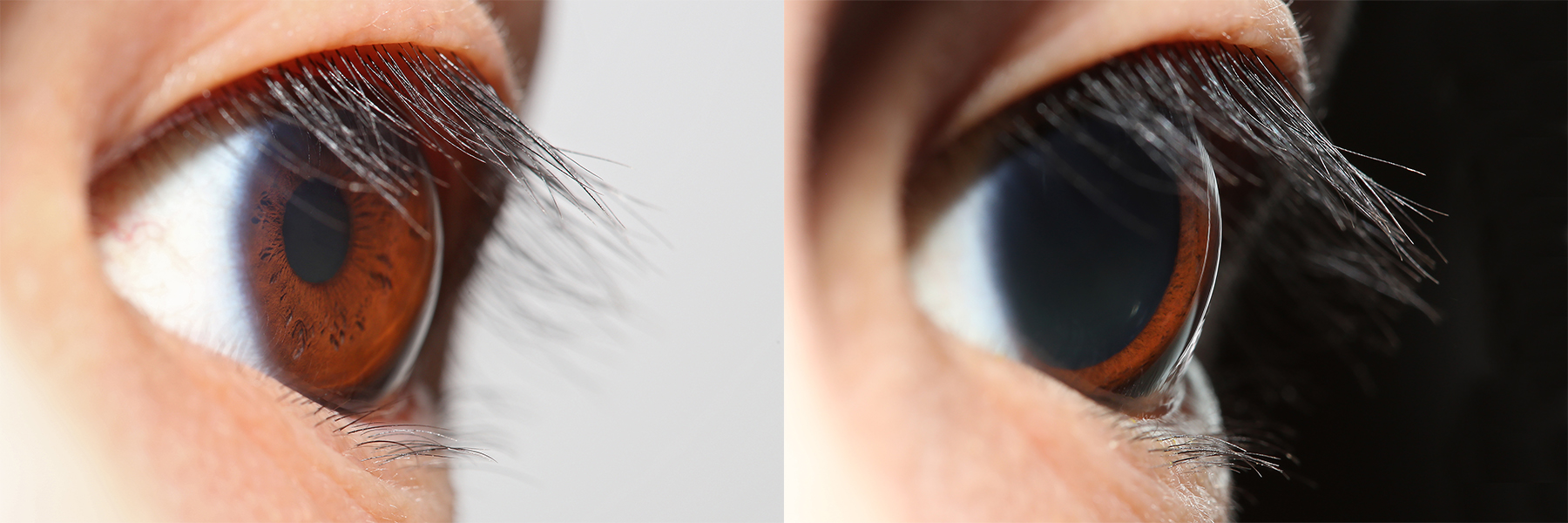
The iris (: irides or irises) is a thin, annular structure in the eye in most mammals and birds that is responsible for controlling the diameter and size of the pupil, and thus the amount of light reaching the retina. In optical terms, the pupil is the eye's aperture, while the iris is the diaphragm (optics), diaphragm. Eye color is defined by the iris. Etymology The word "iris" is derived from the Greek word for "rainbow", also Iris (mythology), its goddess plus messenger of the gods in the ''Iliad'', because of the many eye color, colours of this eye part. Structure The iris consists of two layers: the front pigmented Wikt:fibrovascular, fibrovascular layer known as a stroma of iris, stroma and, behind the stroma, pigmented epithelial cells. The stroma is connected to a sphincter muscle (sphincter pupillae), which contracts the pupil in a circular motion, and a set of dilator muscles (dilator pupillae), which pull the iris radially to enlarge the pupil, pulling it in folds. ... [...More Info...] [...Related Items...] OR: [Wikipedia] [Google] [Baidu] |
Stroma Of Iris
The stroma of the iris is a fibrovascular layer of tissue located at the front of the iris. Structure The stroma is a delicate interlacement of fibres. Some circle the circumference of the iris and the majority radiate toward the pupil. Blood vessels and nerves intersperse this mesh. In dark eyes, the stroma often contains pigment granules. Blue eyes and the eyes of albinos, however, lack pigment. The stroma connects to a sphincter A sphincter is a circular muscle that normally maintains constriction of a natural body passage or orifice and relaxes as required by normal physiological functioning. Sphincters are found in many animals. There are over 60 types in the human bo ... muscle ( sphincter pupillae), which contracts the pupil in a circular motion, and a set of dilator muscles ( dilator pupillae) which pull the iris radially to enlarge the pupil, pulling it in folds. The back surface is covered by a commonly, heavily pigmented epithelial layer that is two cells t ... [...More Info...] [...Related Items...] OR: [Wikipedia] [Google] [Baidu] |
Melanocyte
Melanocytes are melanin-producing neural-crest, neural crest-derived cell (biology), cells located in the bottom layer (the stratum basale) of the skin's epidermis (skin), epidermis, the middle layer of the eye (the uvea), the inner ear, vaginal epithelium, meninges, bones, and heart found in many mammals and birds. Melanin is a dark pigment primarily responsible for skin color. Once synthesized, melanin is contained in special organelles called melanosomes which can be transported to nearby keratinocytes to induce pigmentation. Thus darker skin tones have more melanosomes present than lighter skin tones. Functionally, melanin serves as protection against Ultraviolet, UV radiation. Melanocytes also have a role in the immune system. Function Through a process called melanogenesis, melanocytes produce melanin, which is a pigment found in the human skin, skin, human eye, eyes, hair, nasal cavity, and inner ear. This melanogenesis leads to a long-lasting pigmentation, which i ... [...More Info...] [...Related Items...] OR: [Wikipedia] [Google] [Baidu] |
Melanin
Melanin (; ) is a family of biomolecules organized as oligomers or polymers, which among other functions provide the pigments of many organisms. Melanin pigments are produced in a specialized group of cells known as melanocytes. There are five basic types of melanin: eumelanin, pheomelanin, neuromelanin, allomelanin and pyomelanin. Melanin is produced through a multistage chemical process known as melanogenesis, where the oxidation of the amino acid tyrosine is followed by polymerization. Pheomelanin is a cysteinated form containing poly benzothiazine portions that are largely responsible for the red or yellow tint given to some skin or hair colors. Neuromelanin is found in the brain. Research has been undertaken to investigate its efficacy in treating neurodegenerative disorders such as Parkinson's. Allomelanin and pyomelanin are two types of nitrogen-free melanin. The phenotypic color variation observed in the epidermis and hair of mammals is primarily determi ... [...More Info...] [...Related Items...] OR: [Wikipedia] [Google] [Baidu] |
Heterochromia
Heterochromia is a variation in coloration most often used to describe color differences of the iris, but can also be applied to color variation of hair or skin. Heterochromia is determined by the production, delivery, and concentration of melanin (a pigment). It may be inherited, or caused by genetic mosaicism, chimerism, disease, or injury. It occurs in humans and certain breeds of domesticated animals. Heterochromia of the eye is called heterochromia iridum (heterochromia between the two eyes) or heterochromia iridis (heterochromia within one eye). It can be complete, sectoral, or central. In complete heterochromia, one iris is a different color from the other. In sectoral heterochromia, part of one iris is a different color from its remainder. In central heterochromia, there is a ring around the pupil or possibly spikes of different colors radiating from the pupil. Though multiple causes have been posited, the scientific consensus is that a lack of genetic diversity is ... [...More Info...] [...Related Items...] OR: [Wikipedia] [Google] [Baidu] |
Parasympathetic Nervous System
The parasympathetic nervous system (PSNS) is one of the three divisions of the autonomic nervous system, the others being the sympathetic nervous system and the enteric nervous system. The autonomic nervous system is responsible for regulating the body's unconscious actions. The parasympathetic system is responsible for stimulation of "rest-and-digest" or "feed-and-breed" activities that occur when the body is at rest, especially after eating, including sexual arousal, salivation, lacrimation (tears), urination, digestion, and defecation. Its action is described as being complementary to that of the sympathetic nervous system, which is responsible for stimulating activities associated with the fight-or-flight response. Nerve fibres of the parasympathetic nervous system arise from the central nervous system. Specific nerves include several cranial nerves, specifically the oculomotor nerve, facial nerve, glossopharyngeal nerve, and vagus nerve. Three spinal nerves ... [...More Info...] [...Related Items...] OR: [Wikipedia] [Google] [Baidu] |
Flushing (physiology)
Flushing is to become markedly red in the face and often other areas of the skin, from various physiological conditions. Flushing is generally distinguished from blushing, since blushing is psychosomatic, milder, generally restricted to the face, cheeks or ears, and generally assumed to reflect emotional stress, such as embarrassment, anger, or romantic stimulation. Flushing is also a cardinal symptom of carcinoid syndrome—the syndrome that results from hormones (often serotonin or histamine) being secreted into systemic circulation. Causes * abrupt cessation of physical exertion (resulting in heart output in excess of current muscular need for blood flow) * abdominal cutaneous nerve entrapment syndrome (ACNES), usually in patients who have had abdominal surgery * alcohol flush reaction * antiestrogens such as tamoxifen * atropine poisoning * body contact with warm or hot water (hot tub, bath, shower) * butorphanol reaction with some narcotic analgesics (since but ... [...More Info...] [...Related Items...] OR: [Wikipedia] [Google] [Baidu] |
Superior Tarsal Muscle
The superior tarsal muscle is a smooth muscle adjoining the levator palpebrae superioris muscle muscle that helps to raise the upper eyelid. Structure The superior tarsal muscle originates on the underside of levator palpebrae superioris muscle and inserts on the superior tarsal plate of the eyelid. Nerve supply The superior tarsal muscle receives its innervation from the sympathetic nervous system. Postganglionic sympathetic fibers originate in the superior cervical ganglion, and travel via the internal carotid plexus, where small branches communicate with the oculomotor nerve as it passes through the cavernous sinus. The sympathetic fibres continue to the superior division of the oculomotor nerve The oculomotor nerve, also known as the third cranial nerve, cranial nerve III, or simply CN III, is a cranial nerve that enters the orbit through the superior orbital fissure and innervates extraocular muscles that enable most movements o ..., where they enter th ... [...More Info...] [...Related Items...] OR: [Wikipedia] [Google] [Baidu] |


