|
Hippocampus Proper
The hippocampal subfields are four subfields CA1, CA2, CA3, and CA4 that make up the structure of the hippocampus. Regions described in the hippocampus are the head, body, and tail, and other hippocampal subfields include the dentate gyrus, the presubiculum, and the subiculum. The CA subfields use the initials of Hippocampus#Name, cornu ammonis, an earlier name of the hippocampus. Structure There are four hippocampal subfields, in the hippocampus proper which form a neural circuit called the trisynaptic circuit. CA1 CA1 is the first region in the hippocampal circuit, from which a significant output pathway goes to layer V of the entorhinal cortex. The main output of CA1 is to the subiculum. CA2 CA2 is a small region located between CA1 and CA3. It receives some input from layer II of the entorhinal cortex via the perforant path. Its pyramidal cells are more like those in CA3 than those in CA1. It is often ignored due to its small size. CA3 CA3 receives input from the mossy fi ... [...More Info...] [...Related Items...] OR: [Wikipedia] [Google] [Baidu] |
Hippocampus
The hippocampus (: hippocampi; via Latin from Ancient Greek, Greek , 'seahorse'), also hippocampus proper, is a major component of the brain of humans and many other vertebrates. In the human brain the hippocampus, the dentate gyrus, and the subiculum are components of the hippocampal formation located in the limbic system. The hippocampus plays important roles in the Memory consolidation, consolidation of information from short-term memory to long-term memory, and in spatial memory that enables Navigation#Navigation in spatial cognition, navigation. In humans, and other primates the hippocampus is located in the archicortex, one of the three regions of allocortex, in each cerebral hemisphere, hemisphere with direct neural projections to, and reciprocal indirect projections from the neocortex. The hippocampus, as the medial pallium, is a structure found in all vertebrates. In Alzheimer's disease (and other forms of dementia), the hippocampus is one of the first regions of th ... [...More Info...] [...Related Items...] OR: [Wikipedia] [Google] [Baidu] |
Granule Cell
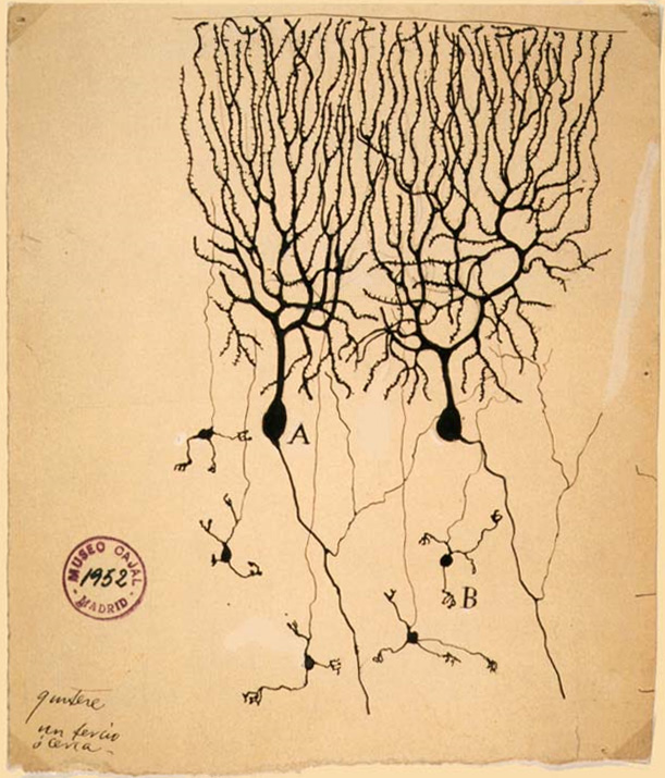
The name granule cell has been used for a number of different types of neurons whose only common feature is that they all have very small cell bodies. Granule cells are found within the granular layer of the cerebellum, the dentate gyrus of the hippocampus, the superficial layer of the dorsal cochlear nucleus, the olfactory bulb, and the cerebral cortex. Cerebellar granule cells account for the majority of neurons in the human brain. These granule cells receive excitatory input from mossy fibers originating from pontine nuclei. Cerebellar granule cells project up through the Purkinje layer into the molecular layer where they branch out into parallel fibers that spread through Purkinje cell dendritic arbors. These parallel fibers form thousands of excitatory granule-cell–Purkinje-cell synapses onto the intermediate and distal dendrites of Purkinje cells using glutamate as a neurotransmitter. Layer 4 granule cells of the cerebral cortex receive inputs from the thala ... [...More Info...] [...Related Items...] OR: [Wikipedia] [Google] [Baidu] |
Dendrites
A dendrite (from Greek δένδρον ''déndron'', "tree") or dendron is a branched cytoplasmic process that extends from a nerve cell that propagates the electrochemical stimulation received from other neural cells to the cell body, or soma, of the neuron from which the dendrites project. Electrical stimulation is transmitted onto dendrites by upstream neurons (usually via their axons) via synapses which are located at various points throughout the dendritic tree. Dendrites play a critical role in integrating these synaptic inputs and in determining the extent to which action potentials are produced by the neuron. Structure and function Dendrites are one of two types of cytoplasmic processes that extrude from the cell body of a neuron, the other type being an axon. Axons can be distinguished from dendrites by several features including shape, length, and function. Dendrites often taper off in shape and are shorter, while axons tend to maintain a constant radius and can b ... [...More Info...] [...Related Items...] OR: [Wikipedia] [Google] [Baidu] |
Axon
An axon (from Greek ἄξων ''áxōn'', axis) or nerve fiber (or nerve fibre: see American and British English spelling differences#-re, -er, spelling differences) is a long, slender cellular extensions, projection of a nerve cell, or neuron, in Vertebrate, vertebrates, that typically conducts electrical impulses known as action potentials away from the Soma (biology), nerve cell body. The function of the axon is to transmit information to different neurons, muscles, and glands. In certain sensory neurons (pseudounipolar neurons), such as those for touch and warmth, the axons are called afferent nerve fibers and the electrical impulse travels along these from the peripheral nervous system, periphery to the cell body and from the cell body to the spinal cord along another branch of the same axon. Axon dysfunction can be the cause of many inherited and acquired neurological disorders that affect both the Peripheral nervous system, peripheral and Central nervous system, central ne ... [...More Info...] [...Related Items...] OR: [Wikipedia] [Google] [Baidu] |
Memory Encoding
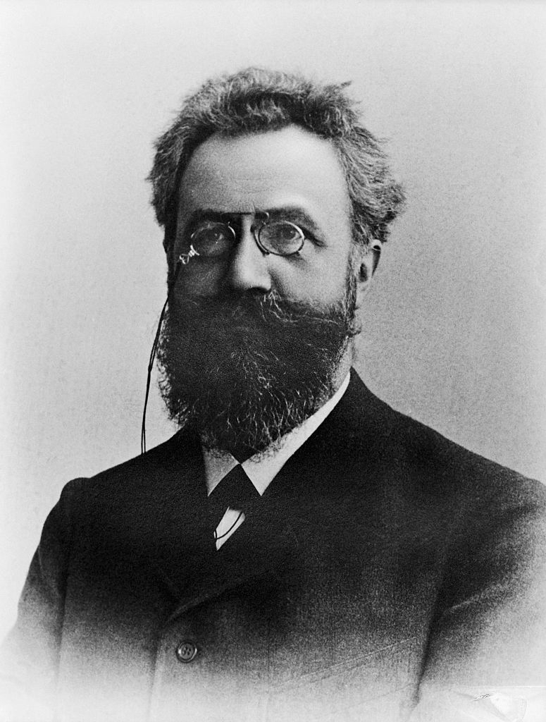
Memory has the ability to encode, store and recall information. Memories give an organism the capability to learn and adapt from previous experiences as well as build relationships. Encoding allows a perceived item of use or interest to be converted into a construct that can be stored within the brain and recalled later from long-term memory. Working memory stores information for immediate use or manipulation, which is aided through hooking onto previously archived items already present in the long-term memory of an individual. History Encoding is still relatively new and unexplored but the origins of encoding date back to age-old philosophers such as Aristotle and Plato. A major figure in the history of encoding is Hermann Ebbinghaus (1850–1909). Ebbinghaus was a pioneer in the field of memory research. Using himself as a subject he studied how we learn and forget information by repeating a list of nonsense syllables to the rhythm of a metronome until they were committed ... [...More Info...] [...Related Items...] OR: [Wikipedia] [Google] [Baidu] |
Action Potentials
An action potential (also known as a nerve impulse or "spike" when in a neuron) is a series of quick changes in voltage across a cell membrane. An action potential occurs when the membrane potential of a specific cell rapidly rises and falls. This depolarization then causes adjacent locations to similarly depolarize. Action potentials occur in several types of excitable cells, which include animal cells like neurons and muscle cells, as well as some plant cells. Certain endocrine cells such as pancreatic beta cells, and certain cells of the anterior pituitary gland are also excitable cells. In neurons, action potentials play a central role in cell–cell communication by providing for—or with regard to saltatory conduction, assisting—the propagation of signals along the neuron's axon toward synaptic boutons situated at the ends of an axon; these signals can then connect with other neurons at synapses, or to motor cells or glands. In other types of cells, their mai ... [...More Info...] [...Related Items...] OR: [Wikipedia] [Google] [Baidu] |
Schaffer Collateral
Schaffer collaterals are axon collaterals given off by CA3 pyramidal cells in the hippocampus. These collaterals project to area CA1 of the hippocampus and are an integral part of memory formation and the emotional network of the Papez circuit, and of the hippocampus, hippocampal trisynaptic loop. It is one of the most studied synapses in the world and named after the Hungarian anatomist-neurologist Károly Schaffer. As a part of the hippocampal structures, Schaffer collaterals develop the limbic system, which plays a critical role in the aspects of learning and memory. The signals of information from the contralateral CA3 region leave via the Schaffer collateral pathways for the CA1 pyramidal neurons. Mature synapses contain fewer Schaffer collateral branches than those synapses that are not fully developed. Many scientists try to use the Schaffer collateral synapse as a sample synapse, a typical excitatory glutamatergic synapse in the Cerebral cortex, cortex that has very well been ... [...More Info...] [...Related Items...] OR: [Wikipedia] [Google] [Baidu] |
Hilus
The dentate gyrus (DG) is one of the subfields of the hippocampus, in the hippocampal formation. The hippocampal formation is located in the temporal lobe of the brain, and includes the hippocampus (including CA1 to CA4) subfields, and other subfields including the dentate gyrus, subiculum, and presubiculum. The dentate gyrus is part of the trisynaptic circuit, a neural circuit of the hippocampus, thought to contribute to the formation of new episodic memories, the spontaneous exploration of novel environments and other functions. The dentate gyrus has toothlike projections from which it is named. The subgranular zone of the dentate gyrus is one of only two major sites of adult neurogenesis in the brain, and is found in many mammals. The other main site is the subventricular zone in the ventricular system. Other sites may include the striatum and the cerebellum. However, whether significant neurogenesis takes place in the adult human dentate gyrus has been a matter of de ... [...More Info...] [...Related Items...] OR: [Wikipedia] [Google] [Baidu] |
Diagonal Band Of Broca
The diagonal band of Broca interconnects the amygdala and the septal area. It is one of the olfactory structures. It is situated upon the inferior aspect of the brain. It forms the medial margin of the anterior perforated substance. It was described by the French neuroanatomist Paul Broca. Structure It consists of fibers that are said to arise in the parolfactory area, the gyrus subcallosus and the anterior perforated substance, and course backward in the longitudinal striae to the dentate gyrus and the hippocampal region. This is a cholinergic bundle of nerve fibers posterior to the anterior perforated substance. It interconnects the subcallosal gyrus in the septal area with the hippocampus and lateral olfactory area. Nuclei Two structures are often described in this brain regions, namely the nuclei of the vertical and horizontal limbs of the diagonal band of Broca (nvlDBB and nhlDBB, respectively). nvlDBB projects to the hippocampal formation through the fornix and ... [...More Info...] [...Related Items...] OR: [Wikipedia] [Google] [Baidu] |
Medial Septum
The medial septal nucleus (MS) is one of the septal nuclei. Neurons in this nucleus give rise to the bulk of efferents from the septal nuclei. A major projection from the medial septal nucleus terminates in the hippocampal formation. It plays a role in the generation of theta waves in the hippocampus. Specifically, the GABAergic cells of the medial septum that act as theta pacemakers target dentate gyrus The dentate gyrus (DG) is one of the subfields of the hippocampus, in the hippocampal formation. The hippocampal formation is located in the temporal lobe of the brain, and includes the hippocampus (including CA1 to CA4) subfields, and other su ..., CA3, and CA1 interneurons. Pacemaking MS interneurons express hyperpolarization-activated cyclic nucleotide-gated (HCN) channels which likely, at least partially, mediate their pacemaker properties. It is composed of GABAergic cells, glutamatergic cells, and cholinergic cells. Each cell-type carries out different functions. In a ... [...More Info...] [...Related Items...] OR: [Wikipedia] [Google] [Baidu] |
Stratum Lucidum Of Hippocampus
The stratum lucidum of the hippocampus is a layer of the hippocampus between the stratum pyramidale and the stratum radiatum. It is the tract of the mossy fiber projections, both inhibitory and excitatory from the granule cells of the dentate gyrus. One mossy fiber may make up to 37 connections to a single pyramidal cell, and innervate around 12 pyramidal cells on top of that. Any given pyramidal cell in the stratum lucidum may get input from as many as 50 granule cells. Location The stratum lucidum is located within the CA3 region of the hippocampus distally to the dentate gyrus and proximally to the CA2 region. It is composed of a densely packed bundle of mossy fibers (unmyelinated) and spiny and aspiny interneurons that lie immediately above the CA3 pyramidal cell layer in the hippocampus, and immediately below the stratum radiatum. Most mossy fiber axons are perpendicular to the CA3 pyramidal region where they project and synapse to either the CA3 pyramidal cells or the str ... [...More Info...] [...Related Items...] OR: [Wikipedia] [Google] [Baidu] |


