|
Grading Of The Tumors Of The Central Nervous System
The concept of grading of the tumors of the central nervous system, agreeing for such the regulation of the "progressiveness" of these neoplasias (from benign and localized tumors to malignant and infiltrating tumors), dates back to 1926 and was introduced by P. Bailey and H. Cushing, in the elaboration of what turned out the first systematic classification of gliomas. In the following, the grading systems present in the current literature are introduced. Then, through a table, the more relevant are compared. ICD-O scale The first edition of the International Classification of Diseases (ICD) dates back to 1893. The current review (ICD-10) dates back to 1994, came into use in the U.S. in 2015, and is revised yearly, being very comprehensive. In 1976 the World Health Organization (WHO) published the first edition of the International Classification of Diseases for Oncology (ICD-O), which is now at the third edition (ICD-O-3, 2000). In this last edition, the Arabic numeral afte ... [...More Info...] [...Related Items...] OR: [Wikipedia] [Google] [Baidu] |
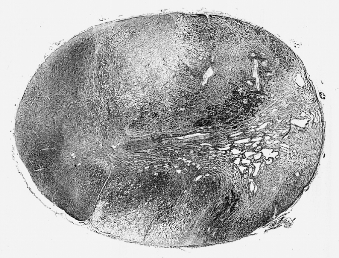
AFIP00405522M-GLIOBLASTOMA ARISING IN ASTROCYTOMA
The Armed Forces Institute of Pathology (AFIP) (1862 – September 15, 2011) was a US government, U.S. government institution concerned with diagnostic consultation, education, and research in the medical specialty of pathology. Overview It was founded in 1862 as the Army Medical Museum in Washington, DC, Washington, D.C., on the grounds of the Walter Reed Army Medical Center (WRAMC). It primarily provided second opinion (medicine), second opinion diagnostic consultations on pathologic specimens such as biopsies from military, veteran, and civilian medical, dental, and veterinary sources. The unique character of the AFIP rested in the expertise of its civilian and military staff of diagnostic pathologists whose daily work consisted of the study of cases that are difficult to diagnose owing to their rarity or their variation from the ordinary. The accumulation of such cases has resulted in a rich repository of lesions, numbering over three million, that have been the basis of ma ... [...More Info...] [...Related Items...] OR: [Wikipedia] [Google] [Baidu] |
Metastasis
Metastasis is a pathogenic agent's spread from an initial or primary site to a different or secondary site within the host's body; the term is typically used when referring to metastasis by a cancerous tumor. The newly pathological sites, then, are metastases (mets). It is generally distinguished from cancer invasion, which is the direct extension and penetration by cancer cells into neighboring tissues. Cancer occurs after cells are genetically altered to proliferate rapidly and indefinitely. This uncontrolled proliferation by mitosis produces a primary heterogeneic tumour. The cells which constitute the tumor eventually undergo metaplasia, followed by dysplasia then anaplasia, resulting in a malignant phenotype. This malignancy allows for invasion into the circulation, followed by invasion to a second site for tumorigenesis. Some cancer cells known as circulating tumor cells acquire the ability to penetrate the walls of lymphatic or blood vessels, after which they are ... [...More Info...] [...Related Items...] OR: [Wikipedia] [Google] [Baidu] |
Pilocytic Micro
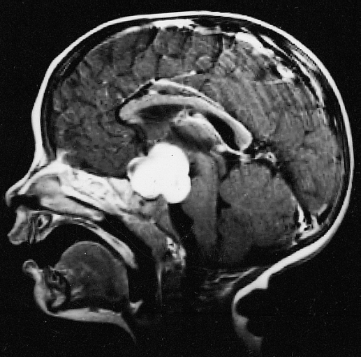
Pilocytic astrocytoma (and its variant pilomyxoid astrocytoma) is a brain tumor that occurs most commonly in children and young adults (in the first 20 years of life). They usually arise in the cerebellum, near the brainstem, in the hypothalamic region, or the optic chiasm, but they may occur in any area where astrocytes are present, including the cerebral hemispheres and the spinal cord. These tumors are usually slow growing and benign, corresponding to WHO malignancy grade 1. Signs and symptoms Children affected by pilocytic astrocytoma can present with different symptoms that might include failure to thrive (lack of appropriate weight gain/ weight loss), headache, nausea, vomiting, irritability, torticollis (tilt neck or wry neck), difficulty to coordinate movements, and visual complaints (including nystagmus). The complaints may vary depending on the location and size of the neoplasm. The most common symptoms are associated with increased intracranial pressure due to the si ... [...More Info...] [...Related Items...] OR: [Wikipedia] [Google] [Baidu] |
Necrosis
Necrosis () is a form of cell injury which results in the premature death of cells in living tissue by autolysis. Necrosis is caused by factors external to the cell or tissue, such as infection, or trauma which result in the unregulated digestion of cell components. In contrast, apoptosis is a naturally occurring programmed and targeted cause of cellular death. While apoptosis often provides beneficial effects to the organism, necrosis is almost always detrimental and can be fatal. Cellular death due to necrosis does not follow the apoptotic signal transduction pathway, but rather various receptors are activated and result in the loss of cell membrane integrity and an uncontrolled release of products of cell death into the extracellular space. This initiates in the surrounding tissue an inflammatory response, which attracts leukocytes and nearby phagocytes which eliminate the dead cells by phagocytosis. However, microbial damaging substances released by leukocytes would cre ... [...More Info...] [...Related Items...] OR: [Wikipedia] [Google] [Baidu] |
Hypervascularity
Hypervascularity is an increased number or concentration of blood vessels. In Graves disease, the thyroid gland is hypervascular, which can help in differentiating the condition from thyroiditis. 90% of thyroid papillary carcinoma Papillary thyroid cancer or papillary thyroid carcinoma is the most common type of thyroid cancer, representing 75 percent to 85 percent of all thyroid cancer cases.Chapter 20 in: 8th edition. It occurs more frequently in women and presents in th ... cases are hypervascular. See also * Angiogenesis References Hematology Oncology Angiology {{circulatory-stub ... [...More Info...] [...Related Items...] OR: [Wikipedia] [Google] [Baidu] |
Cell Growth
Cell growth refers to an increase in the total mass of a cell, including both cytoplasmic, nuclear and organelle volume. Cell growth occurs when the overall rate of cellular biosynthesis (production of biomolecules or anabolism) is greater than the overall rate of cellular degradation (the destruction of biomolecules via the proteasome, lysosome or autophagy, or catabolism). Cell growth is not to be confused with cell division or the cell cycle, which are distinct processes that can occur alongside cell growth during the process of cell proliferation, where a cell, known as the mother cell, grows and divides to produce two daughter cells. Importantly, cell growth and cell division can also occur independently of one another. During early embryonic development ( cleavage of the zygote to form a morula and blastoderm), cell divisions occur repeatedly without cell growth. Conversely, some cells can grow without cell division or without any progression of the cell cycle, su ... [...More Info...] [...Related Items...] OR: [Wikipedia] [Google] [Baidu] |
Endothelial
The endothelium is a single layer of squamous endothelial cells that line the interior surface of blood vessels and lymphatic vessels. The endothelium forms an interface between circulating blood or lymph in the lumen and the rest of the vessel wall. Endothelial cells form the barrier between vessels and tissue and control the flow of substances and fluid into and out of a tissue. Endothelial cells in direct contact with blood are called vascular endothelial cells whereas those in direct contact with lymph are known as lymphatic endothelial cells. Vascular endothelial cells line the entire circulatory system, from the heart to the smallest capillaries. These cells have unique functions that include fluid filtration, such as in the glomerulus of the kidney, blood vessel tone, hemostasis, neutrophil recruitment, and hormone trafficking. Endothelium of the interior surfaces of the heart chambers is called endocardium. An impaired function can lead to serious health issues throug ... [...More Info...] [...Related Items...] OR: [Wikipedia] [Google] [Baidu] |
Mitosis
In cell biology, mitosis () is a part of the cell cycle in which replicated chromosomes are separated into two new nuclei. Cell division by mitosis gives rise to genetically identical cells in which the total number of chromosomes is maintained. Therefore, mitosis is also known as equational division. In general, mitosis is preceded by S phase of interphase (during which DNA replication occurs) and is often followed by telophase and cytokinesis; which divides the cytoplasm, organelles and cell membrane of one cell into two new cells containing roughly equal shares of these cellular components. The different stages of mitosis altogether define the mitotic (M) phase of an animal cell cycle—the division of the mother cell into two daughter cells genetically identical to each other. The process of mitosis is divided into stages corresponding to the completion of one set of activities and the start of the next. These stages are preprophase (specific to plant cells), propha ... [...More Info...] [...Related Items...] OR: [Wikipedia] [Google] [Baidu] |
Atypia
Atypia (from Greek, ''a'' + ''typos'', without type; a condition of being irregular or nonstandard) is a histopathologic term for a structural abnormality in a cell, i.e. it is used to describe atypical cells. Atypia can be caused by an infection or irritation if diagnosed in a Pap smear, for example. In the uterus it is more likely to be precancerous. The related concept of dysplasia refers to an abnormality of development, and includes abnormalities on larger, histopathologic scales. Example features Features that constitute atypia have different definitions for different diseases, but often include the following nucleus abnormalities: *Enlargement *Pleomorphism *Nuclear polychromasia, which means variability in nuclear chromatin content. Polychromasia otherwise refers to a disease of immature red blood cells. *Numerous mitotic figures Examples for Barrett's esophagus In Barrett's esophagus, features that are classified as atypia but not as dysplasia are mainly: *''Nuclear ... [...More Info...] [...Related Items...] OR: [Wikipedia] [Google] [Baidu] |
Anaplastic Astrocytoma
Anaplastic astrocytoma is a rare WHO grade III type of astrocytoma, which is a type of cancer of the brain. In the United States, the annual incidence rate for anaplastic astrocytoma is 0.44 per 100,000 people. Signs and symptoms Initial presenting symptoms most commonly are headache, depressed mental status, focal neurological deficits, and/or seizures. The growth rate and mean interval between onset of symptoms and diagnosis is approximately 1.5–2 years but is highly variable, being intermediate between that of low-grade astrocytomas and glioblastomas. Seizures are less common among patients with anaplastic astrocytomas compared to low-grade lesions. Causes Most high-grade gliomas occur sporadically or without identifiable cause. However, a small proportion (less than 5%) of persons with malignant astrocytoma have a definite or suspected hereditary predisposition. The main hereditary predispositions are mainly neurofibromatosis type I, Li-Fraumeni syndrome, hereditary nonp ... [...More Info...] [...Related Items...] OR: [Wikipedia] [Google] [Baidu] |
Astrocytoma
Astrocytomas are a type of brain tumor. They originate in a particular kind of glial cells, star-shaped brain cells in the cerebrum called astrocytes. This type of tumor does not usually spread outside the brain and spinal cord and it does not usually affect other organs. Astrocytomas are the most common glioma and can occur in most parts of the brain and occasionally in the spinal cord. Within the astrocytomas, two broad classes are recognized in literature, those with: * Narrow zones of infiltration (mostly noninvasive tumors; e.g., pilocytic astrocytoma, subependymal giant cell astrocytoma, pleomorphic xanthoastrocytoma), that often are clearly outlined on diagnostic images * Diffuse zones of infiltration (e.g., high-grade astrocytoma, anaplastic astrocytoma, glioblastoma), that share various features, including the ability to arise at any location in the central nervous system, but with a preference for the cerebral hemispheres; they occur usually in adults, and have an intrins ... [...More Info...] [...Related Items...] OR: [Wikipedia] [Google] [Baidu] |
Glioblastoma Macro
Glioblastoma, previously known as glioblastoma multiforme (GBM), is one of the most aggressive types of cancer that begin within the brain. Initially, signs and symptoms of glioblastoma are nonspecific. They may include headaches, personality changes, nausea, and symptoms similar to those of a stroke. Symptoms often worsen rapidly and may progress to unconsciousness. The cause of most cases of glioblastoma is not known. Uncommon risk factors include genetic disorders, such as neurofibromatosis and Li–Fraumeni syndrome, and previous radiation therapy. Glioblastomas represent 15% of all brain tumors. They can either start from normal brain cells or develop from an existing low-grade astrocytoma. The diagnosis typically is made by a combination of a CT scan, MRI scan, and tissue biopsy. There is no known method of preventing the cancer. Treatment usually involves surgery, after which chemotherapy and radiation therapy are used. The medication temozolomide is frequently used ... [...More Info...] [...Related Items...] OR: [Wikipedia] [Google] [Baidu] |


