|
Gamma Camera
A gamma camera (γ-camera), also called a scintillation camera or Anger camera, is a device used to image gamma radiation emitting radioisotopes, a technique known as scintigraphy. The applications of scintigraphy include early drug development and nuclear medical imaging to view and analyse images of the human body or the distribution of medically injected, inhaled, or ingested radionuclides emitting gamma rays. Imaging techniques Scintigraphy ("scint") is the use of gamma cameras to capture emitted radiation from internal radioisotopes to create two-dimensional images. SPECT (single photon emission computed tomography) imaging, as used in nuclear cardiac stress testing, is performed using gamma cameras. Usually one, two or three detectors or heads, are slowly rotated around the patient's torso. Multi-headed gamma cameras can also be used for positron emission tomography (PET) scanning, provided that their hardware and software can be configured to detect "coincidences" ... [...More Info...] [...Related Items...] OR: [Wikipedia] [Google] [Baidu] |
Lung Scintigraphy Keosys
The lungs are the primary organs of the respiratory system in humans and most other animals, including some snails and a small number of fish. In mammals and most other vertebrates, two lungs are located near the backbone on either side of the heart. Their function in the respiratory system is to extract oxygen from the air and transfer it into the bloodstream, and to release carbon dioxide from the bloodstream into the atmosphere, in a process of gas exchange. Respiration is driven by different muscular systems in different species. Mammals, reptiles and birds use their different muscles to support and foster breathing. In earlier tetrapods, air was driven into the lungs by the pharyngeal muscles via buccal pumping, a mechanism still seen in amphibians. In humans, the main muscle of respiration that drives breathing is the diaphragm. The lungs also provide airflow that makes vocal sounds including human speech possible. Humans have two lungs, one on the left and one on th ... [...More Info...] [...Related Items...] OR: [Wikipedia] [Google] [Baidu] |
Samuel Curran
Sir Samuel Crowe Curran (23 May 1912 – 15 February 1998), FRS, FRSE, was a physicist and the first Principal and Vice-Chancellor of the University of Strathclyde – the first of the new technical universities in Britain. He is the inventor of the scintillation counter, the proportional counter, and the proximity fuze. To date, Curran remains the longest serving principal and vice chancellor of the University of Strathclyde, holding the post for 16 years, not counting his previous five years as principal of the Royal College of Science and Technology. Life Samuel Curran was born on 23 May 1912 at Ballymena in Northern Ireland, the son of John Hamilton Curran (from Kinghorn in Fife), and his wife, Sarah Carson Crowe (some sources state Sarah Owen Crowe). The family moved to Scotland soon after for his father to work as foreman of a steelworks near Wishaw. His brother Robert Curran, later a famous pathologist, was born soon after. He had two other brothers, Hamilton an ... [...More Info...] [...Related Items...] OR: [Wikipedia] [Google] [Baidu] |
Technetium-99m
Technetium-99m (99mTc) is a metastable nuclear isomer of technetium-99 (itself an isotope of technetium), symbolized as 99mTc, that is used in tens of millions of medical diagnostic procedures annually, making it the most commonly used medical radioisotope in the world. Technetium-99m is used as a radioactive tracer and can be detected in the body by medical equipment (gamma cameras). It is well suited to the role, because it emits readily detectable gamma rays with a photon energy of 140 keV (these 8.8 pm photons are about the same wavelength as emitted by conventional X-ray diagnostic equipment) and its half-life for gamma emission is 6.0058 hours (meaning 93.7% of it decays to 99Tc in 24 hours). The relatively "short" physical half-life of the isotope and its biological half-life of 1 day (in terms of human activity and metabolism) allows for scanning procedures which collect data rapidly but keep total patient radiation exposure low. The same characteristic ... [...More Info...] [...Related Items...] OR: [Wikipedia] [Google] [Baidu] |
Pulse Height Analyzer
A pulse-height analyzer (PHA) is an instrument that accepts electronic pulses of varying heights from particle and event detectors, digitizes the pulse heights, and saves the number of pulses of each height in registers or channels, thus recording a pulse-height spectrum or pulse-height distribution used for later pulse-height analysis. PHAs are used in nuclear- and elementary-particle physics research. A PHA is a specific modification to multichannel analyzers. A pulse-height analyzer is also integrated into particle counters or used as a discrete module to calibrate particle counters. See also * Nuclear electronics Experimental particle physics {{nuclear-stub ... [...More Info...] [...Related Items...] OR: [Wikipedia] [Google] [Baidu] |
Photomultiplier
A photomultiplier is a device that converts incident photons into an electrical signal. Kinds of photomultiplier include: * Photomultiplier tube, a vacuum tube converting incident photons into an electric signal. Photomultiplier tubes (PMTs for short) are members of the class of vacuum tubes, and more specifically vacuum phototubes, which are extremely sensitive detectors of light in the ultraviolet, visible, and near-infrared ranges of the electromagnetic spectrum. ** Magnetic photomultiplier, developed by the Soviets in the 1930s. ** Electrostatic photomultiplier, a kind of photomultiplier tube demonstrated by Jan Rajchman of RCA Laboratories in Princeton, NJ in the late 1930s which became the standard for all future commercial photomultipliers. The first mass-produced photomultiplier, the Type 931, was of this design and is still commercially produced today. * Silicon photomultiplier, a solid-state device converting incident photons into an electric signal. Silicon photomu ... [...More Info...] [...Related Items...] OR: [Wikipedia] [Google] [Baidu] |
Vacuum Tube
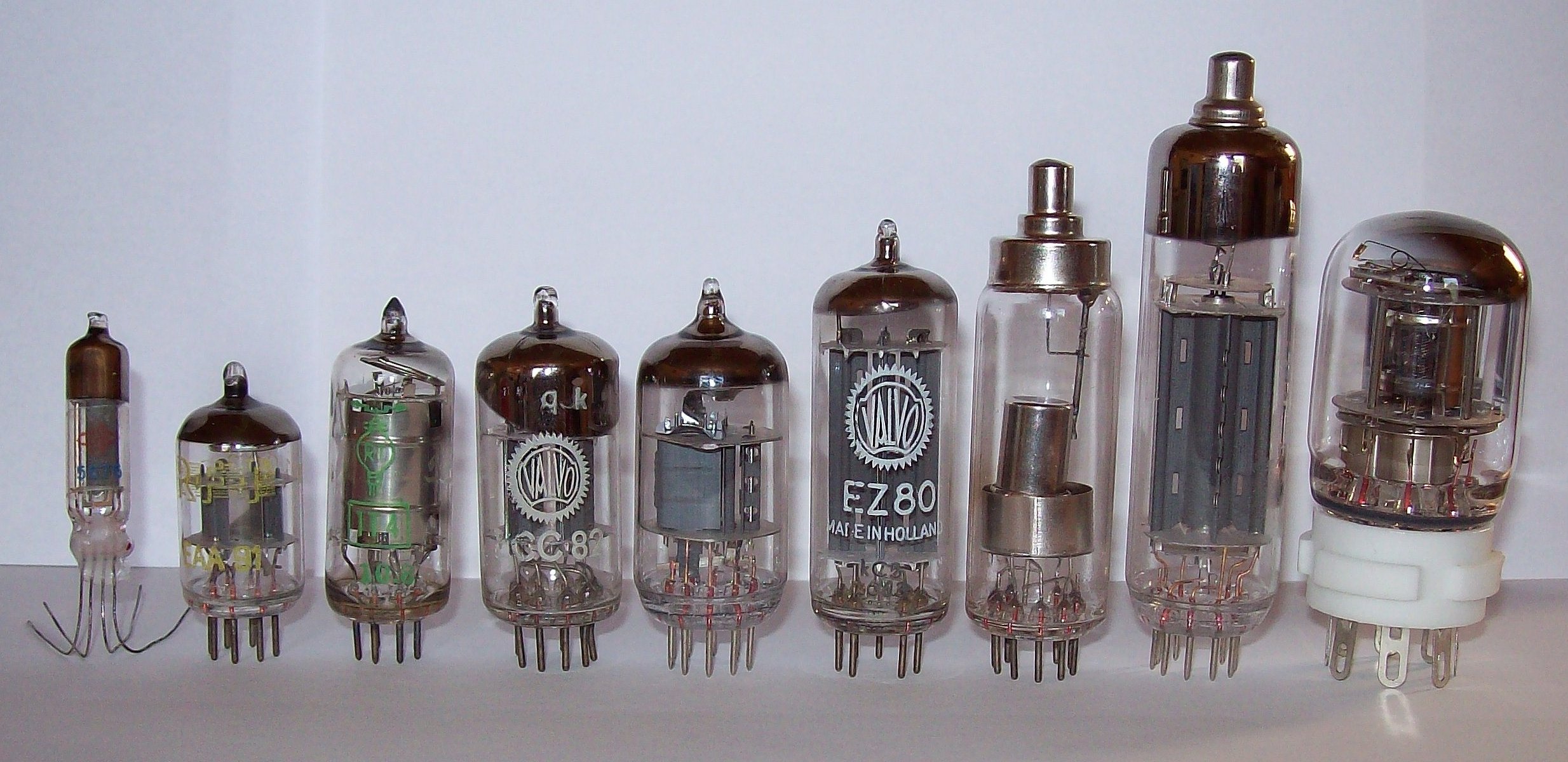
A vacuum tube, electron tube, valve (British usage), or tube (North America), is a device that controls electric current flow in a high vacuum between electrodes to which an electric potential difference has been applied. The type known as a thermionic tube or thermionic valve utilizes thermionic emission of electrons from a hot cathode for fundamental electronic functions such as signal amplification and current rectification. Non-thermionic types such as a vacuum phototube, however, achieve electron emission through the photoelectric effect, and are used for such purposes as the detection of light intensities. In both types, the electrons are accelerated from the cathode to the anode by the electric field in the tube. The simplest vacuum tube, the diode (i.e. Fleming valve), invented in 1904 by John Ambrose Fleming, contains only a heated electron-emitting cathode and an anode. Electrons can only flow in one direction through the device—from the cathode to t ... [...More Info...] [...Related Items...] OR: [Wikipedia] [Google] [Baidu] |
Hal Anger
Hal Oscar Anger (May 20, 1920 – October 31, 2005) was an American electrical engineer and biophysicist at Donner Laboratory, University of California, Berkeley, known for his invention of the gamma camera. In all, Anger held 15 patents, many of them for work at the Ernest O. Lawrence Radiation Laboratory. Anger received several awards in recognition of his inventions and their contributions to the field of nuclear medicine. Anger died in Berkeley, California. Career In 1957, he invented the scintillation camera, known also as the gamma camera or Anger camera. Anger also developed the well counter A well counter is a device used for measuring radioactivity in small samples. It usually employs a sodium iodide crystal detector. It was invented in 1951 by Hal Anger, who is also well known for inventing the scintillation camera.Gottschalk, Ale ..., widely used in laboratory tests to measure radioactivity in samples. Anger also developed a multi-plane tomographic radiation scann ... [...More Info...] [...Related Items...] OR: [Wikipedia] [Google] [Baidu] |
Collimated And Penetration
A collimated beam of light or other electromagnetic radiation has parallel rays, and therefore will spread minimally as it propagates. A perfectly collimated light beam, with no divergence, would not disperse with distance. However, diffraction prevents the creation of any such beam. Light can be approximately collimated by a number of processes, for instance by means of a collimator. Perfectly collimated light is sometimes said to be ''focused at infinity''. Thus, as the distance from a point source increases, the spherical wavefronts become flatter and closer to plane waves, which are perfectly collimated. Other forms of electromagnetic radiation can also be collimated. In radiology, X-rays are collimated to reduce the volume of the patient's tissue that is irradiated, and to remove stray photons that reduce the quality of the x-ray image ("film fog"). In scintigraphy, a gamma ray collimator is used in front of a detector to allow only photons perpendicular to the surface to be ... [...More Info...] [...Related Items...] OR: [Wikipedia] [Google] [Baidu] |
Compton Effect
Compton scattering, discovered by Arthur Holly Compton, is the scattering of a high frequency photon after an interaction with a charged particle, usually an electron. If it results in a decrease in energy (increase in wavelength) of the photon (which may be an X-ray or gamma ray photon), it is called the Compton effect. Part of the energy of the photon is transferred to the recoiling electron. Inverse Compton scattering occurs when a charged particle transfers part of its energy to a photon. Introduction Compton scattering is an example of elastic scattering of light by a free charged particle, where the wavelength of the scattered light is different from that of the incident radiation. In Compton's original experiment (see Fig. 1), the energy of the X ray photon (≈17 keV) was significantly larger than the binding energy of the atomic electron, so the electrons could be treated as being free after scattering. The amount by which the light's wavelength changes is called the ... [...More Info...] [...Related Items...] OR: [Wikipedia] [Google] [Baidu] |
Photoelectric Effect
The photoelectric effect is the emission of electrons when electromagnetic radiation, such as light, hits a material. Electrons emitted in this manner are called photoelectrons. The phenomenon is studied in condensed matter physics, and solid state and quantum chemistry to draw inferences about the properties of atoms, molecules and solids. The effect has found use in electronic devices specialized for light detection and precisely timed electron emission. The experimental results disagree with classical electromagnetism, which predicts that continuous light waves transfer energy to electrons, which would then be emitted when they accumulate enough energy. An alteration in the intensity of light would theoretically change the kinetic energy of the emitted electrons, with sufficiently dim light resulting in a delayed emission. The experimental results instead show that electrons are dislodged only when the light exceeds a certain frequency—regardless of the light's intensity o ... [...More Info...] [...Related Items...] OR: [Wikipedia] [Google] [Baidu] |
Radiopharmaceutical
Radiopharmaceuticals, or medicinal radiocompounds, are a group of pharmaceutical drugs containing radioactive isotopes. Radiopharmaceuticals can be used as diagnostic and therapeutic agents. Radiopharmaceuticals emit radiation themselves, which is different from contrast media which absorb or alter external electromagnetism or ultrasound. Radiopharmacology is the branch of pharmacology that specializes in these agents. The main group of these compounds are the radiotracers used to diagnose dysfunction in body tissues. While not all medical isotopes are radioactive, radiopharmaceuticals are the oldest and still most common such drugs. Drug nomenclature As with other pharmaceutical drugs, there is standardization of the drug nomenclature for radiopharmaceuticals, although various standards coexist. The International Nonproprietary Names (INNs), United States Pharmacopeia (USP) names, and IUPAC names for these agents are usually similar other than trivial style difference ... [...More Info...] [...Related Items...] OR: [Wikipedia] [Google] [Baidu] |
Scintillation (physics)
Scintillation is the physical process where a material, called scintillator, emits UV or visible light under excitation from high energy photons (X-rays or γ-rays) or energetic particles,(such as electrons, alpha particles, neutrons or ions). See scintillator and scintillation counter for practical applications. Overview The process of scintillation is one of luminescence whereby light of a characteristic spectrum is emitted following the absorption of radiation. The scintillation process can be summarized in three main stages (A) conversion, (B) transport and energy transfer to the luminescence center, and (C) luminescence. The emitted radiation is usually less energetic than the absorbed radiation, hence generally scintillation is a down-conversion process. Conversion processes The first stage of scintillation, conversion, is the process where the energy from the incident radiation is absorbed by the scintillator and highly energetic electrons and holes are created in the ... [...More Info...] [...Related Items...] OR: [Wikipedia] [Google] [Baidu] |
.jpg)


