|
General Somatic Efferent Fibers
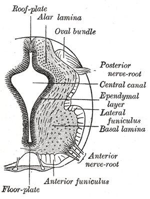
The general (spinal) somatic efferent neurons (GSE, somatomotor, or somatic motor fibers) arise from motor neuron cell bodies in the ventral horns of the gray matter within the spinal cord. They exit the spinal cord through the ventral roots, carrying motor impulses to skeletal muscle through a neuromuscular junction. Of the somatic efferent neurons, there exist subtypes. * Alpha motor neurons (α) target extrafusal muscle fibers. * Gamma motor neurons (γ) target intrafusal muscle fibres Cranial nerves also supply their own somatic efferent neurons to the extraocular muscles and some of the muscles of the tongue. See also * Nerve fiber * Efferent nerve * General visceral efferent fiber (GVE) * Special visceral efferent fiber (SVE) References Peripheral nervous system {{Portal bar, Anatomy ... [...More Info...] [...Related Items...] OR: [Wikipedia] [Google] [Baidu] |
General Somatic Afferent Fibers
The general somatic afferent fibers (GSA or somatic sensory fibers) are afferent fibers that arise from neurons in sensory ganglia and are found in all the spinal nerves, except occasionally the first cervical. General somatic afferents conduct impulses of Pain#Nociceptive, pain, touch and temperature from the surface of the body through the dorsal roots to the spinal cord, and impulses of muscle sense, tendon sense and joint sense from the deeper structures. See also * Afferent nerve * General visceral afferent fiber (GVA) * Special somatic afferent fiber (SSA) * Special visceral afferent fiber (SVA) References Spinal cord {{Portal bar, Anatomy ... [...More Info...] [...Related Items...] OR: [Wikipedia] [Google] [Baidu] |
Gamma Motor Neurons
A gamma motor neuron (γ motor neuron), also called gamma motoneuron, or fusimotor neuron, is a type of lower motor neuron that takes part in the process of muscle contraction, and represents about 30% of ( Aγ) fibers going to the muscle. Like alpha motor neurons, their cell bodies are located in the anterior grey column of the spinal cord. They receive input from the reticular formation of the pons in the brainstem. Their axons are smaller than those of the alpha motor neurons, with a diameter of only 5 μm. Unlike the alpha motor neurons, gamma motor neurons do not directly adjust the lengthening or shortening of muscles. However, their role is important in keeping muscle spindles taut, thereby allowing the continued firing of alpha neurons, leading to muscle contraction. These neurons also play a role in adjusting the sensitivity of muscle spindles. The presence of myelination in gamma motor neurons allows a conduction velocity of 4 to 24 meters per second, signifi ... [...More Info...] [...Related Items...] OR: [Wikipedia] [Google] [Baidu] |
General Visceral Efferent Fiber
General visceral efferent fibers (GVE), visceral efferents or autonomic efferents are the efferent nerve fibers of the autonomic nervous system (also known as the ''visceral efferent nervous system'') that provide motor innervation to smooth muscle, cardiac muscle, and glands (contrast with special visceral efferent (SVE) fibers) through postganglionic varicosities. GVE fibers may be either sympathetic or parasympathetic. Cranial and sacral spinal fibers are parasympathetic GVE fibers, while thoracic and lumbar spinal cord give rise to sympathetic GVE fibers. The cranial nerves containing GVE fibers include the oculomotor nerve (CN III), the facial nerve (CN VII), the glossopharyngeal nerve (CN IX) and the vagus nerve (CN X).Mehta, Samir et al. Step-Up: A High-Yield, Systems-Based Review for the USMLE Step 1. Baltimore, MD: LWW, 2003. Additional images File:Gray840.png, Sympathetic connections of the ciliary and superior cervical ganglia. File:Gray839.png, Autonomic nervous ... [...More Info...] [...Related Items...] OR: [Wikipedia] [Google] [Baidu] |
Efferent Nerve
A motor nerve, or efferent nerve, is a nerve that contains exclusively efferent nerve fibers and transmits motor signals from the central nervous system (CNS) to the effector organs (muscles and glands), as opposed to sensory nerves, which transfer signals from sensory receptors in the periphery to the CNS. This is different from the motor neuron, which includes a cell body and branching of dendrites, while the nerve is made up of a bundle of axons. In the strict sense, a "motor nerve" can refer exclusively to the connection to muscles, excluding other organs. The vast majority of nerves contain both sensory and motor fibers and are therefore called mixed nerves. Structure and function Motor nerve fibers transduce signals from the CNS to peripheral neurons of proximal muscle tissue. Motor nerve axon terminals innervate skeletal and smooth muscle, as they are heavily involved in muscle control. Motor nerves tend to be rich in acetylcholine vesicles because the motor nerve, a bu ... [...More Info...] [...Related Items...] OR: [Wikipedia] [Google] [Baidu] |
Nerve Fiber
An axon (from Greek ἄξων ''áxōn'', axis) or nerve fiber (or nerve fibre: see spelling differences) is a long, slender projection of a nerve cell, or neuron, in vertebrates, that typically conducts electrical impulses known as action potentials away from the nerve cell body. The function of the axon is to transmit information to different neurons, muscles, and glands. In certain sensory neurons ( pseudounipolar neurons), such as those for touch and warmth, the axons are called afferent nerve fibers and the electrical impulse travels along these from the periphery to the cell body and from the cell body to the spinal cord along another branch of the same axon. Axon dysfunction can be the cause of many inherited and acquired neurological disorders that affect both the peripheral and central neurons. Nerve fibers are classed into three typesgroup A nerve fibers, group B nerve fibers, and group C nerve fibers. Groups A and B are myelinated, and group C are unmyelinated. T ... [...More Info...] [...Related Items...] OR: [Wikipedia] [Google] [Baidu] |
Tongue
The tongue is a Muscle, muscular organ (anatomy), organ in the mouth of a typical tetrapod. It manipulates food for chewing and swallowing as part of the digestive system, digestive process, and is the primary organ of taste. The tongue's upper surface (dorsum) is covered by taste buds housed in numerous lingual papillae. It is sensitive and kept moist by saliva and is richly supplied with nerves and blood vessels. The tongue also serves as a natural means of cleaning the teeth. A major function of the tongue is to enable speech in humans and animal communication, vocalization in other animals. The human tongue is divided into two parts, an oral cavity, oral part at the front and a pharynx, pharyngeal part at the back. The left and right sides are also separated along most of its length by a vertical section of connective tissue, fibrous tissue (the lingual septum) that results in a groove, the median sulcus, on the tongue's surface. There are two groups of glossal muscles. The f ... [...More Info...] [...Related Items...] OR: [Wikipedia] [Google] [Baidu] |
Extraocular Muscles
The extraocular muscles, or extrinsic ocular muscles, are the seven extrinsic muscles of the eye in human eye, humans and other animals. Six of the extraocular muscles, the four recti muscles, and the superior oblique muscle, superior and inferior oblique muscles, control Eye movement, movement of the eye. The other muscle, the Levator palpebrae superioris muscle, levator palpebrae superioris, controls eyelid elevation and depression, elevation. The actions of the six muscles responsible for eye movement depend on the position of the eye at the time of muscle contraction. The ciliary muscle, pupillary sphincter muscle and pupillary dilator muscle sometimes are called intrinsic ocular muscles or intraocular muscles. Structure Since only a small part of the eye called the Fovea centralis, fovea provides sharp vision, the eye must move to follow a target. Eye movements must be precise and fast. This is seen in scenarios like reading, where the reader must shift gaze constantly. Alt ... [...More Info...] [...Related Items...] OR: [Wikipedia] [Google] [Baidu] |
Intrafusal Muscle Fibre
Intrafusal muscle fibers are skeletal muscle fibers that serve as specialized sensory organs (proprioceptors). They detect the amount and rate of change in length of a muscle.Casagrand, Janet (2008) ''Action and Movement: Spinal Control of Motor Units and Spinal Reflexes.'' University of Colorado, Boulder. They constitute the muscle spindle, and are innervated by both sensory (afferent) and motor (efferent) fibers. Intrafusal muscle fibers are not to be confused with extrafusal muscle fibers, which contract, generating skeletal movement and are innervated by alpha motor neurons. Structure Types There are two types of intrafusal muscle fibers: nuclear bag fibers and nuclear chain fibers. They bear two types of sensory ending, known as annulospiral and flower-spray endings. Both ends of these fibers contract, but the central region only stretches and does not contract. Intrafusal muscle fibers are walled off from the rest of the muscle by an outer connective tissue sheath ... [...More Info...] [...Related Items...] OR: [Wikipedia] [Google] [Baidu] |
Extrafusal Muscle Fiber
Extrafusal muscle fibers are the standard skeletal muscle fibers that are innervated by alpha motor neurons and generate tension by contracting, thereby allowing for skeletal movement. They make up the large mass of skeletal striated muscle tissue and are attached to bone by fibrous tissue extensions (tendons). Each alpha motor neuron and the extrafusal muscle fibers innervated by it make up a motor unit. The connection between the alpha motor neuron and the extrafusal muscle fiber is a neuromuscular junction, where the neuron's signal, the action potential, is transduced to the muscle fiber by the neurotransmitter acetylcholine. Extrafusal muscle fibers are not to be confused with intrafusal muscle fibers, which are innervated by sensory nerve endings in central noncontractile parts and by gamma motor neurons in contractile ends and thus serve as a sensory proprioceptor. Extrafusal muscle fibers can be generated in vitro (in a dish) from pluripotent stem cells through d ... [...More Info...] [...Related Items...] OR: [Wikipedia] [Google] [Baidu] |
General Visceral Efferent Fibers
General visceral efferent fibers (GVE), visceral efferents or autonomic efferents are the efferent nerve fibers of the autonomic nervous system (also known as the ''visceral efferent nervous system'') that provide motor innervation to smooth muscle, cardiac muscle, and glands (contrast with special visceral efferent (SVE) fibers) through postganglionic varicosities. GVE fibers may be either sympathetic or parasympathetic. Cranial and sacral spinal fibers are parasympathetic GVE fibers, while thoracic and lumbar spinal cord give rise to sympathetic GVE fibers. The cranial nerves containing GVE fibers include the oculomotor nerve (CN III), the facial nerve (CN VII), the glossopharyngeal nerve (CN IX) and the vagus nerve (CN X).Mehta, Samir et al. Step-Up: A High-Yield, Systems-Based Review for the USMLE Step 1. Baltimore, MD: LWW, 2003. Additional images File:Gray840.png, Sympathetic connections of the ciliary and superior cervical ganglia. File:Gray839.png, Autonomic nervou ... [...More Info...] [...Related Items...] OR: [Wikipedia] [Google] [Baidu] |
Alpha Motor Neurons
Alpha (α) motor neurons (also called alpha motoneurons), are large, multipolar lower motor neurons of the brainstem and spinal cord. They innervate extrafusal muscle fibers of skeletal muscle and are directly responsible for initiating their contraction. Alpha motor neurons are distinct from gamma motor neurons, which innervate intrafusal muscle fibers of muscle spindles. While their cell bodies are found in the central nervous system (CNS), α motor neurons are also considered part of the somatic nervous system—a branch of the peripheral nervous system (PNS)—because their axons extend into the periphery to innervate skeletal muscles. An alpha motor neuron and the muscle fibers it innervates comprise a motor unit. A motor neuron pool contains the cell bodies of all the alpha motor neurons involved in contracting a single muscle. Location Alpha motor neurons (α-MNs) innervating the head and neck are found in the brainstem; the remaining α-MNs innervate the rest of the b ... [...More Info...] [...Related Items...] OR: [Wikipedia] [Google] [Baidu] |
Neuromuscular Junction
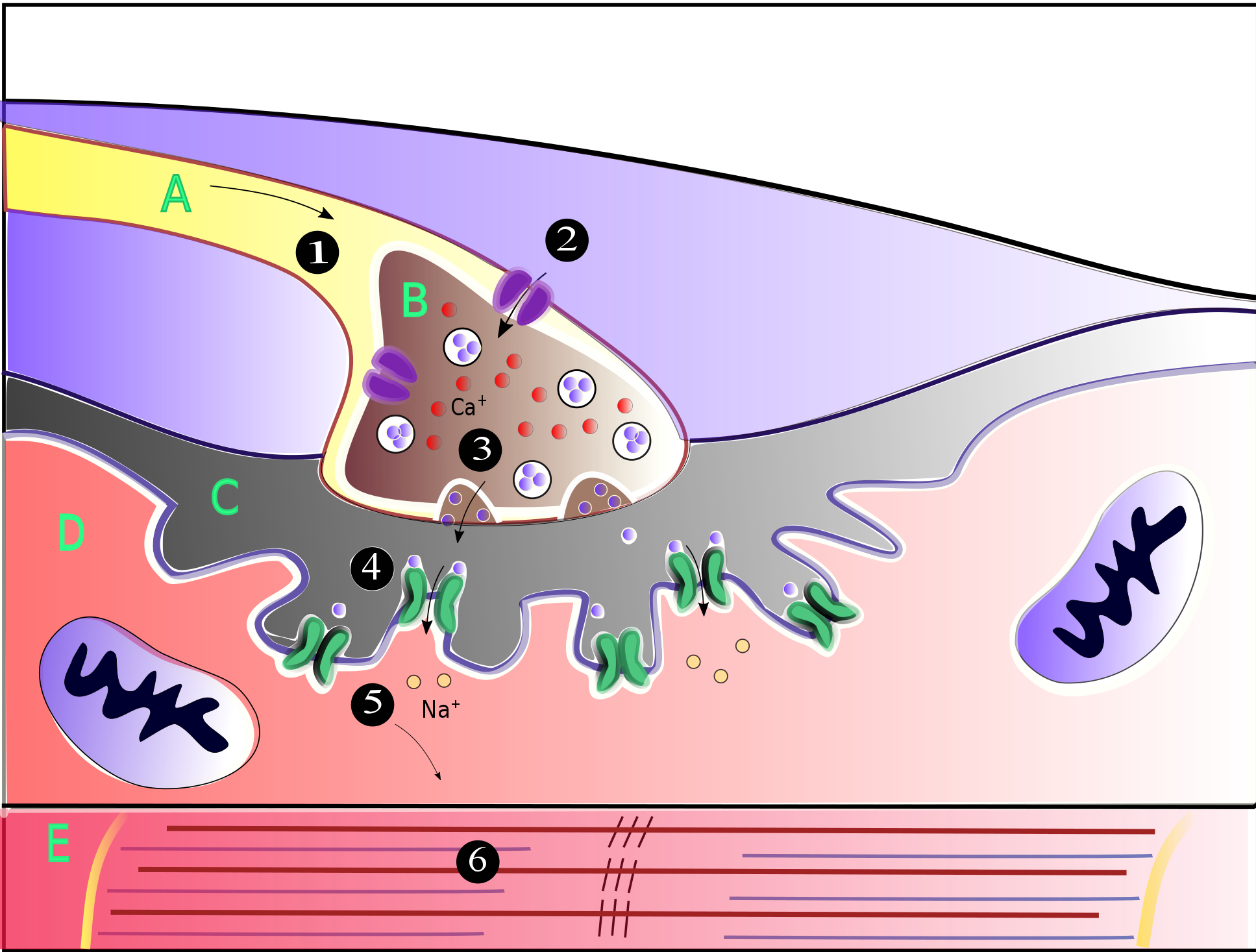
A neuromuscular junction (or myoneural junction) is a chemical synapse between a motor neuron and a muscle fiber. It allows the motor neuron to transmit a signal to the muscle fiber, causing muscle contraction. Muscles require innervation to function—and even just to maintain muscle tone, avoiding atrophy. In the neuromuscular system, nerves from the central nervous system and the peripheral nervous system are linked and work together with muscles. Synaptic transmission at the neuromuscular junction begins when an action potential reaches the presynaptic terminal of a motor neuron, which activates voltage-gated calcium channels to allow calcium ions to enter the neuron. Calcium ions bind to sensor proteins (synaptotagmins) on synaptic vesicles, triggering vesicle fusion with the cell membrane and subsequent neurotransmitter release from the motor neuron into the synaptic cleft. In vertebrates, motor neurons release acetylcholine (ACh), a small molecule neurotransmitter, which ... [...More Info...] [...Related Items...] OR: [Wikipedia] [Google] [Baidu] |


