|
Foramen Secundum
The foramen secundum, or ostium secundum is a in the septum primum, a precursor to the interatrial septum of the human heart. It is not the same as the foramen ovale, which is an opening in the septum secundum. Development The foramen secundum () is formed from small perforations that develop in the septum primum. The septum primum is a that grows down between the single primitive atrium of the developing heart to separate it into left and right atria.Schoenwolf, Gary C., and William J. Larsen. Larsen's Human Embryology. 4th ed. Philadelphia: Churchill Livingstone/Elsevier, 2009. 363. Print. Closure Once a baby is born, blood should flow through the lungs, which now function to provide oxygen to the blood. The foramen secundum and foramen ovale act as a shunt where blood bypasses the lungs and does not become oxygenated. To provide proper blood flow as a newborn, the foramen secundum and foramen ovale must close at birth. Since the lungs now require a significant amount ... [...More Info...] [...Related Items...] OR: [Wikipedia] [Google] [Baidu] |
Right Ventricle
A ventricle is one of two large chambers toward the bottom of the heart that collect and expel blood towards the peripheral beds within the body and lungs. The blood pumped by a ventricle is supplied by an atrium, an adjacent chamber in the upper heart that is smaller than a ventricle. Interventricular means between the ventricles (for example the interventricular septum), while intraventricular means within one ventricle (for example an intraventricular block). In a four-chambered heart, such as that in humans, there are two ventricles that operate in a double circulatory system: the right ventricle pumps blood into the pulmonary circulation to the lungs, and the left ventricle pumps blood into the systemic circulation through the aorta. Structure Ventricles have thicker walls than atria and generate higher blood pressures. The physiological load on the ventricles requiring pumping of blood throughout the body and lungs is much greater than the pressure generated by the atria t ... [...More Info...] [...Related Items...] OR: [Wikipedia] [Google] [Baidu] |
Systole (medicine)
Systole ( ) is the part of the cardiac cycle during which some chambers of the heart contract after refilling with blood. The term originates, via New Latin, from Ancient Greek (''sustolē''), from (''sustéllein'' 'to contract'; from ''sun'' 'together' + ''stéllein'' 'to send'), and is similar to the use of the English term ''to squeeze''. The mammalian heart has four chambers: the left atrium above the left ventricle (lighter pink, see graphic), which two are connected through the mitral (or bicuspid) valve; and the right atrium above the right ventricle (lighter blue), connected through the tricuspid valve. The atria are the receiving blood chambers for the circulation of blood and the ventricles are the discharging chambers. In late ventricular diastole, the atrial chambers contract and send blood to the larger, lower ventricle chambers. This flow fills the ventricles with blood, and the resulting pressure closes the valves to the atria. The ventricles now perform i ... [...More Info...] [...Related Items...] OR: [Wikipedia] [Google] [Baidu] |
Mitral Valve Prolapse
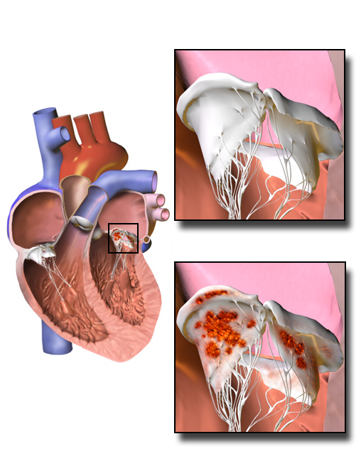
Mitral valve prolapse (MVP) is a valvular heart disease characterized by the displacement of an abnormally thickened mitral valve leaflet into the left atrium during systole. It is the primary form of myxomatous degeneration of the valve. There are various types of MVP, broadly classified as classic and nonclassic. In severe cases of classic MVP, complications include mitral regurgitation, infective endocarditis, congestive heart failure, and, in rare circumstances, cardiac arrest. The diagnosis of MVP depends upon echocardiography, which uses ultrasound to visualize the mitral valve. MVP is the most common valvular abnormality and is estimated to affect 2–3% of the population and 1 in 40 people might have it. The condition was first described by John Brereton Barlow in 1966. It was subsequently termed ''mitral valve prolapse'' by J. Michael Criley. Although mid-systolic click (sound of prolapsing mitral leaflet) and systolic murmur have been noticed earlier with stethoscope ... [...More Info...] [...Related Items...] OR: [Wikipedia] [Google] [Baidu] |
Mitral Regurgitation
Mitral regurgitation (MR), also known as mitral insufficiency or mitral incompetence, is a form of valvular heart disease in which the mitral valve is insufficient and does not close properly when the heart pumps out blood.Mitral valve regurgitation at Mount Sinai Hospital It is the abnormal leaking of blood backwards – regurgitation from the , through the mitral valve, into the |
Cardiac Dysrhythmia
Arrhythmias, also known as cardiac arrhythmias, heart arrhythmias, or dysrhythmias, are irregularities in the heartbeat, including when it is too fast or too slow. A resting heart rate that is too fast – above 100 beats per minute in adults – is called tachycardia, and a resting heart rate that is too slow – below 60 beats per minute – is called bradycardia. Some types of arrhythmias have no symptoms. Symptoms, when present, may include palpitations or feeling a pause between heartbeats. In more serious cases, there may be lightheadedness, passing out, shortness of breath or chest pain. While most cases of arrhythmia are not serious, some predispose a person to complications such as stroke or heart failure. Others may result in sudden death. Arrhythmias are often categorized into four groups: extra beats, supraventricular tachycardias, ventricular arrhythmias and bradyarrhythmias. Extra beats include premature atrial contractions, premature ventricular con ... [...More Info...] [...Related Items...] OR: [Wikipedia] [Google] [Baidu] |
Prophylactic
Preventive healthcare, or prophylaxis, consists of measures taken for the purposes of disease prevention.Hugh R. Leavell and E. Gurney Clark as "the science and art of preventing disease, prolonging life, and promoting physical and mental health and efficiency. Leavell, H. R., & Clark, E. G. (1979). Preventive Medicine for the Doctor in his Community (3rd ed.). Huntington, NY: Robert E. Krieger Publishing Company. Disease and disability are affected by environmental factors, genetic predisposition, disease agents, and lifestyle choices, and are dynamic processes which begin before individuals realize they are affected. Disease prevention relies on anticipatory actions that can be categorized as primal, primary, secondary, and tertiary prevention. Each year, millions of people die of preventable deaths. A 2004 study showed that about half of all deaths in the United States in 2000 were due to preventable behaviors and exposures. Leading causes included cardiovascular disease ... [...More Info...] [...Related Items...] OR: [Wikipedia] [Google] [Baidu] |
Infective Endocarditis
Infective endocarditis is an infection of the inner surface of the heart, usually the valves. Signs and symptoms may include fever, small areas of bleeding into the skin, heart murmur, feeling tired, and low red blood cell count. Complications may include backward blood flow in the heart, heart failure – the heart struggling to pump a sufficient amount of blood to meet the body's needs, abnormal electrical conduction in the heart, stroke, and kidney failure. The cause is typically a bacterial infection and less commonly a fungal infection. Risk factors include valvular heart disease, including rheumatic disease, congenital heart disease, artificial valves, hemodialysis, intravenous drug use, and electronic pacemakers. The bacteria most commonly involved are streptococci or staphylococci. Diagnosis is suspected based on symptoms and supported by blood cultures or ultrasound of the heart. There is also a noninfective form of endocarditis. The usefulness of antibiotic ... [...More Info...] [...Related Items...] OR: [Wikipedia] [Google] [Baidu] |
Femoral Vein
In the human body, the femoral vein is a blood vessel that accompanies the femoral artery in the femoral sheath. It begins at the adductor hiatus (an opening in the adductor magnus muscle) as the continuation of the popliteal vein. It ends at the inferior margin of the inguinal ligament where it becomes the external iliac vein. The femoral vein bears valves which are mostly bicuspid and whose number is variable between individuals and often between left and right leg. Structure Segments *The common femoral vein is the segment of the femoral vein between the branching point of the deep femoral vein and the inferior margin of the inguinal ligament.Page 590 in: *The subsartorial vein or superficial femoral vein are designations ... [...More Info...] [...Related Items...] OR: [Wikipedia] [Google] [Baidu] |
Dacron
Polyethylene terephthalate (or poly(ethylene terephthalate), PET, PETE, or the obsolete PETP or PET-P), is the most common thermoplastic polymer resin of the polyester family and is used in fibres for clothing, containers for liquids and foods, and thermoforming for manufacturing, and in combination with glass fibre for engineering resins. In 2016, annual production of PET was 56 million tons. The biggest application is in fibres (in excess of 60%), with bottle production accounting for about 30% of global demand. In the context of textile applications, PET is referred to by its common name, polyester, whereas the acronym ''PET'' is generally used in relation to packaging. Polyester makes up about 18% of world polymer production and is the fourth-most-produced polymer after polyethylene (PE), polypropylene (PP) and polyvinyl chloride (PVC). PET consists of repeating (C10H8O4) units. PET is commonly recycled, and has the digit 1 (♳) as its resin identification code (RIC). Th ... [...More Info...] [...Related Items...] OR: [Wikipedia] [Google] [Baidu] |
Pericardium
The pericardium, also called pericardial sac, is a double-walled sac containing the heart and the roots of the great vessels. It has two layers, an outer layer made of strong connective tissue (fibrous pericardium), and an inner layer made of serous membrane (serous pericardium). It encloses the pericardial cavity, which contains pericardial fluid, and defines the middle mediastinum. It separates the heart from interference of other structures, protects it against infection and blunt trauma, and lubricates the heart's movements. The English name originates from the Ancient Greek prefix "''peri-''" (περί; "around") and the suffix "''-cardion''" (κάρδιον; "heart"). Anatomy The pericardium is a tough fibroelastic sac which covers the heart from all sides except at the cardiac root (where the great vessels join the heart) and the bottom (where only the serous pericardium exists to cover the upper surface of the central tendon of diaphragm). The fibrous pericardiu ... [...More Info...] [...Related Items...] OR: [Wikipedia] [Google] [Baidu] |
General Anesthesia
General anaesthesia (UK) or general anesthesia (US) is a medically induced loss of consciousness that renders the patient unarousable even with painful stimuli. This effect is achieved by administering either intravenous or inhalational general anaesthetic medications, which often act in combination with an analgesic and neuromuscular blocking agent. Spontaneous ventilation is often inadequate during the procedure and intervention is often necessary to protect the airway. General anaesthesia is generally performed in an operating theater to allow surgical procedures that would otherwise be intolerably painful for a patient, or in an intensive care unit or emergency department to facilitate endotracheal intubation and mechanical ventilation in critically ill patients. A variety of drugs may be administered, with the overall goal of achieving unconsciousness, amnesia, analgesia, loss of reflexes of the autonomic nervous system, and in some cases paralysis of skeletal muscles. The ... [...More Info...] [...Related Items...] OR: [Wikipedia] [Google] [Baidu] |
Acyanotic Heart Defect
An acyanotic heart defect, is a class of congenital heart defects. In these, blood is shunted (flows) from the left side of the heart to the right side of the heart, most often due to a structural defect (hole) in the interventricular septum. People often retain normal levels of oxyhemoglobin saturation in systemic circulation. This term is outdated, because a person with an acyanotic heart defect may show cyanosis (turn blue due to insufficient oxygen in the blood). Signs and symptoms Presentation is the following: * Shortness of breath * Congested cough * Diaphoresis * Fatigue * Frequent respiratory infections * Machine-like heart murmur * Tachycardia * Tachypnea * Respiratory distress * Mild cyanosis (in right sided heart failure) * Poor growth and development (from increased energy spent on breathing) Complications This condition can cause congestive heart failure. Diagnosis Types Left to right shunting heart defects include: * Ventricular septal defect (VSD) (30% o ... [...More Info...] [...Related Items...] OR: [Wikipedia] [Google] [Baidu] |

.png)


