|
Fuchs Spot
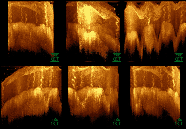
The Fuchs spot (also known as Förster-Fuchs' Spot) is a degeneration of the macula in cases of high myopia. It is named after the two persons who first described it: Ernst Fuchs, who described a pigmented lesion in 1901, and Forster, who described subretinal neovascularization in 1862. It occurs due to proliferation of retinal pigment epithelium associated with choroidal hemorrhage. The size of the spots are proportionate to the severity of the pathological myopia. Symptoms First signs of a Fuchs spot are distorted sight of straight lines near the fovea, which some days later turn to the typical well-circumscribed patches after absorption of haemorrhage, and a pigmented scar remains. As in macular degeneration, central sight is affected. Atrophy leads to the loss of two or more lines of the Snellen chart A Snellen chart is an eye chart that can be used to measure visual acuity. Snellen charts are named after the Dutch ophthalmologist Herman Snellen, who developed the ch ... [...More Info...] [...Related Items...] OR: [Wikipedia] [Google] [Baidu] |
Optical Coherence Tomography
Optical coherence tomography (OCT) is an imaging technique that uses low-coherence light to capture micrometer-resolution, two- and three-dimensional images from within optical scattering media (e.g., biological tissue). It is used for medical imaging and industrial nondestructive testing (NDT). Optical coherence tomography is based on low-coherence interferometry, typically employing near-infrared light. The use of relatively long wavelength light allows it to penetrate into the scattering medium. Confocal microscopy, another optical technique, typically penetrates less deeply into the sample but with higher resolution. Depending on the properties of the light source (superluminescent diodes, ultrashort pulsed lasers, and supercontinuum lasers have been employed), optical coherence tomography has achieved sub-micrometer resolution (with very wide-spectrum sources emitting over a ~100 nm wavelength range). Optical coherence tomography is one of a class of optical tom ... [...More Info...] [...Related Items...] OR: [Wikipedia] [Google] [Baidu] |
Macula
The macula (/ˈmakjʊlə/) or macula lutea is an oval-shaped pigmented area in the center of the retina of the human eye and in other animals. The macula in humans has a diameter of around and is subdivided into the umbo, foveola, foveal avascular zone, fovea, parafovea, and perifovea areas. The anatomical macula at a size of is much larger than the clinical macula which, at a size of , corresponds to the anatomical fovea. The macula is responsible for the central, high-resolution, color vision that is possible in good light; and this kind of vision is impaired if the macula is damaged, for example in macular degeneration. The clinical macula is seen when viewed from the pupil, as in ophthalmoscopy or retinal photography. The term macula lutea comes from Latin ''macula'', "spot", and ''lutea'', "yellow". Structure The macula is an oval-shaped pigmented area in the center of the retina of the human eye and other animal eyes. Its center is shifted slightly away from ... [...More Info...] [...Related Items...] OR: [Wikipedia] [Google] [Baidu] |
Myopia
Near-sightedness, also known as myopia and short-sightedness, is an eye disease where light focuses in front of, instead of on, the retina. As a result, distant objects appear blurry while close objects appear normal. Other symptoms may include headaches and eye strain. Severe near-sightedness is associated with an increased risk of retinal detachment, cataracts, and glaucoma. The underlying mechanism involves the length of the eyeball growing too long or less commonly the lens being too strong. It is a type of refractive error. Diagnosis is by eye examination. Tentative evidence indicates that the risk of near-sightedness can be decreased by having young children spend more time outside. This decrease in risk may be related to natural light exposure. Near-sightedness can be corrected with eyeglasses, contact lenses, or a refractive surgery. Eyeglasses are the easiest and safest method of correction. Contact lenses can provide a wider field of vision, but are associate ... [...More Info...] [...Related Items...] OR: [Wikipedia] [Google] [Baidu] |
Ernst Fuchs (doctor)
Ernst Fuchs (14 June 1851, in Vienna – 21 November 1930, in Vienna) was an Austrian ophthalmologist, physician and researcher. Overview The importance of Fuchs' life achievement must surely be based on his discovery and description of numerous ocular diseases and abnormalities. As a result of more than 250 scientific publications, the name Ernst Fuchs became well known throughout the world. Although his name is commonly recognized in conjunction with various corneal and anterior segment disorders, Ernst Fuchs' contribution to ophthalmology exceeds purely the delineation of ocular diseases and the detailed description of signs. Fuchs' collection of microscopic samples laid the foundation for anatomical and pathological understanding of blood vessels, muscles, and most other tissues of the eye. Additionally, Fuchs was able to pass on his unique knowledge, educating ophthalmologists at an international level. His Textbook of Ophthalmology was, for many decades, the most extensive ... [...More Info...] [...Related Items...] OR: [Wikipedia] [Google] [Baidu] |
Carl Friedrich Richard Förster
Carl Friedrich Richard Förster (15 November 1825 – 7 July 1902) was a German ophthalmologist born in the town of Lissa (today Leszno, Poland). He received his medical doctorate in 1849, and for most of his career was associated with the University of Breslau. Two of his assistants at Breslau were Hermann Wilbrand (1851–1935) and Hermann Rudolph Aubert (1826–1892). Förster is remembered for conducting tests of visual acuity, and for his investigations of indirect vision (peripheral vision) and visual performance across the visual field together with Hermann Aubert. The linear variation of visual performance that they described is sometimes referred to as the Aubert-Foerster law. From Förster's research, the eponymous terms of "Förster perimeter" and "Förster's shift" are derived. A Förster perimeter was an instrument used to measure an individual's field of vision. He also devised a specialized photometer to determine the smallest amount of light that will permit an o ... [...More Info...] [...Related Items...] OR: [Wikipedia] [Google] [Baidu] |
Choroidal Neovascularization
Choroidal neovascularization (CNV) is the creation of new blood vessels in the choroid layer of the eye. Choroidal neovascularization is a common cause of neovascular degenerative maculopathy (i.e. 'wet' macular degeneration) commonly exacerbated by extreme myopia, malignant myopic degeneration, or age-related developments. Causes CNV can occur rapidly in individuals with defects in Bruch's membrane, the innermost layer of the choroid. It is also associated with excessive amounts of vascular endothelial growth factor (VEGF). As well as in wet macular degeneration, CNV can also occur frequently with the rare genetic disease pseudoxanthoma elasticum and rarely with the more common optic disc drusen. CNV has also been associated with extreme myopia or malignant myopic degeneration, where in choroidal neovascularization occurs primarily in the presence of cracks within the retinal (specifically) macular tissue known as lacquer cracks. Symptoms CNV can create a sudden deterioration ... [...More Info...] [...Related Items...] OR: [Wikipedia] [Google] [Baidu] |
Retinal Pigment Epithelium
The pigmented layer of retina or retinal pigment epithelium (RPE) is the pigmented cell layer just outside the neurosensory retina that nourishes retinal visual cells, and is firmly attached to the underlying choroid and overlying retinal visual cells. History The RPE was known in the 18th and 19th centuries as the pigmentum nigrum, referring to the observation that the RPE is dark (black in many animals, brown in humans); and as the tapetum nigrum, referring to the observation that in animals with a tapetum lucidum, in the region of the tapetum lucidum the RPE is not pigmented. Anatomy The RPE is composed of a single layer of hexagonal cells that are densely packed with pigment granules. When viewed from the outer surface, these cells are smooth and hexagonal in shape. When seen in section, each cell consists of an outer non-pigmented part containing a large oval nucleus and an inner pigmented portion which extends as a series of straight thread-like processes between the r ... [...More Info...] [...Related Items...] OR: [Wikipedia] [Google] [Baidu] |
Fovea Centralis
The fovea centralis is a small, central pit composed of closely packed cones in the eye. It is located in the center of the macula lutea of the retina. The fovea is responsible for sharp central vision (also called foveal vision), which is necessary in humans for activities for which visual detail is of primary importance, such as reading and driving. The fovea is surrounded by the ''parafovea'' belt and the ''perifovea'' outer region. The parafovea is the intermediate belt, where the ganglion cell layer is composed of more than five layers of cells, as well as the highest density of cones; the perifovea is the outermost region where the ganglion cell layer contains two to four layers of cells, and is where visual acuity is below the optimum. The perifovea contains an even more diminished density of cones, having 12 per 100 micrometres versus 50 per 100 micrometres in the most central fovea. That, in turn, is surrounded by a larger peripheral area, which delivers highly com ... [...More Info...] [...Related Items...] OR: [Wikipedia] [Google] [Baidu] |
Bleeding
Bleeding, hemorrhage, haemorrhage or blood loss, is blood escaping from the circulatory system from damaged blood vessels. Bleeding can occur internally, or externally either through a natural opening such as the mouth, nose, ear, urethra, vagina or anus, or through a puncture in the skin. Hypovolemia is a massive decrease in blood volume, and death by excessive loss of blood is referred to as exsanguination. Typically, a healthy person can endure a loss of 10–15% of the total blood volume without serious medical difficulties (by comparison, blood donation typically takes 8–10% of the donor's blood volume). The stopping or controlling of bleeding is called hemostasis and is an important part of both first aid and surgery. Types * Upper head ** Intracranial hemorrhage – bleeding in the skull. ** Cerebral hemorrhage – a type of intracranial hemorrhage, bleeding within the brain tissue itself. ** Intracerebral hemorrhage – bleeding in the brain caused by the rupture o ... [...More Info...] [...Related Items...] OR: [Wikipedia] [Google] [Baidu] |
Macular Degeneration
Macular degeneration, also known as age-related macular degeneration (AMD or ARMD), is a medical condition which may result in blurred or no vision in the center of the visual field. Early on there are often no symptoms. Over time, however, some people experience a gradual worsening of vision that may affect one or both eyes. While it does not result in complete blindness, loss of central vision can make it hard to recognize faces, drive, read, or perform other activities of daily life. Visual hallucinations may also occur. Macular degeneration typically occurs in older people. Genetic factors and smoking also play a role. It is due to damage to the macula of the retina. Diagnosis is by a complete eye exam. The severity is divided into early, intermediate, and late types. The late type is additionally divided into "dry" and "wet" forms with the dry form making up 90% of cases. The difference between the two forms is the change of macula. Those with dry form AMD have drusen, ... [...More Info...] [...Related Items...] OR: [Wikipedia] [Google] [Baidu] |
Atrophy
Atrophy is the partial or complete wasting away of a part of the body. Causes of atrophy include mutations (which can destroy the gene to build up the organ), poor nourishment, poor circulation, loss of hormonal support, loss of nerve supply to the target organ, excessive amount of apoptosis of cells, and disuse or lack of exercise or disease intrinsic to the tissue itself. In medical practice, hormonal and nerve inputs that maintain an organ or body part are said to have ''trophic'' effects. A diminished muscular trophic condition is designated as ''atrophy''. Atrophy is reduction in size of cell, organ or tissue, after attaining its normal mature growth. In contrast, hypoplasia is the reduction in the cellular numbers of an organ, or tissue that has not attained normal maturity. Atrophy is the general physiological process of reabsorption and breakdown of tissues, involving apoptosis. When it occurs as a result of disease or loss of trophic support because of other diseas ... [...More Info...] [...Related Items...] OR: [Wikipedia] [Google] [Baidu] |
Snellen Chart
A Snellen chart is an eye chart that can be used to measure visual acuity. Snellen charts are named after the Dutch ophthalmologist Herman Snellen, who developed the chart in 1862. Many ophthalmologists and vision scientists now use an improved chart known as the LogMAR chart. History Snellen developed charts using symbols based in a 5×5 unit grid. The experimental charts developed in 1861 used abstract symbols. Snellen's charts published in 1862 used alphanumeric capitals in the 5×5 grid. The original chart shows A, C, E, G, L, N, P, R, T, 5, V, Z, B, D, 4, F, H, K, O, S, 3, U, Y, A, C, E, G, L, 2. Description The normal Snellen chart is printed with eleven lines of block letters. The first line consists of one very large letter, which may be one of several letters, for example E, H, or N. Subsequent rows have increasing numbers of letters that decrease in size. A person taking the test covers one eye from 6 metres or 20 feet away, and reads aloud the letters of each row ... [...More Info...] [...Related Items...] OR: [Wikipedia] [Google] [Baidu] |


