|
Enucleation Of The Eye
Enucleation is the removal of the eye that leaves the eye muscles and remaining orbital contents intact. This type of ocular surgery is indicated for a number of ocular tumors, in eyes that have sustained severe trauma, and in eyes that are otherwise blind and painful. Self-enucleation or auto-enucleation (oedipism) and other forms of serious self-inflicted eye injury are an extremely rare form of severe self-harm that usually results from mental illnesses involving acute psychosis. The name comes from Oedipus of Greek mythology, who gouged out his own eyes. Classification There are three types of eye removal: * Evisceration – removal of the iris, cornea, and internal eye contents, but with the sclera and attached extraocular muscles left behind * Enucleation of the eye - removal of the eyeball, but with the eyelids and adjacent structures of the eye socket remaining. An intraocular tumor excision requires an enucleation, not an evisceration. * Exenteration – removal of ... [...More Info...] [...Related Items...] OR: [Wikipedia] [Google] [Baidu] |
Eye Muscles
The extraocular muscles (extrinsic ocular muscles), are the seven extrinsic muscles of the human eye. Six of the extraocular muscles, the four recti muscles, and the superior and inferior oblique muscles, control movement of the eye and the other muscle, the levator palpebrae superioris, controls eyelid elevation. The actions of the six muscles responsible for eye movement depend on the position of the eye at the time of muscle contraction. Structure Since only a small part of the eye called the fovea provides sharp vision, the eye must move to follow a target. Eye movements must be precise and fast. This is seen in scenarios like reading, where the reader must shift gaze constantly. Although under voluntary control, most eye movement is accomplished without conscious effort. Precisely how the integration between voluntary and involuntary control of the eye occurs is a subject of continuing research."eye, human."Encyclopædia Britannica from Encyclopædia Britannica 2006 Ultima ... [...More Info...] [...Related Items...] OR: [Wikipedia] [Google] [Baidu] |
Uveal Melanoma
Uveal melanoma is a type of eye cancer in the uvea of the eye. It is traditionally classed as originating in the iris, choroid, and ciliary body, but can also be divided into class I (low metastatic risk) and class II (high metastatic risk). Symptoms include blurred vision, loss of vision or photopsia, but there may be no symptoms. Tumors arise from the pigment cells ( melanocytes) that reside within the uvea and give color to the eye. These melanocytes are distinct from the retinal pigment epithelium cells underlying the retina that do not form melanomas. When eye melanoma is spread to distant parts of the body, the five-year survival rate is about 15%.Eye Cancer Survival Rates American Cancer Society, Last Medical Review: December 9, 2014 Last Revised: February 5, 2016 It is the ... [...More Info...] [...Related Items...] OR: [Wikipedia] [Google] [Baidu] |
Ocularist
An ocularist specializes in the fabrication and fitting of ocular prostheses for people who have lost an eye or eyes due to trauma or illness.{{Cite journal , last1=Khandekar , first1=Rajiv , last2=Changal , first2=Nusrat , last3=AlRasheed , first3=Waleed , date=2020 , title=Ocularists the less known mid eye care professionals and their contribution in eye health care , journal=Saudi Journal of Ophthalmology , language=en , volume=34 , issue=3 , pages=195–197 , doi=10.4103/1319-4534.310403 , pmid=34085013 , pmc=8081081 , issn=1319-4534 The fabrication process for a custom made eye typically includes taking an impression of the eye socket, shaping a plastic shell, painting the iris, and then fitting the ocular prostheses. Prefabricated ocular prostheses with different colored iris are also available. An ocularist may select the stock eye that is most closely matched to patient's iris color. However, due to better adaptation, comfort, and aesthetics, custom-made ocular prostheses ar ... [...More Info...] [...Related Items...] OR: [Wikipedia] [Google] [Baidu] |
Ocular Prosthesis
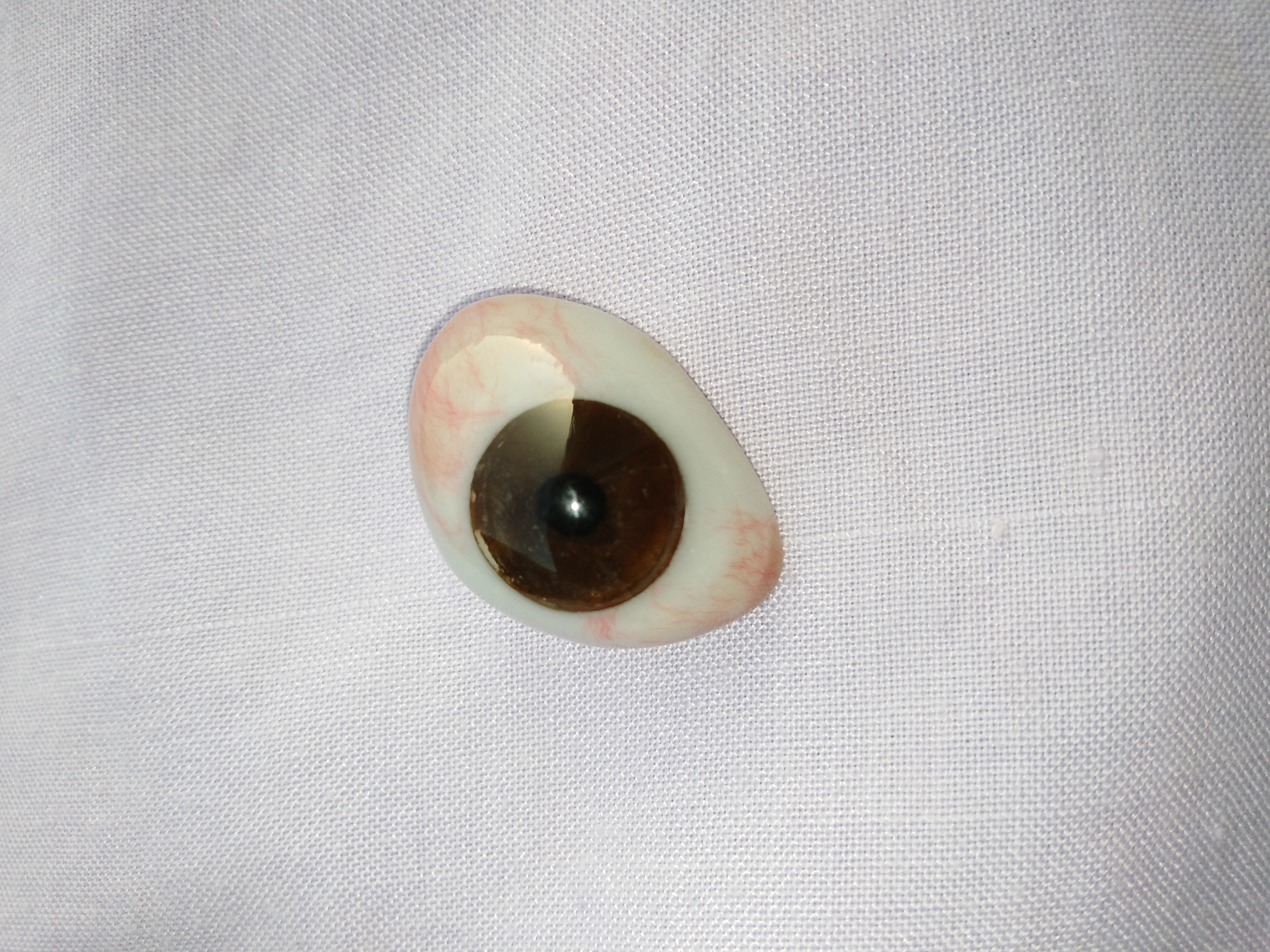
An ocular prosthesis, artificial eye or glass eye is a type of craniofacial prosthesis that replaces an absent natural eye following an enucleation, evisceration, or orbital exenteration. The prosthesis fits over an orbital implant and under the eyelids. Though often referred to as a glass eye, the ocular prosthesis roughly takes the shape of a convex shell and is made of medical grade plastic acrylic. A few ocular prostheses today are made of cryolite glass. A variant of the ocular prosthesis is a very thin hard shell known as a scleral shell which can be worn over a damaged or eviscerated eye. Makers of ocular prosthetics are known as ocularists. An ocular prosthesis does ''not'' provide vision; this would be a visual prosthesis. Someone with an ocular prosthesis is altogether blind on the affected side and has monocular (one sided) vision. History The earliest known evidence of the use of ocular prosthesis is that of a woman found in Shahr-I Sokhta, Iran dating back t ... [...More Info...] [...Related Items...] OR: [Wikipedia] [Google] [Baidu] |
Hydroxylapatite
Hydroxyapatite, also called hydroxylapatite (HA), is a naturally occurring mineral form of calcium apatite with the formula Ca5(PO4)3(OH), but it is usually written Ca10(PO4)6(OH)2 to denote that the crystal unit cell comprises two entities. Hydroxyapatite is the hydroxyl endmember of the complex apatite group. The OH− ion can be replaced by fluoride, chloride or carbonate, producing fluorapatite or chlorapatite. It crystallizes in the hexagonal crystal system. Pure hydroxyapatite powder is white. Naturally occurring apatites can, however, also have brown, yellow, or green colorations, comparable to the discolorations of dental fluorosis. Up to 50% by volume and 70% by weight of human bone is a modified form of hydroxyapatite, known as bone mineral. Carbonated calcium-deficient hydroxyapatite is the main mineral of which dental enamel and dentin are composed. Hydroxyapatite crystals are also found in pathological calcifications such as those found in breast tumors, as w ... [...More Info...] [...Related Items...] OR: [Wikipedia] [Google] [Baidu] |
Conjunctiva
The conjunctiva is a thin mucous membrane that lines the inside of the eyelids and covers the sclera (the white of the eye). It is composed of non-keratinized, stratified squamous epithelium with goblet cells, stratified columnar epithelium and stratified cuboidal epithelium (depending on the zone). The conjunctiva is highly vascularised, with many microvessels easily accessible for imaging studies. Structure The conjunctiva is typically divided into three parts: Blood supply Blood to the bulbar conjunctiva is primarily derived from the ophthalmic artery. The blood supply to the palpebral conjunctiva (the eyelid) is derived from the external carotid artery. However, the circulations of the bulbar conjunctiva and palpebral conjunctiva are linked, so both bulbar conjunctival and palpebral conjunctival vessels are supplied by both the ophthalmic artery and the external carotid artery, to varying extents. Nerve supply Sensory innervation of the conjunctiva is divided int ... [...More Info...] [...Related Items...] OR: [Wikipedia] [Google] [Baidu] |
Tenon's Capsule
Tenon's capsule (), also known as the Tenon capsule, fascial sheath of the eyeball () or the fascia bulbi, is a thin membrane which envelops the eyeball from the optic nerve to the corneal limbus, separating it from the orbital fat and forming a socket in which it moves. The inner surface of Tenon's capsule is smooth and is separated from the outer surface of the sclera by the periscleral lymph space. This lymph space is continuous with the subdural and subarachnoid cavities and is traversed by delicate bands of connective tissue which extend between the capsule and the sclera. The capsule is perforated behind by the ciliary vessels and nerves and fuses with the sheath of the optic nerve and with the sclera around the entrance of the optic nerve. In front it adheres to the conjunctiva, and both structures are attached to the ciliary region of the eyeball. The structure was named after Jacques-René Tenon (1724–1816), a French surgeon and pathologist. Structure Relations ... [...More Info...] [...Related Items...] OR: [Wikipedia] [Google] [Baidu] |
Keratoplasty
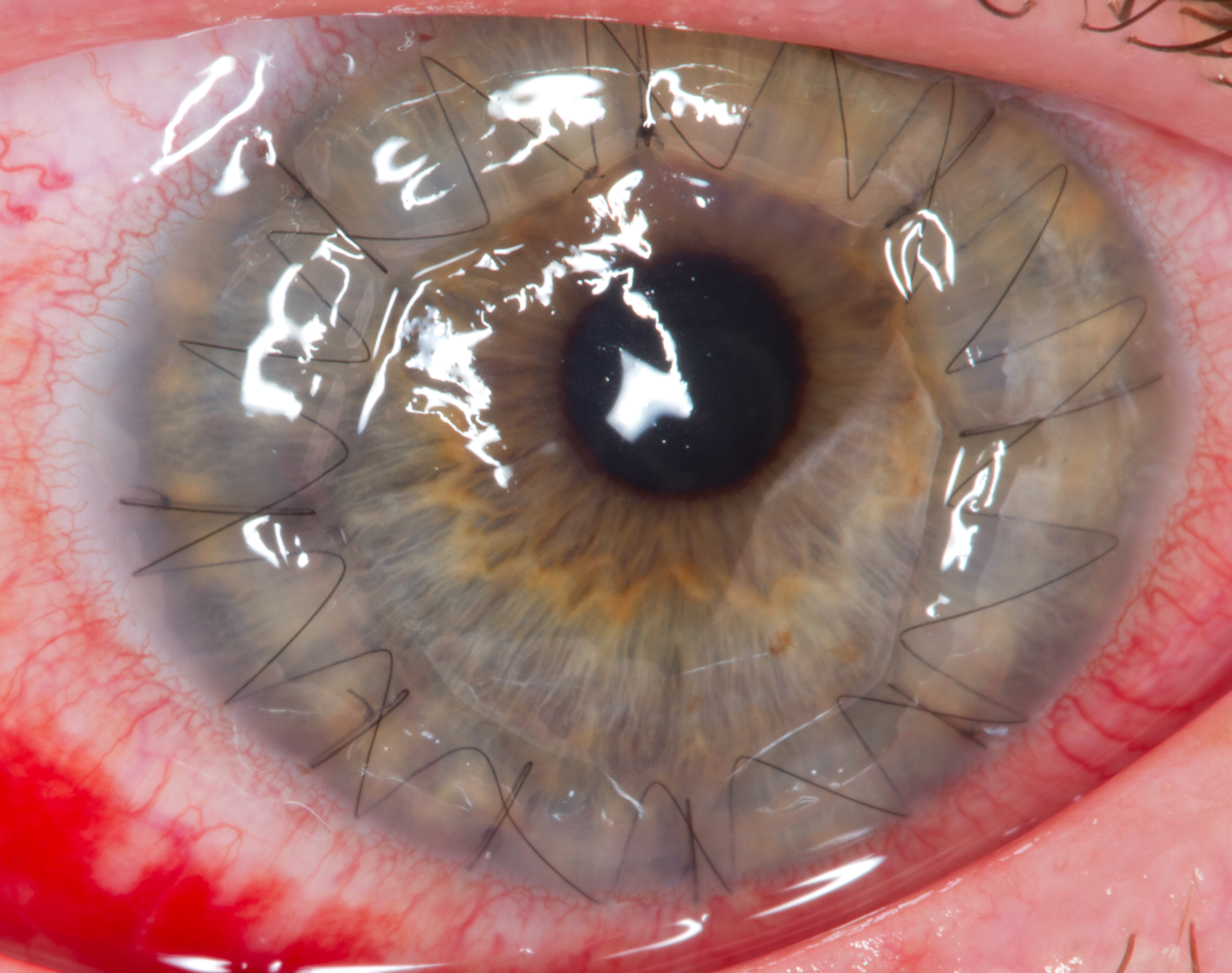
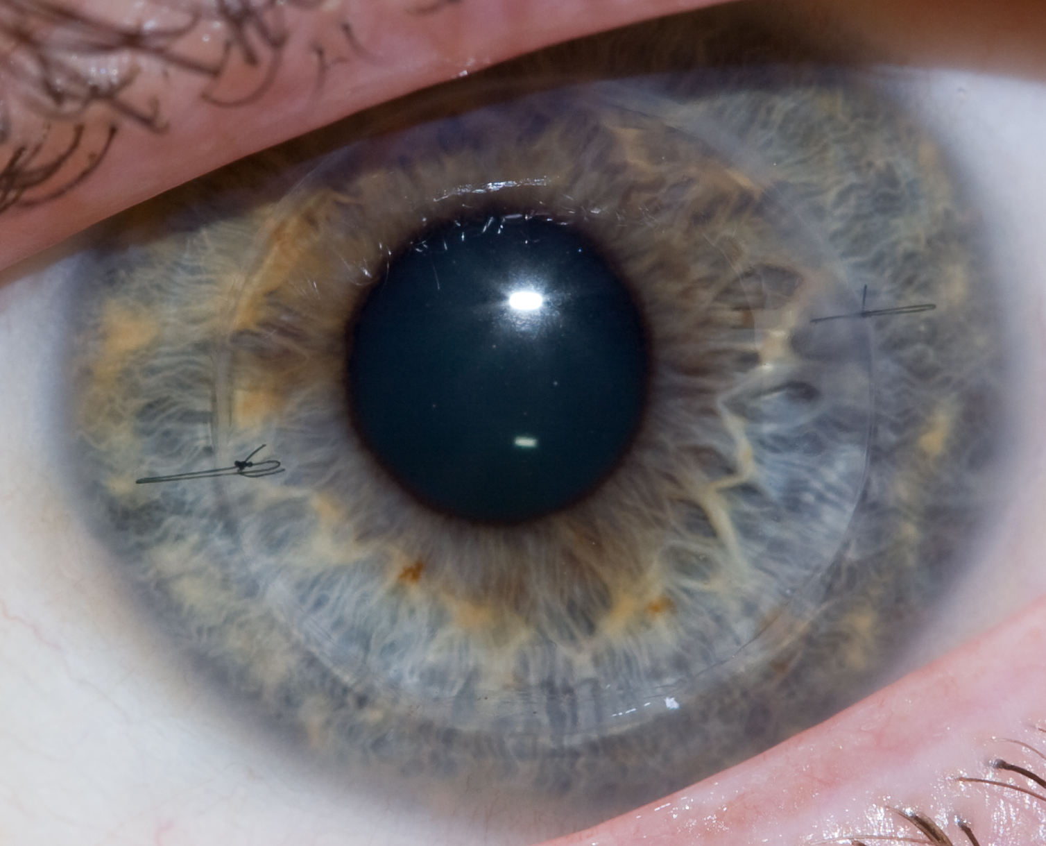
Corneal transplantation, also known as corneal grafting, is a surgical procedure where a damaged or diseased cornea is replaced by donated corneal tissue (the graft). When the entire cornea is replaced it is known as penetrating keratoplasty and when only part of the cornea is replaced it is known as lamellar keratoplasty. Keratoplasty simply means surgery to the cornea. The graft is taken from a recently deceased individual with no known diseases or other factors that may affect the chance of survival of the donated tissue or the health of the recipient. The cornea is the transparent front part of the eye that covers the iris, pupil and anterior chamber. The surgical procedure is performed by ophthalmologists, physicians who specialize in eyes, and is often done on an outpatient basis. Donors can be of any age, as is shown in the case of Janis Babson, who donated her eyes after dying at the age of 10. Corneal transplantation is performed when medicines, keratoconus conserva ... [...More Info...] [...Related Items...] OR: [Wikipedia] [Google] [Baidu] |
Corneal Transplant
Corneal transplantation, also known as corneal grafting, is a surgical procedure where a damaged or diseased cornea is replaced by donated corneal tissue (the graft). When the entire cornea is replaced it is known as penetrating keratoplasty and when only part of the cornea is replaced it is known as lamellar keratoplasty. Keratoplasty simply means surgery to the cornea. The graft is taken from a recently deceased individual with no known diseases or other factors that may affect the chance of survival of the donated tissue or the health of the recipient. The cornea is the transparent front part of the eye that covers the iris, pupil and anterior chamber. The surgical procedure is performed by ophthalmologists, physicians who specialize in eyes, and is often done on an outpatient basis. Donors can be of any age, as is shown in the case of Janis Babson, who donated her eyes after dying at the age of 10. Corneal transplantation is performed when medicines, keratoconus conser ... [...More Info...] [...Related Items...] OR: [Wikipedia] [Google] [Baidu] |
Congenital Cystic Eye
Congenital cystic eye (also known as ''CCE'' or ''cystic eyeball'') is an extremely rare ocular malformation where the eye fails to develop correctly ''in utero'' and is replaced by benign, fluid-filled tissue. Its incidence is unknown, due to the very small number of cases reported. An audit by Duke-Elder of the medical literature from 1880 to 1963 discovered only 28 cases. The term was coined in 1937 by the renowned ophthalmologist Ida Mann. Embryologically, the defect is thought to occur around day 35 of gestation, when the vesicle fails to invaginate. Dysgenesis of the vesicle later in development may result in coloboma A coloboma (from the Greek , meaning defect) is a hole in one of the structures of the eye, such as the iris, retina, choroid, or optic disc. The hole is present from birth and can be caused when a gap called the choroid fissure, which is presen ..., a separate and less severe malformation of the ocular structures. CCE is almost always unilateral, bu ... [...More Info...] [...Related Items...] OR: [Wikipedia] [Google] [Baidu] |
Blindness
Visual impairment, also known as vision impairment, is a medical definition primarily measured based on an individual's better eye visual acuity; in the absence of treatment such as correctable eyewear, assistive devices, and medical treatment– visual impairment may cause the individual difficulties with normal daily tasks including reading and walking. Low vision is a functional definition of visual impairment that is chronic, uncorrectable with treatment or correctable lenses, and impacts daily living. As such low vision can be used as a disability metric and varies based on an individual's experience, environmental demands, accommodations, and access to services. The American Academy of Ophthalmology defines visual impairment as the best-corrected visual acuity of less than 20/40 in the better eye, and the World Health Organization defines it as a presenting acuity of less than 6/12 in the better eye. The term blindness is used for complete or nearly complete vision loss. In ... [...More Info...] [...Related Items...] OR: [Wikipedia] [Google] [Baidu] |
Sympathetic Ophthalmia
Sympathetic ophthalmia (SO), also called spared eye injury, is a diffuse granulomatous inflammation of the uveal layer of both eyes following trauma to one eye. It can leave the affected person completely blind. Symptoms may develop from days to several years after a penetrating eye injury. It typically results from a delayed hypersensitivity reaction. Signs and symptoms Eye floaters and loss of accommodation are among the earliest symptoms. The disease may progress to severe inflammation of the uveal layer of the eye (uveitis) with pain and sensitivity of the eyes to light. The affected eye often remains relatively painless while the inflammatory disease spreads through the uvea, where characteristic focal infiltrates in the choroid named Dalén–Fuchs nodules can be seen. The retina, however, usually remains uninvolved, although perivascular cuffing of the retinal vessels with inflammatory cells may occur. Swelling of the optic disc (papilledema), secondary glaucoma, viti ... [...More Info...] [...Related Items...] OR: [Wikipedia] [Google] [Baidu] |


