|
Dupuytren's Contracture
Dupuytren's contracture (also called Dupuytren's disease, Morbus Dupuytren, Viking disease, palmar fibromatosis and Celtic hand) is a condition in which one or more fingers become progressively bent in a flexed position. It is named after Guillaume Dupuytren, who first described the underlying mechanism of action, followed by the first successful operation in 1831 and publication of the results in ''The Lancet'' in 1834. It usually begins as small, hard nodules just under the skin of the palm, then worsens over time until the fingers can no longer be fully straightened. While typically not painful, some aching or itching may be present. The ring finger followed by the little and middle fingers are most commonly affected. It can affect one or both hands. The condition can interfere with activities such as preparing food, writing, putting the hand in a tight pocket, putting on gloves, or shaking hands. The cause is unknown but might have a genetic component. Risk factors include ... [...More Info...] [...Related Items...] OR: [Wikipedia] [Google] [Baidu] |
Rheumatology
Rheumatology (Greek ''ῥεῦμα'', ''rheûma'', flowing current) is a branch of medicine devoted to the diagnosis and management of disorders whose common feature is inflammation in the bones, muscles, joints, and internal organs. Rheumatology covers more than 100 different complex diseases, collectively known as rheumatic diseases, which includes many forms of arthritis as well as lupus and Sjögren's syndrome. Doctors who have undergone formal training in rheumatology are called rheumatologists. Many of these diseases are now known to be disorders of the immune system, and rheumatology has significant overlap with immunology, the branch of medicine that studies the immune system. Rheumatologist A rheumatologist is a physician who specializes in the field of medical sub-specialty called rheumatology. A rheumatologist holds a board certification after specialized training. In the United States, training in this field requires four years undergraduate school, four year ... [...More Info...] [...Related Items...] OR: [Wikipedia] [Google] [Baidu] |
Connective Tissue
Connective tissue is one of the four primary types of animal tissue, along with epithelial tissue, muscle tissue, and nervous tissue. It develops from the mesenchyme derived from the mesoderm the middle embryonic germ layer. Connective tissue is found in between other tissues everywhere in the body, including the nervous system. The three meninges, membranes that envelop the brain and spinal cord are composed of connective tissue. Most types of connective tissue consists of three main components: elastic and collagen fibers, ground substance, and cells. Blood, and lymph are classed as specialized fluid connective tissues that do not contain fiber. All are immersed in the body water. The cells of connective tissue include fibroblasts, adipocytes, macrophages, mast cells and leucocytes. The term "connective tissue" (in German, ''Bindegewebe'') was introduced in 1830 by Johannes Peter Müller. The tissue was already recognized as a distinct class in the 18th century. ... [...More Info...] [...Related Items...] OR: [Wikipedia] [Google] [Baidu] |
Proximal Interphalangeal
The interphalangeal joints of the hand are the hinge joints between the phalanges of the fingers that provide flexion towards the palm of the hand. There are two sets in each finger (except in the thumb, which has only one joint): * "proximal interphalangeal joints" (PIJ or PIP), those between the first (also called proximal) and second (intermediate) phalanges * "distal interphalangeal joints" (DIJ or DIP), those between the second (intermediate) and third (distal) phalanges Anatomically, the proximal and distal interphalangeal joints are very similar. There are some minor differences in how the palmar plates are attached proximally and in the segmentation of the flexor tendon sheath, but the major differences are the smaller dimension and reduced mobility of the distal joint. Joint structure The PIP joint exhibits great lateral stability. Its transverse diameter is greater than its antero-posterior diameter and its thick collateral ligaments are tight in all positions du ... [...More Info...] [...Related Items...] OR: [Wikipedia] [Google] [Baidu] |
Tenosynovitis
Tenosynovitis is the inflammation of the fluid-filled sheath (called the synovium) that surrounds a tendon, typically leading to joint pain, swelling, and stiffness. Tenosynovitis can be either infectious or noninfectious. Common clinical manifestations of noninfectious tenosynovitis include de Quervain tendinopathy and stenosing tenosynovitis (more commonly known as trigger finger) Signs and symptoms Infectious tenosynovitis occurs between 2.5% and 9.4% of all hand infections. Kanavel's cardinal signs is used to diagnose infectious tenosynovitis. They are: tenderness to touch along the flexor aspect of the finger, fusiform enlargement of the affected finger, the finger being held in slight flexion at rest, and severe pain with passive extension. Fever may also be present but is uncommon. Pathogenesis Infectious tenosynovitis is the infection of closed synovial sheaths in the flexor tendons of the fingers. It is usually caused by trauma, but bacteria can spread from other site ... [...More Info...] [...Related Items...] OR: [Wikipedia] [Google] [Baidu] |
Contracture
In pathology, a contracture is a permanent shortening of a muscle or joint. It is usually in response to prolonged hypertonic spasticity in a concentrated muscle area, such as is seen in the tightest muscles of people with conditions like spastic cerebral palsy, but can also be due to the congenital abnormal development of muscles and connective tissue in the womb. Contractures develop when normally elastic tissues such as muscles or tendons are replaced by inelastic tissues (fibrosis). This results in the shortening and hardening of these tissues, ultimately causing rigidity, joint deformities and a total loss of movement around the joint. Most of the physical therapy, occupational therapy and other exercise regimens targeted towards people with spasticity focuses on trying to prevent contractures from happening in the first place. However, research on sustained traction of connective tissue in approaches such as adaptive yoga has demonstrated that contracture can be reduced, a ... [...More Info...] [...Related Items...] OR: [Wikipedia] [Google] [Baidu] |
Metacarpophalangeal
The metacarpophalangeal joints (MCP) are situated between the metacarpal bones and the proximal phalanges of the fingers. These joints are of the condyloid kind, formed by the reception of the rounded heads of the metacarpal bones into shallow cavities on the proximal ends of the proximal phalanges. Being condyloid, they allow the movements of flexion, extension, abduction, adduction and circumduction at the joint. Structure Ligaments Each joint has: * palmar ligaments of metacarpophalangeal articulations * collateral ligaments of metacarpophalangeal articulations Dorsal surfaces The dorsal surfaces of these joints are covered by the expansions of the Extensor tendons, together with some loose areolar tissue which connects the deep surfaces of the tendons to the bones. Function The movements which occur in these joints are flexion, extension, adduction, abduction, and circumduction; the movements of abduction and adduction are very limited, and cannot be performed while th ... [...More Info...] [...Related Items...] OR: [Wikipedia] [Google] [Baidu] |
Tendon
A tendon or sinew is a tough, high-tensile-strength band of dense fibrous connective tissue that connects muscle to bone. It is able to transmit the mechanical forces of muscle contraction to the skeletal system without sacrificing its ability to withstand significant amounts of tension. Tendons are similar to ligaments; both are made of collagen. Ligaments connect one bone to another, while tendons connect muscle to bone. Structure Histologically, tendons consist of dense regular connective tissue. The main cellular component of tendons are specialized fibroblasts called tendon cells (tenocytes). Tenocytes synthesize the extracellular matrix of tendons, abundant in densely packed collagen fibers. The collagen fibers are parallel to each other and organized into tendon fascicles. Individual fascicles are bound by the endotendineum, which is a delicate loose connective tissue containing thin collagen fibrils and elastic fibres. Groups of fascicles are bounded by the epitenon, ... [...More Info...] [...Related Items...] OR: [Wikipedia] [Google] [Baidu] |
Flexor
A flexor is a muscle that flexes a joint. In anatomy, flexion (from the Latin verb ''flectere'', to bend) is a joint movement that decreases the angle between the bones that converge at the joint. For example, one’s elbow joint flexes when one brings their hand closer to the shoulder. Flexion is typically instigated by muscle contraction of a flexor. Flexors Upper limb *of the humerus bone (the bone in the upper arm) at the shoulder **Pectoralis major **Anterior deltoid **Coracobrachialis **Biceps brachii * of the forearm at the elbow ** Brachialis **Brachioradialis **Biceps brachii *of carpus (the carpal bones) at the wrist **flexor carpi radialis **flexor carpi ulnaris **palmaris longus *of the hand **flexor pollicis longus muscle **flexor pollicis brevis muscle **flexor digitorum profundus muscle **flexor digitorum superficialis muscle Lower limb Hip The hip flexors are (in descending order of importance to the action of flexing the hip joint):Platzer (2004), p 246 *Coll ... [...More Info...] [...Related Items...] OR: [Wikipedia] [Google] [Baidu] |
National Health Service (England)
The National Health Service (NHS) is the Publicly funded health care, publicly funded healthcare system in England, and one of the four National Health Service systems in the United Kingdom. It is the second largest single-payer healthcare system in the world after the Brazilian Sistema Único de Saúde. Primarily funded by the government from general taxation (plus a small amount from National Insurance contributions), and overseen by the Department of Health and Social Care, the NHS provides healthcare to all legal English residents and residents from other regions of the UK, with most services free at the point of use for most people. The NHS also conducts research through the National Institute for Health and Care Research (NIHR). Free healthcare at the point of use comes from the core principles at the founding of the National Health Service. The 1942 Beveridge cross-party report established the principles of the NHS which was implemented by the Attlee ministry, Labour ... [...More Info...] [...Related Items...] OR: [Wikipedia] [Google] [Baidu] |
Nordic Race
The Nordic race was a racial concept which originated in 19th century anthropology. It was considered a race or one of the putative sub-races into which some late-19th to mid-20th century anthropologists divided the Caucasian race, claiming that its ancestral homelands were Northwestern and Northern Europe, particularly to populations such as Anglo-Saxons, Germanic peoples, Balts, Baltic Finns, Northern French, and certain Celts and Slavs. The supposed physical traits of the Nordics included light eyes, light skin, tall stature, and dolichocephalic skull; their psychological traits were deemed to be truthfulness, equitability, a competitive spirit, naivete, reservedness, and individualism. In the early 20th century, the belief that the Nordic race constituted the superior branch of the Caucasian race gave rise to the ideology of Nordicism. With the rise of modern genetics, the concept of distinct human races in a biological sense has become obsolete. In 2019, the American Ass ... [...More Info...] [...Related Items...] OR: [Wikipedia] [Google] [Baidu] |
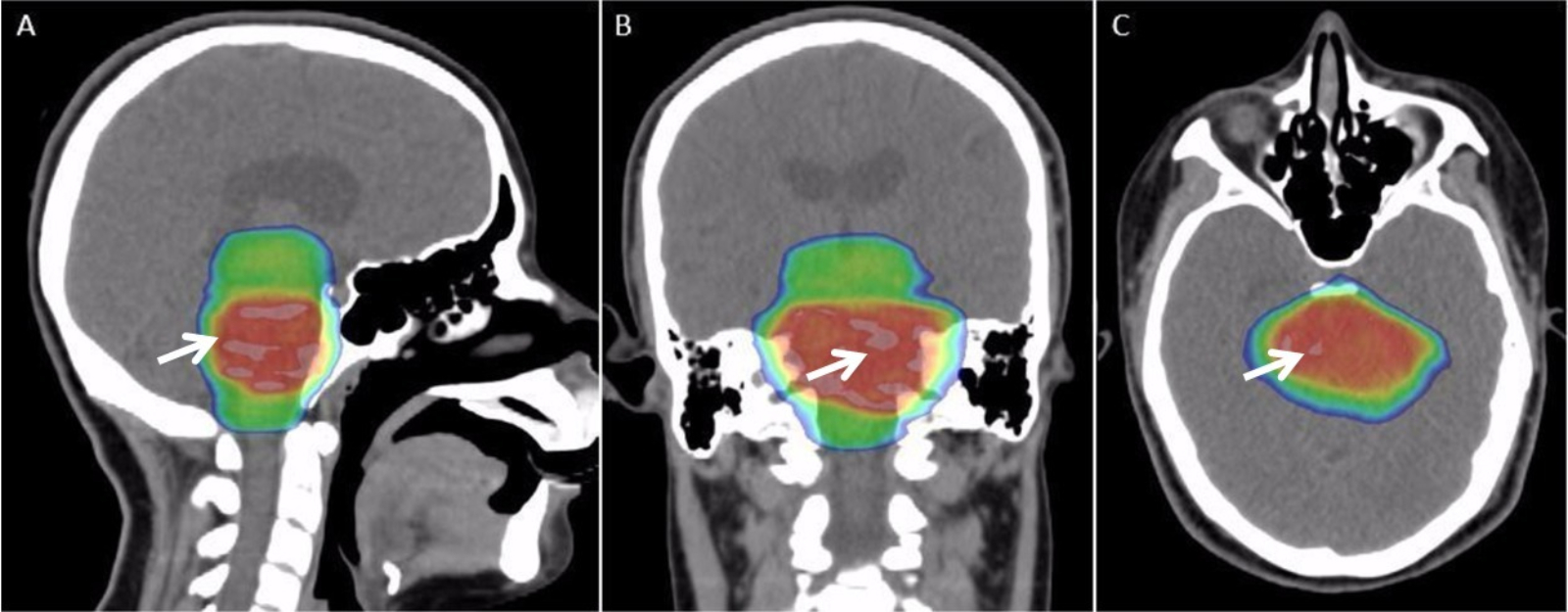
Radiation Therapy
Radiation therapy or radiotherapy, often abbreviated RT, RTx, or XRT, is a therapy using ionizing radiation, generally provided as part of cancer treatment to control or kill malignant cells and normally delivered by a linear accelerator. Radiation therapy may be curative in a number of types of cancer if they are localized to one area of the body. It may also be used as part of adjuvant therapy, to prevent tumor recurrence after surgery to remove a primary malignant tumor (for example, early stages of breast cancer). Radiation therapy is synergistic with chemotherapy, and has been used before, during, and after chemotherapy in susceptible cancers. The subspecialty of oncology concerned with radiotherapy is called radiation oncology. A physician who practices in this subspecialty is a radiation oncologist. Radiation therapy is commonly applied to the cancerous tumor because of its ability to control cell growth. Ionizing radiation works by damaging the DNA of cancerous tissue ... [...More Info...] [...Related Items...] OR: [Wikipedia] [Google] [Baidu] |


Aug2005.jpg)

