|
Dentate Gyrus
The dentate gyrus (DG) is one of the subfields of the hippocampus, in the hippocampal formation. The hippocampal formation is located in the temporal lobe of the brain, and includes the hippocampus (including CA1 to CA4) subfields, and other subfields including the dentate gyrus, subiculum, and presubiculum. The dentate gyrus is part of the trisynaptic circuit, a neural circuit of the hippocampus, thought to contribute to the formation of new episodic memories, the spontaneous exploration of novel environments and other functions. The dentate gyrus has toothlike projections from which it is named. The subgranular zone of the dentate gyrus is one of only two major sites of adult neurogenesis in the brain, and is found in many mammals. The other main site is the subventricular zone in the ventricular system. Other sites may include the striatum and the cerebellum. However, whether significant neurogenesis takes place in the adult human dentate gyrus has been a matter of d ... [...More Info...] [...Related Items...] OR: [Wikipedia] [Google] [Baidu] |
Hippocampal
The hippocampus (: hippocampi; via Latin from Greek , 'seahorse'), also hippocampus proper, is a major component of the brain of humans and many other vertebrates. In the human brain the hippocampus, the dentate gyrus, and the subiculum are components of the hippocampal formation located in the limbic system. The hippocampus plays important roles in the consolidation of information from short-term memory to long-term memory, and in spatial memory that enables navigation. In humans, and other primates the hippocampus is located in the archicortex, one of the three regions of allocortex, in each hemisphere with direct neural projections to, and reciprocal indirect projections from the neocortex. The hippocampus, as the medial pallium, is a structure found in all vertebrates. In Alzheimer's disease (and other forms of dementia), the hippocampus is one of the first regions of the brain to be damaged; short-term memory loss and disorientation are included among the e ... [...More Info...] [...Related Items...] OR: [Wikipedia] [Google] [Baidu] |
Ventricular System
In neuroanatomy, the ventricular system is a set of four interconnected cavities known as cerebral ventricles in the brain. Within each ventricle is a region of choroid plexus which produces the circulating cerebrospinal fluid (CSF). The ventricular system is continuous with the central canal of the spinal cord from the fourth ventricle, allowing for the flow of CSF to circulate. All of the ventricular system and the central canal of the spinal cord are lined with ependyma, a specialised form of epithelium connected by tight junctions that make up the blood–cerebrospinal fluid barrier. Structure The system comprises four ventricles: * lateral ventricles right and left (one for each hemisphere) * third ventricle * fourth ventricle There are several foramina, openings acting as channels, that connect the ventricles. The interventricular foramina (also called the foramina of Monro) connect the lateral ventricles to the third ventricle through which the cerebrospinal ... [...More Info...] [...Related Items...] OR: [Wikipedia] [Google] [Baidu] |
Entorhinal Cortex
The entorhinal cortex (EC) is an area of the brain's allocortex, located in the medial temporal lobe, whose functions include being a widespread network hub for memory, navigation, and the perception of time.Integrating time from experience in the lateral entorhinal cortex Albert Tsao, Jørgen Sugar, Li Lu, Cheng Wang, James J. Knierim, May-Britt Moser & Edvard I. Moser Naturevolume 561, pages57–62 (2018) The EC is the main interface between the hippocampus and neocortex. The EC-hippocampus system plays an important role in declarative (autobiographical/episodic/semantic) memories and in particular spatial memories including memory formation, memory consolidation, and memory optimization in sleep. The EC is also responsible for the pre-processing (familiarity) of the input signals in the reflex nictitating membrane response of classical trace conditioning; the association of impulses from the eye and the ear occurs in the entorhinal cortex. Anatomy The entorhina ... [...More Info...] [...Related Items...] OR: [Wikipedia] [Google] [Baidu] |
Stellate Cell
Stellate cells are neurons in the central nervous system, named for their star-like shape formed by dendritic processes radiating from the cell body. These cells play significant roles in various brain functions, including inhibition in the cerebellum and excitation in the cortex, and are involved in synaptic plasticity and neurovascular coupling. Morphology Stellate cells are characterized by their star-shaped dendritic trees. Dendrites can vary between neurons, with stellate cells being either spiny or aspinous. In contrast, pyramidal cells, which are also found in the cerebral cortex, are always spiny and pyramid-shaped. The classification of neurons often depends on the presence or absence of dendritic spines: those with spines are classified as spiny, while those without are classified as aspinous. Types and locations Cerebellar Many stellate cells are GABAergic and are located in the molecular layer of the cerebellum. Most common stellate cells are the inhibito ... [...More Info...] [...Related Items...] OR: [Wikipedia] [Google] [Baidu] |
Uncus
The uncus is an anterior extremity of the parahippocampal gyrus. It is separated from the apex of the temporal lobe by a sulcus called the rhinal sulcus. Although superficially continuous with the hippocampal gyrus, the uncus forms morphologically a part of the rhinencephalon. An important landmark that crosses the inferior surface of the uncus is the band of Giacomini or ''tail'' of the dentate gyrus. The term comes from the Latin word uncus, meaning ''hook'', and it was coined by Félix Vicq-d'Azyr (1748–1794). Clinical significance The part of the olfactory cortex that is on the temporal lobe covers the area of the uncus, which leads into the two significant clinical aspects: herniations and seizures * Herniations of the brain can occur if increased intracranial pressure due to a tumor, hemorrhage, or edema pushes the uncus over the tentorial notch against the brainstem and related cranial nerves. This can compress the oculomotor nerve (CN III). This causes pr ... [...More Info...] [...Related Items...] OR: [Wikipedia] [Google] [Baidu] |
Septal Area
The septal area (medial olfactory area), consisting of the lateral septum and medial septum, is an area in the lower, posterior part of the medial surface of the frontal lobe, and refers to the nearby septum pellucidum. The septal nuclei are located in this area. The septal nuclei are composed of medium-size neurons which are classified into dorsal, ventral, medial, and caudal groups. The septal nuclei receive reciprocal connections from the olfactory bulb, hippocampus, amygdala, hypothalamus, midbrain, habenula, cingulate gyrus, and thalamus. The septal nuclei are essential in generating the theta rhythm of the hippocampus. The septal area (medial olfactory area) has no relation to the sense of smell, but it is considered a pleasure zone in animals. The septal nuclei play a role in reward and reinforcement along with the nucleus accumbens. In the 1950s, Olds & Milner showed that rats with electrodes implanted in this area will self-stimulate repeatedly (e.g., press a bar to rec ... [...More Info...] [...Related Items...] OR: [Wikipedia] [Google] [Baidu] |
Pyramidal Neuron
Pyramidal cells, or pyramidal neurons, are a type of multipolar neuron found in areas of the brain including the cerebral cortex, the hippocampus, and the amygdala. Pyramidal cells are the primary excitation units of the mammalian prefrontal cortex and the corticospinal tract. One of the main structural features of the pyramidal neuron is the conic shaped soma (biology), soma, or cell body, after which the neuron is named. Other key structural features of the pyramidal cell are a single axon, a large apical dendrite, multiple basal dendrites, and the presence of dendritic spines. Pyramidal neurons are also one of two cell types where the pathognomonic, characteristic Medical sign, sign, Negri bodies, are found in post-mortem rabies infection. Pyramidal neurons were first discovered and studied by Santiago Ramón y Cajal. Since then, studies on pyramidal neurons have focused on topics ranging from neuroplasticity to cognition. Structure One of the main structural features of ... [...More Info...] [...Related Items...] OR: [Wikipedia] [Google] [Baidu] |
Dendrite
A dendrite (from Ancient Greek language, Greek δένδρον ''déndron'', "tree") or dendron is a branched cytoplasmic process that extends from a nerve cell that propagates the neurotransmission, electrochemical stimulation received from other neural cells to the cell body, or soma (biology), soma, of the neuron from which the dendrites project. Electrical stimulation is transmitted onto dendrites by upstream neurons (usually via their axons) via synapses which are located at various points throughout the dendritic tree. Dendrites play a critical role in integrating these synaptic inputs and in determining the extent to which action potentials are produced by the neuron. Structure and function Dendrites are one of two types of cytoplasmic processes that extrude from the cell body of a neuron, the other type being an axon. Axons can be distinguished from dendrites by several features including shape, length, and function. Dendrites often taper off in shape and are shorter, ... [...More Info...] [...Related Items...] OR: [Wikipedia] [Google] [Baidu] |
Synapse
In the nervous system, a synapse is a structure that allows a neuron (or nerve cell) to pass an electrical or chemical signal to another neuron or a target effector cell. Synapses can be classified as either chemical or electrical, depending on the mechanism of signal transmission between neurons. In the case of electrical synapses, neurons are coupled bidirectionally with each other through gap junctions and have a connected cytoplasmic milieu. These types of synapses are known to produce synchronous network activity in the brain, but can also result in complicated, chaotic network level dynamics. Therefore, signal directionality cannot always be defined across electrical synapses. Chemical synapses, on the other hand, communicate through neurotransmitters released from the presynaptic neuron into the synaptic cleft. Upon release, these neurotransmitters bind to specific receptors on the postsynaptic membrane, inducing an electrical or chemical response in the target neuron ... [...More Info...] [...Related Items...] OR: [Wikipedia] [Google] [Baidu] |
Mossy Fiber (hippocampus)
Mossy may refer to: Places *Mossy, West Virginia, unincorporated community in Fayette County, West Virginia, United States Given names *Mossy Cade (born 1961), former professional American football player *Mossy Lawler (born 1980), rugby union player *Mossy Murphy, retired Irish sportsperson *Tomás Quinn, retired Irish sportsperson *Mossy O'Riordan, Irish sportsperson who played hurling with the Cork senior inter-county team in the 1940s and 1950s *Mossy, a fictional character in ''The Golden Key (MacDonald book), The Golden Key'' by George MacDonald See also *Battle of Mossy Creek, minor battle of the American Civil War, on December 29, 1863 *Mossy fiber (cerebellum), one of the major inputs to cerebellum *Mossy fiber (hippocampus), pathway to the CA3 region *Mossy forest shrew (''Crocidura musseri''), a species of shrew native to Indonesia *Mossy-nest swiftlet (''Aerodramus salangana''), a species of swift in the family Apodidae *Mossie (other) *Mossi (disambiguati ... [...More Info...] [...Related Items...] OR: [Wikipedia] [Google] [Baidu] |
Granule Cell
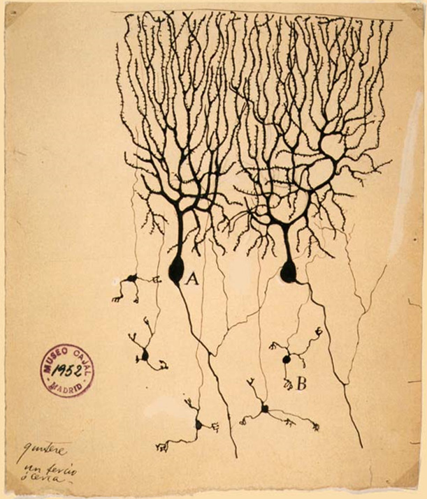
The name granule cell has been used for a number of different types of neurons whose only common feature is that they all have very small cell bodies. Granule cells are found within the granular layer of the cerebellum, the dentate gyrus of the hippocampus, the superficial layer of the dorsal cochlear nucleus, the olfactory bulb, and the cerebral cortex. Cerebellar granule cells account for the majority of neurons in the human brain. These granule cells receive excitatory input from mossy fibers originating from pontine nuclei. Cerebellar granule cells project up through the Purkinje layer into the molecular layer where they branch out into parallel fibers that spread through Purkinje cell dendritic arbors. These parallel fibers form thousands of excitatory granule-cell–Purkinje-cell synapses onto the intermediate and distal dendrites of Purkinje cells using glutamate as a neurotransmitter. Layer 4 granule cells of the cerebral cortex receive inputs from the thala ... [...More Info...] [...Related Items...] OR: [Wikipedia] [Google] [Baidu] |
Archicortex
The archicortex, or archipallium, is the phylogenetically second oldest region of the brain's cerebral cortex (the oldest is the paleocortex). In older species, such as fish, the archipallium makes up most of the cerebrum. Amphibians develop an archipallium and paleopallium. In humans, the archicortex makes up the three cortical layers of the hippocampus. It has fewer cortical layers than both the neocortex, which has six, and the paleocortex, which has either four or five. The archicortex, along with the paleocortex and periallocortex, is a subtype of allocortex. Because the number of cortical layers that make up a type of cortical tissue seems to be directly proportional to both the information-processing capabilities of that tissue and its phylogenetic age, the archicortex is thought to be the oldest and most basic type of cortical tissue. Location The archicortex is most prevalent in the olfactory cortex and the hippocampus, which are responsible for processing smells ... [...More Info...] [...Related Items...] OR: [Wikipedia] [Google] [Baidu] |


