|
DNA Demethylation
For molecular biology in mammals, DNA demethylation causes replacement of 5-methylcytosine (5mC) in a DNA sequence by cytosine (C) (see figure of 5mC and C). DNA demethylation can occur by an active process at the site of a 5mC in a DNA sequence or, in replicating cells, by preventing addition of methyl groups to DNA so that the replicated DNA will largely have cytosine in the DNA sequence (5mC will be diluted out). Methylated cytosine is frequently present in the linear DNA sequence where a cytosine is followed by a guanine in a Directionality (molecular biology), 5' → 3' direction (a CpG site). In mammals, DNA methyltransferases (which add methyl groups to DNA bases) exhibit a strong sequence preference for cytosines at CpG sites. There appear to be more than 20 million CpG dinucleotides in the human genome (see CpG site#Genomic distribution, genomic distribution). In mammals, on average, 70% to 80% of CpG cytosines are methylated, though the level of methylation varies with ... [...More Info...] [...Related Items...] OR: [Wikipedia] [Google] [Baidu] |
DNA Methylation
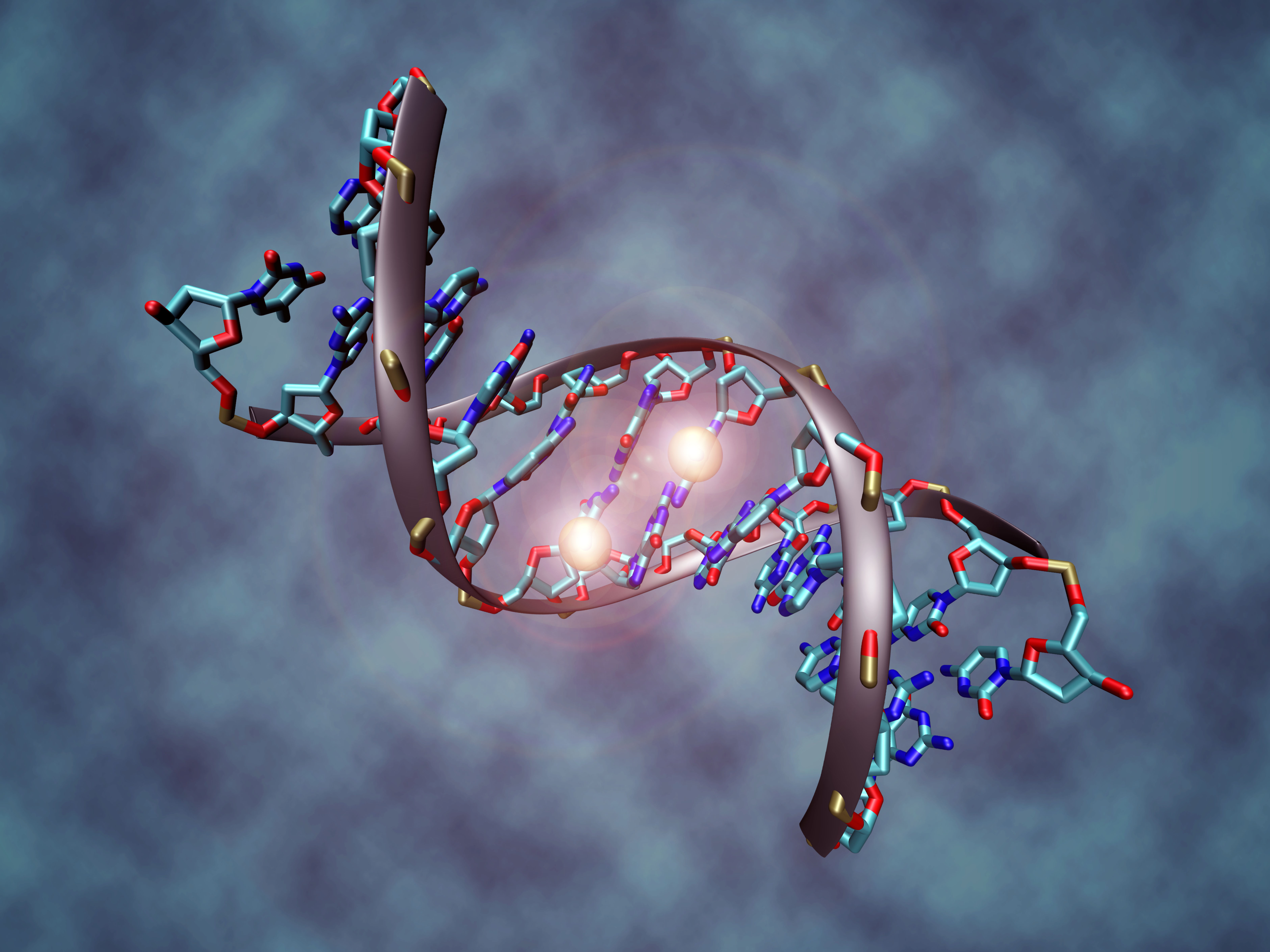
DNA methylation is a biological process by which methyl groups are added to the DNA molecule. Methylation can change the activity of a DNA segment without changing the sequence. When located in a gene promoter (genetics), promoter, DNA methylation typically acts to repress gene Transcription (genetics), transcription. In mammals, DNA methylation is essential for normal development and is associated with a number of key processes including genomic imprinting, X-chromosome inactivation, repression of transposable elements, aging, and carcinogenesis. As of 2016, two nucleobases have been found on which natural, enzymatic DNA methylation takes place: adenine and cytosine. The modified bases are N6-methyladenine,D. B. Dunn, J. D. Smith: "The occurrence of 6-methylaminopurine in deoxyribonucleic acids". In: ''Biochem J.'' 68(4), Apr 1958, S. 627–636. [//www.ncbi.nlm.nih.gov/pubmed/13522672?dopt=Abstract PMID 13522672]. . 5-methylcytosineB. F. Vanyushin, S. G. Tkacheva, A. N. Belozers ... [...More Info...] [...Related Items...] OR: [Wikipedia] [Google] [Baidu] |
Sperm
Sperm (: sperm or sperms) is the male reproductive Cell (biology), cell, or gamete, in anisogamous forms of sexual reproduction (forms in which there is a larger, female reproductive cell and a smaller, male one). Animals produce motile sperm with a tail known as a flagellum, which are known as spermatozoa, while some red algae and fungi produce non-motile sperm cells, known as spermatia. Flowering plants contain non-motile sperm inside pollen, while some more basal plants like ferns and some gymnosperms have motile sperm. Sperm cells form during the process known as spermatogenesis, which in amniotes (reptiles and mammals) takes place in the seminiferous tubules of the testicles. This process involves the production of several successive sperm cell precursors, starting with spermatogonia, which Cellular differentiation, differentiate into spermatocytes. The spermatocytes then undergo meiosis, reducing their Ploidy, chromosome number by half, which produces spermatids. The sper ... [...More Info...] [...Related Items...] OR: [Wikipedia] [Google] [Baidu] |
Gonadal Ridge
In embryology, the genital ridge (genital fold or gonadal ridge) is the developmental precursor to the gonads. The genital ridge initially consists mainly of mesenchyme and cells of underlying mesonephric origin. Once oogonia enter this area they attempt to associate with these somatic cells. Development proceeds and the oogonia become fully surrounded by a layer of cells (pre-granulosa cells). The genital ridge appears at approximately five weeks, and gives rise to the sex cords. Associated genes Genes associated with the developing gonad can be categorized into those that form the sexually indifferent gonad, those that determine whether the indifferent gonad will differentiate as male or female, and those that promote differentiation into male or female parts. Genes that form the sexually indifferent gonad are '' SF1'' and ''WT1''. Genes that determine sex are '' SRY'', ''SOX9'', and '' DAX1''. Genes driving the differentiation into male or female structures are ''SF1'', ... [...More Info...] [...Related Items...] OR: [Wikipedia] [Google] [Baidu] |
Somatic Cell
In cellular biology, a somatic cell (), or vegetal cell, is any biological cell forming the body of a multicellular organism other than a gamete, germ cell, gametocyte or undifferentiated stem cell. Somatic cells compose the body of an organism and divide through mitosis. In contrast, gametes derive from meiosis within the germ cells of the germline and they fuse during sexual reproduction. Stem cells also can divide through mitosis, but are different from somatic in that they differentiate into diverse specialized cell types. In mammals, somatic cells make up all the internal organs, skin, bones, blood and connective tissue, while mammalian germ cells give rise to spermatozoa and ova which fuse during fertilization to produce a cell called a zygote, which divides and differentiates into the cells of an embryo. There are approximately 220 types of somatic cell in the human body. Theoretically, these cells are not germ cells (the source of gametes); they transmit their mut ... [...More Info...] [...Related Items...] OR: [Wikipedia] [Google] [Baidu] |
Embryo
An embryo ( ) is the initial stage of development for a multicellular organism. In organisms that reproduce sexually, embryonic development is the part of the life cycle that begins just after fertilization of the female egg cell by the male sperm cell. The resulting fusion of these two cells produces a single-celled zygote that undergoes many cell divisions that produce cells known as blastomeres. The blastomeres (4-cell stage) are arranged as a solid ball that when reaching a certain size, called a morula, (16-cell stage) takes in fluid to create a cavity called a blastocoel. The structure is then termed a blastula, or a blastocyst in mammals. The mammalian blastocyst hatches before implantating into the endometrial lining of the womb. Once implanted the embryo will continue its development through the next stages of gastrulation, neurulation, and organogenesis. Gastrulation is the formation of the three germ layers that will form all of the different parts of t ... [...More Info...] [...Related Items...] OR: [Wikipedia] [Google] [Baidu] |
Germ Cell
A germ cell is any cell that gives rise to the gametes of an organism that reproduces sexually. In many animals, the germ cells originate in the primitive streak and migrate via the gut of an embryo to the developing gonads. There, they undergo meiosis, followed by cellular differentiation into mature gametes, either eggs or sperm. Unlike animals, plants do not have germ cells designated in early development. Instead, germ cells can arise from somatic cells in the adult, such as the floral meristem of flowering plants. Introduction Multicellular eukaryotes are made of two fundamental cell types: germ and somatic cells. Germ cells produce gametes and are the only cells that can undergo meiosis as well as mitosis. Somatic cells are all the other cells that form the building blocks of the body and they only divide by mitosis. The lineage of germ cells is called the germline. Germ cell specification begins during cleavage in many animals or in the epiblast during gastr ... [...More Info...] [...Related Items...] OR: [Wikipedia] [Google] [Baidu] |
Epiblast
In amniote embryonic development, the epiblast (also known as the primitive ectoderm) is one of two distinct cell layers arising from the inner cell mass in the mammalian blastocyst, or from the blastula in reptiles and birds. It drives the embryo proper through its differentiation into the three primary germ layers, ectoderm, mesoderm and endoderm, during gastrulation. The amniotic ectoderm and extraembryonic mesoderm also originate from the epiblast. The other layer of the inner cell mass, the hypoblast, gives rise to the yolk sac. The layer surrounding the inner cell mass, the trophectoderm, gives rise to the chorion. Discovery of the epiblast The epiblast was first discovered by Christian Heinrich Pander (1794-1865), a Baltic German biologist and embryologist. With the help of anatomist Ignaz Döllinger (1770–1841) and draftsman Eduard Joseph d'Alton (1772-1840), Pander observed thousands of chicken eggs under a microscope, and ultimately discovered and describe ... [...More Info...] [...Related Items...] OR: [Wikipedia] [Google] [Baidu] |
Blastocyst
The blastocyst is a structure formed in the early embryonic development of mammals. It possesses an inner cell mass (ICM) also known as the ''embryoblast'' which subsequently forms the embryo, and an outer layer of trophoblast cells called the trophectoderm. This layer surrounds the inner cell mass and a fluid-filled cavity or lumen known as the blastocoel. In the late blastocyst, the trophectoderm is known as the trophoblast. The trophoblast gives rise to the chorion and amnion, the two fetal membranes that surround the embryo. The placenta derives from the embryonic chorion (the portion of the chorion that develops villi) and the underlying uterine tissue of the mother. The corresponding structure in non-mammalian animals is an undifferentiated ball of cells called the blastula. In humans, blastocyst formation begins about five days after fertilization when a fluid-filled cavity opens up in the morula, the early embryonic stage of a ball of 16 cells. The blastocyst ... [...More Info...] [...Related Items...] OR: [Wikipedia] [Google] [Baidu] |
Morula
In embryology, cleavage is the division of cells in the early development of the embryo, following fertilization. The zygotes of many species undergo rapid cell cycles with no significant overall growth, producing a cluster of cells the same size as the original zygote. The different cells derived from cleavage are called blastomeres and form a compact mass called the morula. Cleavage ends with the formation of the blastula, or of the blastocyst in mammals. Depending mostly on the concentration of yolk in the egg, the cleavage can be holoblastic (total or complete cleavage) or meroblastic (partial or incomplete cleavage). The pole of the egg with the highest concentration of yolk is referred to as the vegetal pole while the opposite is referred to as the animal pole. Cleavage differs from other forms of cell division in that it increases the number of cells and nuclear mass without increasing the cytoplasmic mass. This means that with each successive subdivision, there is ro ... [...More Info...] [...Related Items...] OR: [Wikipedia] [Google] [Baidu] |
DNMT3B
DNA (cytosine-5)-methyltransferase 3 beta, is an enzyme that in humans in encoded by the DNMT3B gene. Mutation in this gene are associated with immunodeficiency, centromere instability and facial anomalies syndrome. Function CpG methylation is an epigenetic modification that is important for embryonic development, imprinting, and X-chromosome inactivation. Studies in mice have demonstrated that DNA methylation is required for mammalian development. This gene encodes a DNA methyltransferase which is thought to function in ''de novo'' methylation, rather than maintenance methylation. The protein localizes primarily to the nucleus and its expression is developmentally regulated. Eight alternatively spliced transcript variants have been described. The full length sequences of variants 4 and 5 have not been determined. Clinical significance Immunodeficiency-centromeric instability-facial anomalies (ICF) syndrome is a result of defects in lymphocyte maturation resulting from ... [...More Info...] [...Related Items...] OR: [Wikipedia] [Google] [Baidu] |
DNA (cytosine-5)-methyltransferase 3A
DNA (cytosine-5)-methyltransferase 3A (DNMT3A) is an enzyme that catalyzes the transfer of methyl groups to specific CpG structures in DNA, a process called DNA methylation. The enzyme is encoded in humans by the ''DNMT3A'' gene. This enzyme is responsible for ''de novo'' DNA methylation. Such function is to be distinguished from maintenance DNA methylation which ensures the fidelity of replication of inherited epigenetic patterns. DNMT3A forms part of the family of DNA methyltransferase enzymes, which consists of the protagonists DNMT1, DNMT3A and DNMT3B. While ''de novo'' DNA methylation modifies the information passed on by the parent to the progeny, it enables key epigenetic modifications essential for processes such as cellular differentiation and embryonic development, transcriptional regulation, heterochromatin formation, X-inactivation, imprinting and genome stability. ''DNMT3a'' is the gene most commonly found mutated in clonal hematopoiesis, a common aging-related ph ... [...More Info...] [...Related Items...] OR: [Wikipedia] [Google] [Baidu] |
DNMT1
DNA (cytosine-5)-methyltransferase 1 (Dnmt1) is an enzyme that catalyzes the transfer of methyl groups to specific CpG sites in DNA, a process called DNA methylation. In humans, it is encoded by the ''DNMT1'' gene In biology, the word gene has two meanings. The Mendelian gene is a basic unit of heredity. The molecular gene is a sequence of nucleotides in DNA that is transcribed to produce a functional RNA. There are two types of molecular genes: protei .... Dnmt1 forms part of the family of DNA methyltransferase enzymes, which consists primarily of DNMT1, DNMT3A, and DNMT3B. Function This enzyme is responsible for maintaining DNA methylation, which ensures the fidelity of this epigenetic patterns across cell divisions. In line with this role, it has a strong preference towards methylating CpGs on hemimethylated DNA. However, DNMT1 can catalyze de novo DNA methylation in specific genomic contexts, including transposable elements and paternal imprint control regions. ... [...More Info...] [...Related Items...] OR: [Wikipedia] [Google] [Baidu] |


