|
Claude Syndrome
Claude's syndrome is a form of brainstem stroke syndrome characterized by the presence of an ipsilateral oculomotor nerve palsy, contralateral hemiparesis, contralateral ataxia, and contralateral hemiplegia of the lower face, tongue, and shoulder. Claude's syndrome affects oculomotor nerve, red nucleus and brachium conjunctivum. Cause Claude's syndrome is caused by midbrain infarction as a result of occlusion of a branch of the posterior cerebral artery. This lesion is usually a unilateral infarction of the red nucleus and cerebellar peduncle, affecting several structures in the midbrain including: It is very similar to Benedikt's syndrome. Other causes It has been reported that posterior cerebral artery stenosis can also precipitate Claude's syndrome. Diagnosis History It carries the name of Henri Charles Jules Claude, a French psychiatrist and neurologist, who described the condition in 1912. See also * Wallenberg's syndrome * Moritz Benedikt Moritz Benedikt also sp ... [...More Info...] [...Related Items...] OR: [Wikipedia] [Google] [Baidu] |
Brainstem Stroke Syndrome
A brainstem stroke syndrome falls under the broader category of stroke syndromes, or specific symptoms caused by vascular injury to an area of brain (for example, the lacunar syndromes). As the brainstem contains numerous cranial nuclei and white matter tracts, a stroke in this area can have a number of unique symptoms depending on the particular blood vessel that was injured and the group of cranial nerves and tracts that are no longer perfused. Symptoms of a brainstem stroke frequently include sudden vertigo and ataxia, with or without weakness. Brainstem stroke can also cause diplopia, slurred speech and decreased level of consciousness. A more serious outcome is locked-in syndrome. Syndromes * The midbrain syndromes (Significant overlap between these three syndromes) ** Superior alternating hemiplegia or Weber's syndrome ** Paramedian midbrain syndrome or Benedikt's syndrome ** Claude's syndrome * Medial pontine syndrome or Middle alternating hemiplegia or Foville's synd ... [...More Info...] [...Related Items...] OR: [Wikipedia] [Google] [Baidu] |
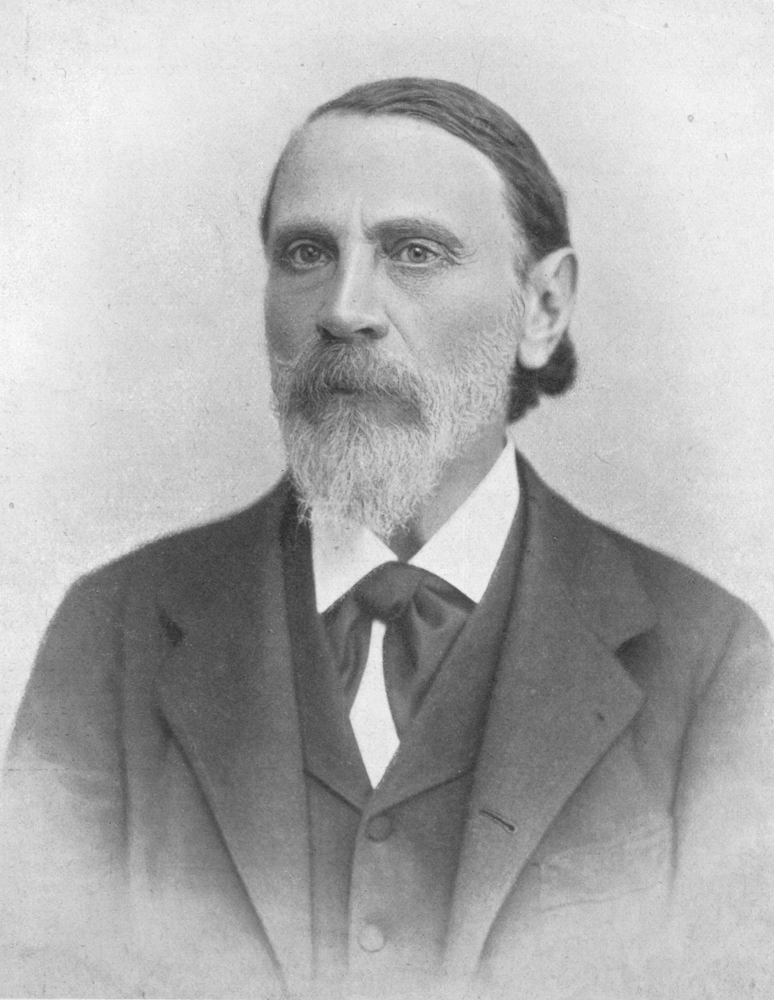
Moritz Benedikt
Moritz Benedikt also spelt Moriz (4 July 1835, in Eisenstadt, Sopron County – 14 April 1920, in Vienna) was a Hungarian-Austrian neurologist who was a native of Eisenstadt. He was an instructor and professor of neurology at the University of Vienna. Benedikt was a physician with the Austrian army during the Second Italian War of Independence (1859) and the Austro-Prussian War. Benedikt was a specialist in the fields of electrotherapeutics and neuropathology. His name is lent to the eponymous " Benedikt's syndrome", a disease characterized by ipsilateral oculomotor paralysis with contralateral tremor and hemiparesis caused by a lesion involving the red nucleus and corticospinal tract in the midbrain tegmentum. Benedikt is remembered today for his controversial research in criminal anthropology. He performed numerous cephalometric studies, and postulated that there were specific differences between "normal" and "criminal brains". He explained his research on the subject in ... [...More Info...] [...Related Items...] OR: [Wikipedia] [Google] [Baidu] |
Wallenberg's Syndrome
Lateral medullary syndrome is a neurological disorder causing a range of symptoms due to ischemia in the lateral part of the medulla oblongata in the brainstem. The ischemia is a result of a blockage most commonly in the vertebral artery or the posterior inferior cerebellar artery. Lateral medullary syndrome is also called Wallenberg's syndrome, posterior inferior cerebellar artery (PICA) syndrome and vertebral artery syndrome. Signs and symptoms This syndrome is characterized by sensory deficits that affect the trunk and extremities contralaterally (opposite to the lesion), and sensory deficits of the face and cranial nerves ipsilaterally (same side as the lesion). Specifically a loss of pain and temperature sensation if the lateral spinothalamic tract is involved. The cross body finding is the chief symptom from which a diagnosis can be made. Patients often have difficulty walking or maintaining balance (ataxia), or difference in temperature of an object based on which side of ... [...More Info...] [...Related Items...] OR: [Wikipedia] [Google] [Baidu] |
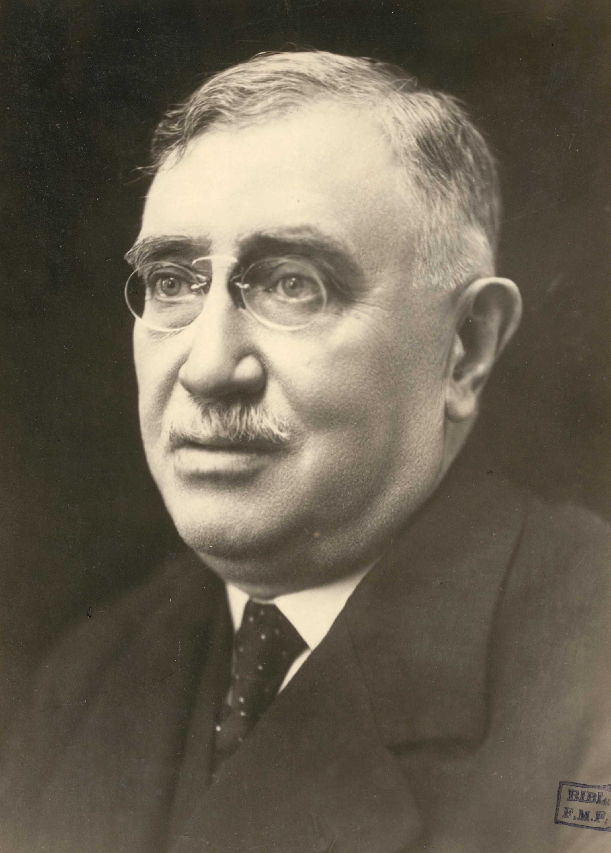
Henri Claude
Henri Charles Jules Claude (31 March 1869 – 29 November 1945) was a French psychiatrist and neurologist born in Paris. He studied medicine under Charles-Joseph Bouchard (1837-1915), and was an assistant to Fulgence Raymond (1844-1910) at the Salpêtrière Hospital. From 1922 until 1939, he served as chair of mental illness and brain diseases at the Sainte-Anne Hospital Center, Hôpital Sainte-Anne in Paris, where he was succeeded by Maxime Laignel-Lavastine. Henri Claude played a leading role in introducing Sigmund Freud, Freudian theories of psychoanalysis into French psychiatry. He was responsible for the creation of the first laboratory of psychotherapy and psychoanalysis at the school of medicine at the University of Paris. His name is lent to the eponymous "Claude syndrome", which is a midbrain syndrome characterized by Oculomotor nerve palsy, oculomotor palsy on the side of the lesion and ataxia on the opposite side. Also "Claude's hyperkinesia, hyperkinesis sign" is na ... [...More Info...] [...Related Items...] OR: [Wikipedia] [Google] [Baidu] |
Stenosis
A stenosis (from Ancient Greek στενός, "narrow") is an abnormal narrowing in a blood vessel or other tubular organ or structure such as foramina and canals. It is also sometimes called a stricture (as in urethral stricture). ''Stricture'' as a term is usually used when narrowing is caused by contraction of smooth muscle (e.g. achalasia, prinzmetal angina); ''stenosis'' is usually used when narrowing is caused by lesion that reduces the space of lumen (e.g. atherosclerosis). The term coarctation is another synonym, but is commonly used only in the context of aortic coarctation. Restenosis is the recurrence of stenosis after a procedure. Types The resulting syndrome depends on the structure affected. Examples of vascular stenotic lesions include: * Intermittent claudication (peripheral artery stenosis) * Angina ( coronary artery stenosis) * Carotid artery stenosis which predispose to (strokes and transient ischaemic episodes) * Renal artery stenosis The types of sten ... [...More Info...] [...Related Items...] OR: [Wikipedia] [Google] [Baidu] |
Benedikt's Syndrome
Benedikt syndrome, also called Benedikt's syndrome or paramedian midbrain syndrome, is a rare type of posterior circulation stroke of the brain, with a range of neurological symptoms affecting the midbrain, cerebellum and other related structures. Signs and symptoms It is characterized by the presence of an oculomotor nerve (CN III) palsy and cerebellar ataxia including tremor and involuntary choreoathetotic movements. Neuroanatomical structures affected include the oculomotor nucleus, red nucleus, corticospinal tracts and superior cerebellar peduncle decussation. It has a similar cause, morphology, signs and symptoms to Weber's syndrome; the main difference between the two being that Weber's is more associated with hemiplegia (i.e. paralysis), and Benedikt's with hemiataxia (i.e. disturbed coordination of movements). It is also similar to Claude's syndrome, but is distinguishable in that Benedikt's has more predominant tremor and choreoathetotic movements while Claude's is more ... [...More Info...] [...Related Items...] OR: [Wikipedia] [Google] [Baidu] |
Diplopia
Diplopia is the simultaneous perception of two images of a single object that may be displaced horizontally or vertically in relation to each other. Also called double vision, it is a loss of visual focus under regular conditions, and is often voluntary. However, when occurring involuntarily, it results in impaired function of the extraocular muscles, where both eyes are still functional, but they cannot turn to target the desired object. Problems with these muscles may be due to mechanical problems, disorders of the neuromuscular junction, disorders of the cranial nerves ( III, IV, and VI) that innervate the muscles, and occasionally disorders involving the supranuclear oculomotor pathways or ingestion of toxins. Diplopia can be one of the first signs of a systemic disease, particularly to a muscular or neurological process, and it may disrupt a person's balance, movement, or reading abilities. Causes Diplopia has a diverse range of ophthalmologic, infectious, autoimmune, neu ... [...More Info...] [...Related Items...] OR: [Wikipedia] [Google] [Baidu] |
Ipsilateral
Standard anatomical terms of location are used to unambiguously describe the anatomy of animals, including humans. The terms, typically derived from Latin or Greek roots, describe something in its standard anatomical position. This position provides a definition of what is at the front ("anterior"), behind ("posterior") and so on. As part of defining and describing terms, the body is described through the use of anatomical planes and anatomical axes. The meaning of terms that are used can change depending on whether an organism is bipedal or quadrupedal. Additionally, for some animals such as invertebrates, some terms may not have any meaning at all; for example, an animal that is radially symmetrical will have no anterior surface, but can still have a description that a part is close to the middle ("proximal") or further from the middle ("distal"). International organisations have determined vocabularies that are often used as standard vocabularies for subdisciplines of anatom ... [...More Info...] [...Related Items...] OR: [Wikipedia] [Google] [Baidu] |
Corticobulbar Tract
In neuroanatomy, the corticobulbar (or corticonuclear) tract is a two-neuron white matter motor pathway connecting the motor cortex in the cerebral cortex to the medullary pyramids, which are part of the brainstem's medulla oblongata (also called "bulbar") region, and are primarily involved in carrying the motor function of the non-oculomotor cranial nerves. The corticobulbar tract is one of the pyramidal tracts, the other being the corticospinal tract. Structure The corticobulbar tract originates in the primary motor cortex of the frontal lobe, just superior to the lateral fissure and rostral to the central sulcus in the precentral gyrus (Brodmann area 4). The tract descends through the corona radiata and genu of the internal capsule with a few fibers in the posterior limb of the internal capsule, as it passes from the cortex down to the midbrain. In the midbrain, the internal capsule becomes the cerebral peduncles. The white matter is located in the ventral portion of the c ... [...More Info...] [...Related Items...] OR: [Wikipedia] [Google] [Baidu] |
Corticospinal Tract
The corticospinal tract is a white matter motor pathway starting at the cerebral cortex that terminates on lower motor neurons and interneurons in the spinal cord, controlling movements of the limbs and trunk. There are more than one million neurons in the corticospinal tract, and they become myelinated usually in the first two years of life. The corticospinal tract is one of the pyramidal tracts, the other being the corticobulbar tract. Anatomy The corticospinal tract originates in several parts of the brain, including not just the motor areas, but also the primary somatosensory cortex and premotor areas. Most of the neurons originate in the primary motor cortex (precentral gyrus, Brodmann area 4) or the premotor frontal areas.Purves, D. et al. (2012). Neuroscience: Fifth edition. Sunderland, MA: Sinauer Associates, Inc.Kolb, B. & Whishaw, I. Q. (2014). An introduction to brain and behavior: Fourth edition. New York, NY: Worth Publishers. About 30% of corticospinal neurons origi ... [...More Info...] [...Related Items...] OR: [Wikipedia] [Google] [Baidu] |
Cerebellar Peduncle
Cerebellar peduncles connect the cerebellum to the brain stem. There are six cerebellar peduncles in total, three on each side: * Superior cerebellar peduncle is a paired structure of white matter that connects the cerebellum to the mid-brain. * Middle cerebellar peduncles connect the cerebellum to the pons and are composed entirely of centripetal fibers. * Inferior cerebellar peduncle is a thick rope-like strand that occupies the upper part of the posterior district of the medulla oblongata. The peduncles form the lateral border of the fourth ventricle The fourth ventricle is one of the four connected fluid-filled cavities within the human brain. These cavities, known collectively as the ventricular system, consist of the left and right lateral ventricles, the third ventricle, and the fourth ve ..., and form a distinctive diamond the middle peduncle forming the central corners of the diamond, while the superior and inferior peduncles form the superior and inferior edges, r ... [...More Info...] [...Related Items...] OR: [Wikipedia] [Google] [Baidu] |

