|
Metacarpus
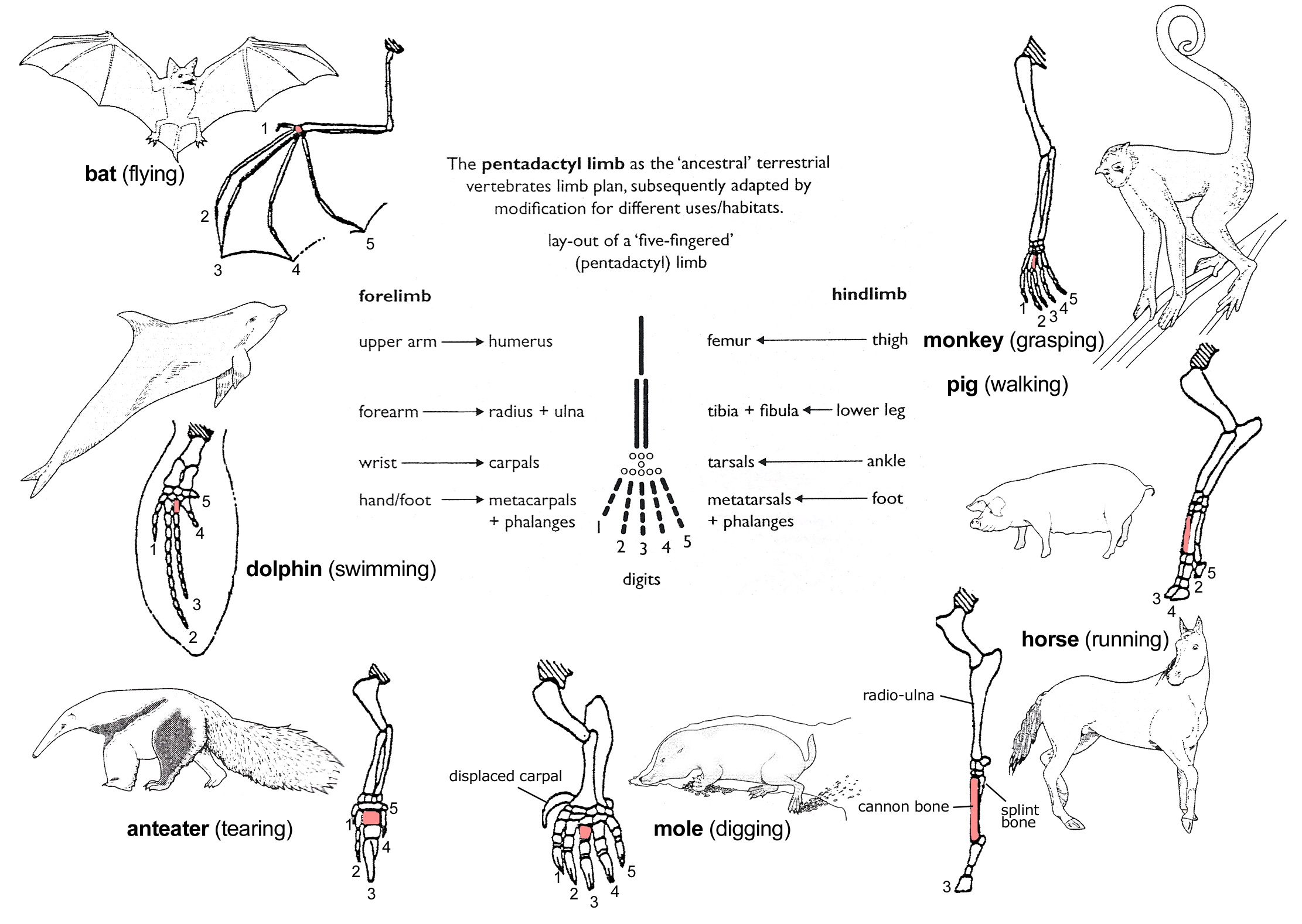
In human anatomy, the metacarpal bones or metacarpus, also known as the "palm bones", are the appendicular skeleton, appendicular bones that form the intermediate part of the hand between the phalanges (fingers) and the carpal bones (wrist, wrist bones), which joint, articulate with the forearm. The metacarpal bones are homologous to the metatarsal bones in the foot. Structure The metacarpals form a transverse arch to which the rigid row of distal carpal bones are fixed. The peripheral metacarpals (those of the thumb and little finger) form the sides of the cup of the palmar gutter and as they are brought together they deepen this concavity. The index metacarpal is the most firmly fixed, while the thumb metacarpal articulates with the trapezium and acts independently from the others. The middle metacarpals are tightly united to the carpus by intrinsic interlocking bone elements at their bases. The ring metacarpal is somewhat more mobile while the fifth metacarpal is semi-indepen ... [...More Info...] [...Related Items...] OR: [Wikipedia] [Google] [Baidu] |
Carpal Bones
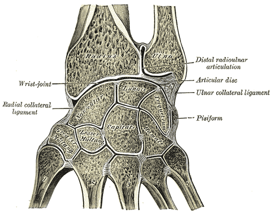
The carpal bones are the eight small bones that make up the wrist (carpus) that connects the hand to the forearm. The terms "carpus" and "carpal" are derived from the Latin wikt:carpus#Latin, carpus and the Greek language, Greek wikt:καρπός#Ancient Greek, καρπός (karpós), meaning "wrist". In human anatomy, the main role of the carpal bones is to joint, articulate with the radius (bone), radial and ulnar heads to form a highly mobile condyloid joint (i.e. wrist joint),Kingston 2000, pp 126-127 to provide attachments for thenar and hypothenar muscles, and to form part of the rigid carpal tunnel which allows the median nerve and tendons of the anterior compartment of the forearm, anterior forearm muscles to be transmitted to the hand and fingers. In tetrapods, the carpus is the sole cluster of bones in the wrist between the radius (bone), radius and ulna and the metacarpus. The bones of the carpus do not belong to individual fingers (or toes in quadrupeds), whereas those ... [...More Info...] [...Related Items...] OR: [Wikipedia] [Google] [Baidu] |
Hand
A hand is a prehensile, multi-fingered appendage located at the end of the forearm or forelimb of primates such as humans, chimpanzees, monkeys, and lemurs. A few other vertebrates such as the Koala#Characteristics, koala (which has two thumb#Opposition and apposition, opposable thumbs on each "hand" and fingerprints extremely similar to human fingerprints) are often described as having "hands" instead of paws on their front limbs. The raccoon is usually described as having "hands" though opposable thumbs are lacking. Some evolutionary anatomists use the term ''hand'' to refer to the appendage of digits on the forelimb more generally—for example, in the context of whether the three Digit (anatomy), digits of the bird hand involved the same Homology (biology), homologous loss of two digits as in the dinosaur hand. The human hand usually has five digits: Finger numbering#Four-finger system, four fingers plus one thumb; however, these are often referred to collectively as Finger ... [...More Info...] [...Related Items...] OR: [Wikipedia] [Google] [Baidu] |
Carpometacarpal Joint
The carpometacarpal (CMC) joints are five joints in the wrist that articulate the distal row of carpal bones and the proximal bases of the five metacarpal bones. The CMC joint of the thumb or the first CMC joint, also known as the trapeziometacarpal (TMC) joint, differs significantly from the other four CMC joints and is therefore described separately. Thumb The carpometacarpal joint of the thumb (''pollex''), also known as the first carpometacarpal joint, or the trapeziometacarpal joint (TMC) because it connects the trapezium to the first metacarpal bone, plays an irreplaceable role in the normal functioning of the thumb. The most important joint connecting the wrist to the metacarpus, osteoarthritis of the TMC is a severely disabling condition; it is up to twenty times more common among elderly women than in the average. Pronation-supination of the first metacarpal is especially important for the action of opposition. The movements of the first CMC are limited by the sha ... [...More Info...] [...Related Items...] OR: [Wikipedia] [Google] [Baidu] |
Foot
The foot (: feet) is an anatomical structure found in many vertebrates. It is the terminal portion of a limb which bears weight and allows locomotion. In many animals with feet, the foot is an organ at the terminal part of the leg made up of one or more segments or bones, generally including claws and/or nails. Etymology The word "foot", in the sense of meaning the "terminal part of the leg of a vertebrate animal" comes from Old English ''fot'', from Proto-Germanic *''fot'' (source also of Old Frisian ''fot'', Old Saxon ''fot'', Old Norse ''fotr'', Danish ''fod'', Swedish ''fot'', Dutch ''voet'', Old High German ''fuoz'', German ''Fuß'', Gothic ''fotus'', all meaning "foot"), from PIE root *''ped-'' "foot". The plural form ''feet'' is an instance of i-mutation. Structure The human foot is a strong and complex mechanical structure containing 26 bones, 33 joints (20 of which are actively articulated), and more than a hundred muscles, tendons, and ligaments.Podiatry Chan ... [...More Info...] [...Related Items...] OR: [Wikipedia] [Google] [Baidu] |
Wrist
In human anatomy, the wrist is variously defined as (1) the carpus or carpal bones, the complex of eight bones forming the proximal skeletal segment of the hand; "The wrist contains eight bones, roughly aligned in two rows, known as the carpal bones." (2) the wrist joint or radiocarpal joint, the joint between the radius and the carpus and; (3) the anatomical region surrounding the carpus including the distal parts of the bones of the forearm and the proximal parts of the metacarpus or five metacarpal bones and the series of joints between these bones, thus referred to as ''wrist joints''. "With the large number of bones composing the wrist (ulna, radius, eight carpas, and five metacarpals), it makes sense that there are many, many joints that make up the structure known as the wrist." This region also includes the carpal tunnel, the anatomical snuff box, bracelet lines, the flexor retinaculum, and the extensor retinaculum. As a consequence of these various definitions, f ... [...More Info...] [...Related Items...] OR: [Wikipedia] [Google] [Baidu] |
Metatarsal Bones
The metatarsal bones or metatarsus (: metatarsi) are a group of five long bones in the midfoot, located between the tarsal bones (which form the heel and the ankle) and the phalanges ( toes). Lacking individual names, the metatarsal bones are numbered from the medial side (the side of the great toe): the first, second, third, fourth, and fifth metatarsal (often depicted with Roman numerals). The metatarsals are analogous to the metacarpal bones of the hand. The lengths of the metatarsal bones in humans are, in descending order, second, third, fourth, fifth, and first. A bovine hind leg has two metatarsals. Structure The five metatarsals are dorsal convex long bones consisting of a shaft or body, a base ( proximally), and a head ( distally).Platzer 2004, p. 220 The body is prismoid in form, tapers gradually from the tarsal to the phalangeal extremity, and is curved longitudinally, so as to be concave below, slightly convex above. The base or posterior extremity is ... [...More Info...] [...Related Items...] OR: [Wikipedia] [Google] [Baidu] |
Carpometacarpal Joint
The carpometacarpal (CMC) joints are five joints in the wrist that articulate the distal row of carpal bones and the proximal bases of the five metacarpal bones. The CMC joint of the thumb or the first CMC joint, also known as the trapeziometacarpal (TMC) joint, differs significantly from the other four CMC joints and is therefore described separately. Thumb The carpometacarpal joint of the thumb (''pollex''), also known as the first carpometacarpal joint, or the trapeziometacarpal joint (TMC) because it connects the trapezium to the first metacarpal bone, plays an irreplaceable role in the normal functioning of the thumb. The most important joint connecting the wrist to the metacarpus, osteoarthritis of the TMC is a severely disabling condition; it is up to twenty times more common among elderly women than in the average. Pronation-supination of the first metacarpal is especially important for the action of opposition. The movements of the first CMC are limited by the sha ... [...More Info...] [...Related Items...] OR: [Wikipedia] [Google] [Baidu] |
Bone
A bone is a rigid organ that constitutes part of the skeleton in most vertebrate animals. Bones protect the various other organs of the body, produce red and white blood cells, store minerals, provide structure and support for the body, and enable mobility. Bones come in a variety of shapes and sizes and have complex internal and external structures. They are lightweight yet strong and hard and serve multiple functions. Bone tissue (osseous tissue), which is also called bone in the uncountable sense of that word, is hard tissue, a type of specialised connective tissue. It has a honeycomb-like matrix internally, which helps to give the bone rigidity. Bone tissue is made up of different types of bone cells. Osteoblasts and osteocytes are involved in the formation and mineralisation of bone; osteoclasts are involved in the resorption of bone tissue. Modified (flattened) osteoblasts become the lining cells that form a protective layer on the bone surface. The mine ... [...More Info...] [...Related Items...] OR: [Wikipedia] [Google] [Baidu] |
Dorsal Interossei Of The Hand
In human anatomy, the dorsal interossei (DI) are four muscles in the back of the hand that act to abduct (spread) the index, middle, and ring fingers away from the hand's midline (ray of middle finger) and assist in flexion at the metacarpophalangeal The metacarpophalangeal joints (MCP) are situated between the metacarpal bones and the proximal phalanges of the fingers. These joints are of the condyloid kind, formed by the reception of the rounded heads of the metacarpal bones into shallow ... joints and extension at the interphalangeal joints of the index, middle and ring fingers. Structure There are four dorsal interossei in each hand. They are specified as 'dorsal' to contrast them with the palmar interossei, which are located on the anterior side of the metacarpals. The dorsal interosseous muscles are bipennate, with each muscle arising by two heads from the adjacent sides of the metacarpal bones, but more extensively from the metacarpal bone of the finger into whic ... [...More Info...] [...Related Items...] OR: [Wikipedia] [Google] [Baidu] |
First Metacarpal Bone
The first metacarpal bone or the metacarpal bone of the thumb is the first bone proximal to the thumb. It is connected to the trapezium of the carpus at the first carpometacarpal joint and to the proximal thumb phalanx at the first metacarpophalangeal joint. Characteristics The first metacarpal bone is short and thick with a shaft thicker and broader than those of the other metacarpal bones. Its narrow shaft connects its widened base and rounded head; the former consisting of a thick cortical bone surrounding the open medullary canal; the latter two consisting of cancellous bone surrounded by a thin cortical shell. Head The head is less rounded and less spherical than those of the other metacarpals, making it better suited for a hinge-like articulation. The distal articular surface is quadrilateral, wide, and flat; thicker and broader transversely and extends much further palmarly than dorsally. On the palmar aspect of the articular surface there is a pair of emin ... [...More Info...] [...Related Items...] OR: [Wikipedia] [Google] [Baidu] |


