|
Lymphatics Of The Head And Neck
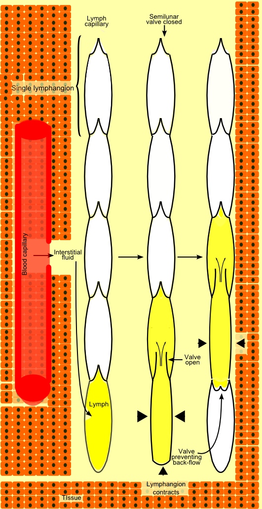
The lymphatic vessels (or lymph vessels or lymphatics) are thin-walled vessels (tubes), structured like blood vessels, that carry lymph. As part of the lymphatic system, lymph vessels are complementary to the cardiovascular system. Lymph vessels are lined by endothelial cells, and have a thin layer of smooth muscle, and adventitia that binds the lymph vessels to the surrounding tissue. Lymph vessels are devoted to the propulsion of the lymph from the lymph capillaries, which are mainly concerned with the absorption of interstitial fluid from the tissues. Lymph capillaries are slightly bigger than their counterpart capillaries of the vascular system. Lymph vessels that carry lymph to a lymph node are called afferent lymph vessels, and those that carry it from a lymph node are called efferent lymph vessels, from where the lymph may travel to another lymph node, may be returned to a vein, or may travel to a larger lymph duct. Lymph ducts drain the lymph into one of the subclavian ve ... [...More Info...] [...Related Items...] OR: [Wikipedia] [Google] [Baidu] |
Lymph Capillaries
Lymph capillaries or lymphatic capillaries are tiny, thin-walled microvessels located in the spaces between cells (except in the central nervous system and non-vascular tissues) which serve to drain and process extracellular fluid. Upon entering the lumen of a lymphatic capillary, the collected fluid is known as lymph. Each lymphatic capillary carries lymph into a lymphatic vessel, which in turn connects to a lymph node, a small bean-shaped gland that filters and monitors the lymphatic fluid for infections. Lymph is ultimately returned to the venous circulation. Lymphatic capillaries are slightly larger in diameter than blood capillaries, and have closed ends (unlike the loop structure of blood capillaries). Lymph capillaries are strategically placed among the blood-related capillaries in order to have efficient and effective uptake from the interstitial fluid during capillary exchange. This intentional formation allows for a more rapid and continuous collection. Their uniq ... [...More Info...] [...Related Items...] OR: [Wikipedia] [Google] [Baidu] |
Subclavian Vein
The subclavian vein is a paired large vein, one on either side of the body, that is responsible for draining blood from the upper extremities, allowing this blood to return to the heart. The left subclavian vein plays a key role in the absorption of lipids, by allowing products that have been carried by lymph in the thoracic duct to enter the bloodstream. The diameter of the subclavian veins is approximately 1–2 cm, depending on the individual. Structure Each subclavian vein is a continuation of the axillary vein and runs from the outer border of the first rib to the medial border of anterior scalene muscle. From here it joins with the internal jugular vein to form the brachiocephalic vein (also known as "innominate vein"). The angle of union is termed the venous angle. The subclavian vein follows the subclavian artery and is separated from the subclavian artery by the insertion of anterior scalene. Thus, the subclavian vein lies anterior to the anterior scalene while ... [...More Info...] [...Related Items...] OR: [Wikipedia] [Google] [Baidu] |
Lymph Vessel
The lymphatic vessels (or lymph vessels or lymphatics) are thin-walled vessels (tubes), structured like blood vessels, that carry lymph. As part of the lymphatic system, lymph vessels are complementary to the cardiovascular system. Lymph vessels are lined by endothelial cells, and have a thin layer of smooth muscle, and adventitia that binds the lymph vessels to the surrounding tissue. Lymph vessels are devoted to the propulsion of the lymph from the lymph capillaries, which are mainly concerned with the absorption of interstitial fluid from the tissues. Lymph capillaries are slightly bigger than their counterpart capillaries of the vascular system. Lymph vessels that carry lymph to a lymph node are called afferent lymph vessels, and those that carry it from a lymph node are called efferent lymph vessels, from where the lymph may travel to another lymph node, may be returned to a vein, or may travel to a larger lymph duct. Lymph ducts drain the lymph into one of the subclavian ... [...More Info...] [...Related Items...] OR: [Wikipedia] [Google] [Baidu] |
Pulse
In medicine, the pulse refers to the rhythmic pulsations (expansion and contraction) of an artery in response to the cardiac cycle (heartbeat). The pulse may be felt ( palpated) in any place that allows an artery to be compressed near the surface of the body close to the skin, such as at the neck ( carotid artery), wrist (radial artery or ulnar artery), at the groin (femoral artery), behind the knee ( popliteal artery), near the ankle joint ( posterior tibial artery), and on foot (dorsalis pedis artery). The pulse is most commonly measured at the wrist or neck for adults and at the brachial artery (inner upper arm between the shoulder and elbow) for infants and very young children. A sphygmograph is an instrument for measuring the pulse. Physiology Claudius Galen was perhaps the first physiologist to describe the pulse. The pulse is an expedient tactile method of determination of systolic blood pressure to a trained observer. Diastolic blood pressure is non-palpable and ... [...More Info...] [...Related Items...] OR: [Wikipedia] [Google] [Baidu] |
Arterial
An artery () is a blood vessel in humans and most other animals that takes oxygenated blood away from the heart in the systemic circulation to one or more parts of the body. Exceptions that carry deoxygenated blood are the pulmonary arteries in the pulmonary circulation that carry blood to the lungs for oxygenation, and the umbilical arteries in the fetal circulation that carry deoxygenated blood to the placenta. It consists of a multi-layered artery wall wrapped into a tube-shaped channel. Arteries contrast with veins, which carry deoxygenated blood back towards the heart; or in the pulmonary and fetal circulations carry oxygenated blood to the lungs and fetus respectively. Structure The anatomy of arteries can be separated into gross anatomy, at the macroscopic level, and microanatomy, which must be studied with a microscope. The arterial system of the human body is divided into systemic arteries, carrying blood from the heart to the whole body, and pulmonary arteries, ... [...More Info...] [...Related Items...] OR: [Wikipedia] [Google] [Baidu] |
Peristalsis
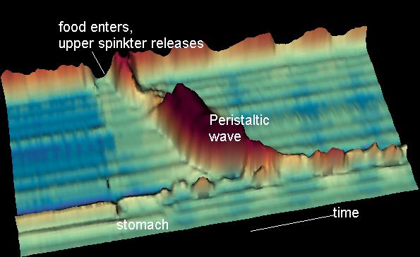
Peristalsis ( , ) is a type of intestinal motility, characterized by symmetry in biology#Radial symmetry, radially symmetrical contraction and relaxation of muscles that propagate in a wave down a tube, in an wikt:anterograde, anterograde direction. Peristalsis is progression of coordinated contraction of involuntary circular muscles, which is preceded by a simultaneous contraction of the longitudinal muscle and relaxation of the circular muscle in the lining of the gut. In much of a digestive tract, such as the human gastrointestinal tract, smooth muscle tissue contracts in sequence to produce a peristaltic wave, which propels a ball of food (called a bolus (digestion), bolus before being transformed into chyme in the stomach) along the tract. The peristaltic movement comprises relaxation of circular smooth muscles, then their contraction behind the chewed material to keep it from moving backward, then longitudinal contraction to push it forward. Earthworms use a similar mec ... [...More Info...] [...Related Items...] OR: [Wikipedia] [Google] [Baidu] |
Lumen (anatomy)
In biology, a lumen (: lumina) is the inside space of a tubular structure, such as an artery or intestine. It comes . It can refer to: *the interior of a vessel, such as the central space in an artery, vein or capillary through which blood flows *the interior of the gastrointestinal tract *the pathways of the bronchi in the lungs *the interior of renal tubules and urinary collecting ducts *the pathways of the female genital tract, starting with a single pathway of the vagina, splitting up in two lumina in the uterus, both of which continue through the fallopian tubes *the fluid-filled cavity forming in the blastocyst during pre-implantation development called the blastocoel In cell biology, lumen is a membrane-defined space that is found inside several organelles, cellular components, or structures, including thylakoid, endoplasmic reticulum, Golgi apparatus, lysosome, mitochondrion, and microtubule. Transluminal procedures ''Transluminal procedures'' are procedures occur ... [...More Info...] [...Related Items...] OR: [Wikipedia] [Google] [Baidu] |
Basement Membrane
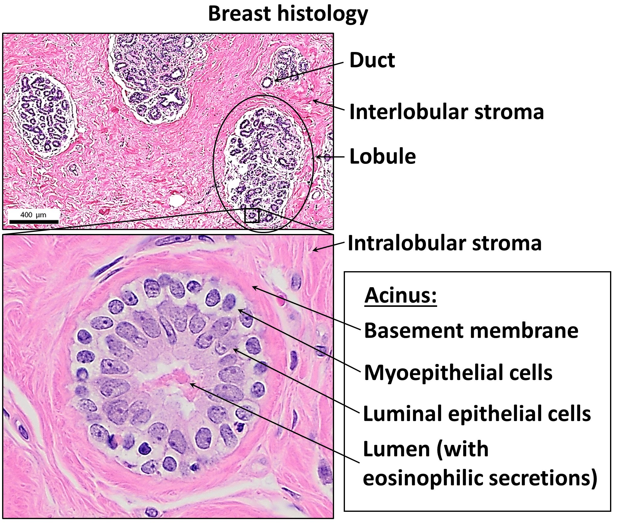
The basement membrane, also known as base membrane, is a thin, pliable sheet-like type of extracellular matrix that provides cell and tissue support and acts as a platform for complex signalling. The basement membrane sits between epithelial tissues including mesothelium and endothelium, and the underlying connective tissue. Structure As seen with the electron microscope, the basement membrane is composed of two layers, the basal lamina and the reticular lamina. The underlying connective tissue attaches to the basal lamina with collagen VII anchoring fibrils and fibrillin microfibrils. The basal lamina layer can further be subdivided into two layers based on their visual appearance in electron microscopy. The lighter-colored layer closer to the epithelium is called the lamina lucida, while the denser-colored layer closer to the connective tissue is called the lamina densa. The electron-dense lamina densa layer is about 30–70 nanometers thick and consists of an und ... [...More Info...] [...Related Items...] OR: [Wikipedia] [Google] [Baidu] |
Epithelium
Epithelium or epithelial tissue is a thin, continuous, protective layer of cells with little extracellular matrix. An example is the epidermis, the outermost layer of the skin. Epithelial ( mesothelial) tissues line the outer surfaces of many internal organs, the corresponding inner surfaces of body cavities, and the inner surfaces of blood vessels. Epithelial tissue is one of the four basic types of animal tissue, along with connective tissue, muscle tissue and nervous tissue. These tissues also lack blood or lymph supply. The tissue is supplied by nerves. There are three principal shapes of epithelial cell: squamous (scaly), columnar, and cuboidal. These can be arranged in a singular layer of cells as simple epithelium, either simple squamous, simple columnar, or simple cuboidal, or in layers of two or more cells deep as stratified (layered), or ''compound'', either squamous, columnar or cuboidal. In some tissues, a layer of columnar cells may appear to be stratified d ... [...More Info...] [...Related Items...] OR: [Wikipedia] [Google] [Baidu] |
Blood Vessel
Blood vessels are the tubular structures of a circulatory system that transport blood throughout many Animal, animals’ bodies. Blood vessels transport blood cells, nutrients, and oxygen to most of the Tissue (biology), tissues of a Body (biology), body. They also take waste and carbon dioxide away from the tissues. Some tissues such as cartilage, epithelium, and the lens (anatomy), lens and cornea of the eye are not supplied with blood vessels and are termed ''avascular''. There are five types of blood vessels: the arteries, which carry the blood away from the heart; the arterioles; the capillaries, where the exchange of water and chemicals between the blood and the tissues occurs; the venules; and the veins, which carry blood from the capillaries back towards the heart. The word ''vascular'', is derived from the Latin ''vas'', meaning ''vessel'', and is mostly used in relation to blood vessels. Etymology * artery – late Middle English; from Latin ''arteria'', from Gree ... [...More Info...] [...Related Items...] OR: [Wikipedia] [Google] [Baidu] |
KC Panchal
KC, Kc and similar may refer to: Places * Kuçovë District, Albania's ISO 3166-2 code * Kansas City metropolitan area, a major metropolitan area of the United States ** Kansas City, Missouri, its principal city ** Kansas City, Kansas, the third-largest city in the state * Karachay-Cherkessia, Russia's ISO 3166-2 code People * Kent Cochrane (1951–2014), Canadian memory disorder patient * KC Concepcion (born 1985), Philippine model, actress, singer, songwriter * Kcee (musician) (born 1979), Nigerian singer, songwriter, performer * K.C. Potter (1939–2024), American academic administrator and LGBT rights activist * Harry Wayne Casey (born 1951), American musician best known for his band KC and the Sunshine Band * Govinda K.C. (born 1957), Nepali orthopaedic surgeon and philanthropic activist * KC or K.C., an abbreviation for the Nepalese surname Khatri Chhetri Arts and entertainment * ''KC'' (album), a 2010 album by KC Concepcion * KC and the Sunshine Band, an American ... [...More Info...] [...Related Items...] OR: [Wikipedia] [Google] [Baidu] |
Subclavian Veins
The subclavian vein is a paired large vein, one on either side of the body, that is responsible for draining blood from the upper extremities, allowing this blood to return to the heart. The left subclavian vein plays a key role in the absorption of lipids, by allowing products that have been carried by lymph in the thoracic duct to enter the bloodstream. The diameter of the subclavian veins is approximately 1–2 cm, depending on the individual. Structure Each subclavian vein is a continuation of the axillary vein and runs from the outer border of the first rib to the medial border of anterior scalene muscle. From here it joins with the internal jugular vein to form the brachiocephalic vein (also known as "innominate vein"). The angle of union is termed the venous angle. The subclavian vein follows the subclavian artery and is separated from the subclavian artery by the insertion of anterior scalene. Thus, the subclavian vein lies anterior to the anterior scalene while th ... [...More Info...] [...Related Items...] OR: [Wikipedia] [Google] [Baidu] |


