|
Chest CT
Computed tomography of the chest or chest CT is a group of computed tomography scan protocols used in medical imaging to evaluate the lungs and search for lung disorders. Contrast agents are sometimes used in CT scans of the chest to accentuate or enhance the differences in radiopacity between vascularized and less vascularized structures, but a standard chest CT scan is usually non-contrasted (i.e. "plain") and relies on different algorithms to produce various series of digitalized images known as ''view'' or "''window''". Modern detail-oriented scans such as high-resolution computed tomography (HRCT) is the gold standard in respiratory medicine and thoracic surgery for investigating disorders of the lung parenchyma (alveoli). Contrasted CT scans of the chest are usually used to confirm diagnosis of for lung cancer and abscesses, as well as to assess lymph node status at the hila and the mediastinum. CT pulmonary angiogram, which uses time-matched ("phased") protocols to asse ... [...More Info...] [...Related Items...] OR: [Wikipedia] [Google] [Baidu] [Amazon] |
Thorax CT Peripheres Brronchialcarcinom Li OF
The thorax (: thoraces or thoraxes) or chest is a part of the anatomy of mammals and other tetrapod animals located between the neck and the abdomen. In insects, crustaceans, and the extinct trilobites, the thorax is one of the three main divisions of the body, each in turn composed of multiple segments. The human thorax includes the thoracic cavity and the thoracic wall. It contains organs including the heart, lungs, and thymus gland, as well as muscles and various other internal structures. The chest may be affected by many diseases, of which the most common symptom is chest pain. Etymology The word thorax comes from the Greek θώραξ ''thṓrax'' "breastplate, cuirass, corslet" via . Humans Structure In humans and other hominids, the thorax is the chest region of the body between the neck and the abdomen, along with its internal organs and other contents. It is mostly protected and supported by the rib cage, spine, and shoulder girdle. Contents The contents of the t ... [...More Info...] [...Related Items...] OR: [Wikipedia] [Google] [Baidu] [Amazon] |
Parenchyma
upright=1.6, Lung parenchyma showing damage due to large subpleural bullae. Parenchyma () is the bulk of functional substance in an animal organ such as the brain or lungs, or a structure such as a tumour. In zoology, it is the tissue that fills the interior of flatworms. In botany, it is some layers in the cross-section of the leaf. Etymology The term ''parenchyma'' is Neo-Latin from the Ancient Greek word meaning 'visceral flesh', and from meaning 'to pour in' from 'beside' + 'in' + 'to pour'. Originally, Erasistratus and other anatomists used it for certain human tissues. Later, it was also applied to plant tissues by Nehemiah Grew. Structure The parenchyma is the ''functional'' parts of an organ, or of a structure such as a tumour in the body. This is in contrast to the stroma, which refers to the ''structural'' tissue of organs or of structures, namely, the connective tissues. Brain The brain parenchyma refers to the functional tissue in th ... [...More Info...] [...Related Items...] OR: [Wikipedia] [Google] [Baidu] [Amazon] |
Diagnostic Radiology
Medical imaging is the technique and process of imaging the interior of a body for clinical analysis and medical intervention, as well as visual representation of the function of some organs or tissues (physiology). Medical imaging seeks to reveal internal structures hidden by the skin and bones, as well as to diagnose and treat disease. Medical imaging also establishes a database of normal anatomy and physiology to make it possible to identify abnormalities. Although imaging of removed organ (anatomy), organs and Tissue (biology), tissues can be performed for medical reasons, such procedures are usually considered part of pathology instead of medical imaging. Measurement and recording techniques that are not primarily designed to produce images, such as electroencephalography (EEG), magnetoencephalography (MEG), electrocardiography (ECG), and others, represent other technologies that produce data susceptible to representation as a parameter graph versus time or maps that contain ... [...More Info...] [...Related Items...] OR: [Wikipedia] [Google] [Baidu] [Amazon] |
Pulmonary Embolism
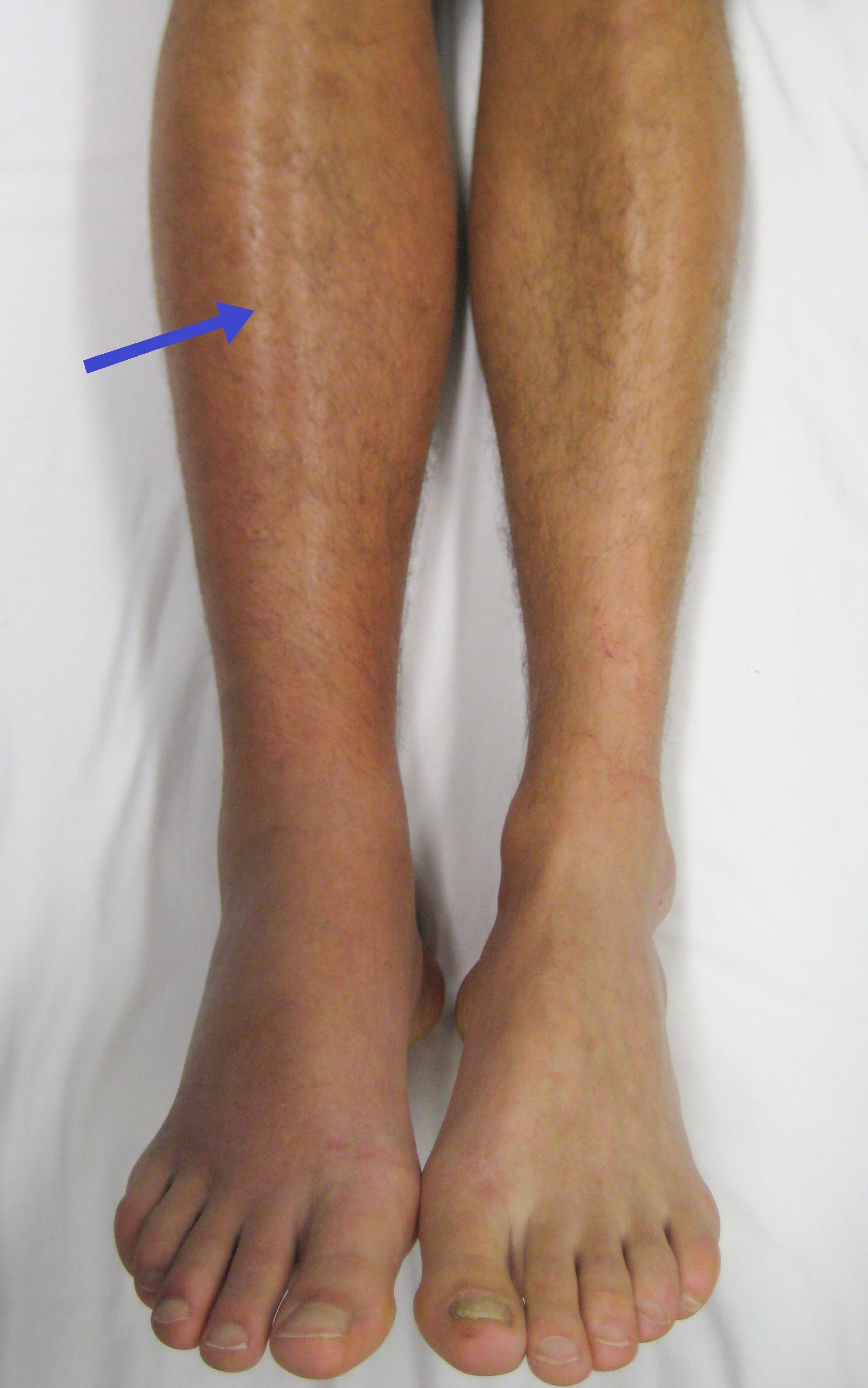
Pulmonary embolism (PE) is a blockage of an pulmonary artery, artery in the lungs by a substance that has moved from elsewhere in the body through the bloodstream (embolism). Symptoms of a PE may include dyspnea, shortness of breath, chest pain particularly upon breathing in, and coughing up blood. Symptoms of a deep vein thrombosis, blood clot in the leg may also be present, such as a erythema, red, warm, swollen, and painful leg. Signs of a PE include low blood oxygen saturation, oxygen levels, tachypnea, rapid breathing, tachycardia, rapid heart rate, and sometimes a mild fever. Severe cases can lead to Syncope (medicine), passing out, shock (circulatory), abnormally low blood pressure, obstructive shock, and cardiac arrest, sudden death. PE usually results from a blood clot in the leg that travels to the lung. The risk of blood clots is increased by advanced age, cancer, prolonged bed rest and immobilization, smoking, stroke, long-haul travel over 4 hours, certain genetics, ... [...More Info...] [...Related Items...] OR: [Wikipedia] [Google] [Baidu] [Amazon] |
Great Veins (other) (two from each lung)
''Great vein'' can be used to refer to any of these veins.
{{disambiguation ...
The term Great veins can refer to either — * The two venae cavae, the superior vena cava, a large diameter, short, vein that carries deoxygenated blood from the upper half of the body to the heart's right atrium, and the inferior vena cava, the large vein that carries deoxygenated blood from the lower half of the body into the right atrium * or to the four pulmonary veins The pulmonary veins are the veins that transfer oxygenated blood from the lungs to the heart. The largest pulmonary veins are the four ''main pulmonary veins'', two from each lung that drain into the left atrium of the heart. The pulmonary ve ... [...More Info...] [...Related Items...] OR: [Wikipedia] [Google] [Baidu] [Amazon] |
Great Arteries
The great arteries are the primary arteries that carry blood away from the heart, which include: * ''Pulmonary artery'': the vessel that carries oxygen-depleted blood from the right ventricle to the lungs. * ''Aorta'': the blood vessel through which oxygenated blood from the left ventricle enters the systemic circulation. Development The great arteries originate from the aortic arches during embryonic development. The aortic arches start as five pairs of symmetrical vessels connecting the heart with the dorsal aorta but then undergo a significant remodelling, in which some of these vessels regress (aortic arches 1 and 2), the 3rd pair of arches contribute to form the common carotids, the right 4th will contribute to the base and central part of the right subclavian artery, while the left 4th will form the central portion of the aortic arch. The 5th pair of vessels only form in some cases without any known contribution to the final structure of the great arteries. The right ... [...More Info...] [...Related Items...] OR: [Wikipedia] [Google] [Baidu] [Amazon] |
CT Pulmonary Angiogram
A CT pulmonary angiogram (CTPA) is a medical diagnostic test that employs Computed tomography angiography, computed tomography (CT) angiography to obtain an image of the pulmonary artery, pulmonary arteries. Its main use is to diagnose pulmonary embolism (PE). It is a preferred choice of imaging in the diagnosis of PE due to its minimally invasive nature for the patient, whose only requirement for the scan is an intravenous line. Modern MDCT (multi-detector CT) scanners are able to deliver images of sufficient resolution within a short time period, such that CTPA has now supplanted previous methods of testing, such as direct pulmonary angiography, as the Gold standard (test), gold standard for diagnosis of pulmonary embolism. The patient receives an intravenous injection of an iodine-containing contrast agent at a high rate using an injector pump. Images are acquired with the maximum intensity of radio-opaque contrast in the pulmonary arteries. This can be done using bolus trackin ... [...More Info...] [...Related Items...] OR: [Wikipedia] [Google] [Baidu] [Amazon] |
Mediastinum
The mediastinum (from ;: mediastina) is the central compartment of the thoracic cavity. Surrounded by loose connective tissue, it is a region that contains vital organs and structures within the thorax, mainly the heart and its vessels, the esophagus, the trachea, the vagus nerve, vagus, phrenic nerve, phrenic and cardiac nerves, the thoracic duct, the thymus and the lymph nodes of the central chest. Anatomy The mediastinum lies within the thorax and is enclosed on the right and left by pulmonary pleurae, pleurae. It is surrounded by the chest wall in front, the lungs to the sides and the Spine (anatomy), spine at the back. It extends from the sternum in front to the vertebral column behind. It contains all the organs of the thorax except the lungs. It is continuous with the loose connective tissue of the neck. The mediastinum can be divided into an upper (or superior) and lower (or inferior) part: * The superior mediastinum starts at the superior thoracic aperture and ends ... [...More Info...] [...Related Items...] OR: [Wikipedia] [Google] [Baidu] [Amazon] |
Root Of The Lung
The root of the lung is a group of structures that emerge at the hilum of each lung, just above the middle of the mediastinal surface and behind the cardiac impression of the lung. It is nearer to the back (posterior border) than the front (anterior border). The root of the lung is connected by the structures that form it to the heart and the trachea. The rib cage is separated from the lung by a two-layered membranous coating, the pleura. The hilum is the large triangular depression where the connection between the parietal pleura (covering the rib cage) and the visceral pleura (covering the lung) is made, and this marks the meeting point between the mediastinum and the pleural cavities. Location The root of the right lung lies behind the superior vena cava and part of the right atrium, and below the azygos vein. That of the left lung passes beneath the aortic arch and in front of the descending aorta; the phrenic nerve, pericardiacophrenic artery and vein, and the ante ... [...More Info...] [...Related Items...] OR: [Wikipedia] [Google] [Baidu] [Amazon] |
Lymph Node
A lymph node, or lymph gland, is a kidney-shaped organ of the lymphatic system and the adaptive immune system. A large number of lymph nodes are linked throughout the body by the lymphatic vessels. They are major sites of lymphocytes that include B and T cells. Lymph nodes are important for the proper functioning of the immune system, acting as filters for foreign particles including cancer cells, but have no detoxification function. In the lymphatic system, a lymph node is a secondary lymphoid organ. A lymph node is enclosed in a fibrous capsule and is made up of an outer cortex and an inner medulla. Lymph nodes become inflamed or enlarged in various diseases, which may range from trivial throat infections to life-threatening cancers. The condition of lymph nodes is very important in cancer staging, which decides the treatment to be used and determines the prognosis. Lymphadenopathy refers to glands that are enlarged or swollen. When inflamed or enlarged, lymph nodes can ... [...More Info...] [...Related Items...] OR: [Wikipedia] [Google] [Baidu] [Amazon] |
Abscess
An abscess is a collection of pus that has built up within the tissue of the body, usually caused by bacterial infection. Signs and symptoms of abscesses include redness, pain, warmth, and swelling. The swelling may feel fluid-filled when pressed. The area of redness often extends beyond the swelling. Carbuncles and boils are types of abscess that often involve hair follicles, with carbuncles being larger. A cyst is related to an abscess, but it contains a material other than pus, and a cyst has a clearly defined wall. Abscesses can also form internally on internal organs and after surgery. They are usually caused by a bacterial infection. Often many different types of bacteria are involved in a single infection. In many areas of the world, the most common bacteria present is ''methicillin-resistant Staphylococcus aureus''. Rarely, parasites can cause abscesses; this is more common in the developing world. Diagnosis of a skin abscess is usually made based on what it looks ... [...More Info...] [...Related Items...] OR: [Wikipedia] [Google] [Baidu] [Amazon] |
Lung Cancer
Lung cancer, also known as lung carcinoma, is a malignant tumor that begins in the lung. Lung cancer is caused by genetic damage to the DNA of cells in the airways, often caused by cigarette smoking or inhaling damaging chemicals. Damaged airway cells gain the ability to multiply unchecked, causing the growth of a tumor. Without treatment, tumors spread throughout the lung, damaging lung function. Eventually lung tumors metastasize, spreading to other parts of the body. Early lung cancer often has no symptoms and can only be detected by medical imaging. As the cancer progresses, most people experience nonspecific respiratory problems: coughing, shortness of breath, or chest pain. Other symptoms depend on the location and size of the tumor. Those suspected of having lung cancer typically undergo a series of imaging tests to determine the location and extent of any tumors. Definitive diagnosis of lung cancer requires a biopsy of the suspected tumor be examined by a patholo ... [...More Info...] [...Related Items...] OR: [Wikipedia] [Google] [Baidu] [Amazon] |


