|
Anatomical Planes
An anatomical plane is a hypothetical plane used to transect the body, in order to describe the location of structures or the direction of movements. In human anatomy and non-human anatomy, four principal planes are used: the median plane, sagittal plane, coronal plane, and transverse plane. * The median plane or midsagittal plane passes through the middle of the body, dividing it into left and right halves. * A para sagittal plane is any plane that runs parallel to the median plane, also dividing the body into left and right sections. * The dorsal plane divides the body into dorsal (towards the backbone) and ventral (towards the belly) parts. In human anatomy coronal plane is preferred, or sometimes the frontal plane, and the description may reference splitting the body into front and back parts, but this phrasing is not as clear for animals with a horizontal spine like quadrupeds or fish. * The transverse plane, also called the axial plane or horizontal plane, is perpe ... [...More Info...] [...Related Items...] OR: [Wikipedia] [Google] [Baidu] |
Human And Goat Anatomical Planes
Humans (''Homo sapiens'') or modern humans are the most common and widespread species of primate, and the last surviving species of the genus ''Homo''. They are Hominidae, great apes characterized by their Prehistory of nakedness and clothing#Evolution of hairlessness, hairlessness, bipedality, bipedalism, and high Human intelligence, intelligence. Humans have large Human brain, brains, enabling more advanced cognitive skills that facilitate successful adaptation to varied environments, development of sophisticated tools, and formation of complex social structures and civilizations. Humans are Sociality, highly social, with individual humans tending to belong to a Level of analysis, multi-layered network of distinct social groups — from families and peer groups to corporations and State (polity), political states. As such, social interactions between humans have established a wide variety of Value theory, values, norm (sociology), social norms, languages, and traditions (co ... [...More Info...] [...Related Items...] OR: [Wikipedia] [Google] [Baidu] |
Interspinous Plane
The interspinous plane (Planum interspinale) is an anatomical transverse plane A transverse plane is a plane that is rotated 90° from two other planes. Anatomy The transverse plane is an anatomical plane that is perpendicular to the sagittal plane and the dorsal plane. It is also called the axial plane or horizonta ... that passes through the anterior superior iliac spines. It separates the lateral lumbar region from the inguinal region and the umbilical region from the pubic region. References {{anatomy-stub Anatomical planes ... [...More Info...] [...Related Items...] OR: [Wikipedia] [Google] [Baidu] |
Superficial Anatomy
Surface anatomy (also called superficial anatomy and visual anatomy) is the study of the external features of the body of an animal.Seeley (2003) chap.1 p.2 In birds, this is termed ''topography''. Surface anatomy deals with anatomical features that can be studied by sight, without dissection. As such, it is a branch of gross anatomy, along with endoscopic and radiological anatomy.Standring (2008) ''Introduction'', ''Anatomical nomenclature'', p.2 Surface anatomy is a descriptive science. In particular, in the case of human surface anatomy, these are the form and proportions of the human body and the surface landmarks which correspond to deeper structures hidden from view, both in static pose and in motion. In addition, the science of surface anatomy includes the theories and systems of body proportions and related artistic canons. The study of surface anatomy is the basis for depicting the human body in classical art. Some pseudo-sciences such as physiognomy, phrenology ... [...More Info...] [...Related Items...] OR: [Wikipedia] [Google] [Baidu] |
Axillary Lines
The axillary lines are the anterior axillary line, midaxillary line and the posterior axillary line. The anterior axillary line is a coronal line on the anterior torso marked by the anterior axillary fold. It's the imaginary line that runs down from the point midway between the middle of the clavicle and the lateral end of the clavicle. The V5 ECG lead is placed on the left anterior axillary line, horizontally even with V4. The midaxillary line is a coronal line on the torso between the anterior and posterior axillary lines. It is a landmark used in thoracentesis, and the V6 electrode of the 10 electrode ECG. The posterior axillary line is a coronal line on the posterior torso marked by the posterior axillary fold. Additional images File:Axillary lines.png, The left side of the thorax with lines labeled See also * List of anatomical lines {{short description, None Anatomical lines, or "reference lines," are theoretical lines drawn through anatomical structures an ... [...More Info...] [...Related Items...] OR: [Wikipedia] [Google] [Baidu] |
Right-hand Rule
In mathematics and physics, the right-hand rule is a Convention (norm), convention and a mnemonic, utilized to define the orientation (vector space), orientation of Cartesian coordinate system, axes in three-dimensional space and to determine the direction of the cross product of two Euclidean vector, vectors, as well as to establish the direction of the force on a Electric current, current-carrying conductor in a magnetic field. The various right- and left-hand rules arise from the fact that the three axes of three-dimensional space have two possible orientations. This can be seen by holding your hands together with palms up and fingers curled. If the curl of the fingers represents a movement from the first or x-axis to the second or y-axis, then the third or z-axis can point along either right thumb or left thumb. History The right-hand rule dates back to the 19th century when it was implemented as a way for identifying the positive direction of coordinate axes in three dime ... [...More Info...] [...Related Items...] OR: [Wikipedia] [Google] [Baidu] |
Cartesian Coordinate System
In geometry, a Cartesian coordinate system (, ) in a plane (geometry), plane is a coordinate system that specifies each point (geometry), point uniquely by a pair of real numbers called ''coordinates'', which are the positive and negative numbers, signed distances to the point from two fixed perpendicular oriented lines, called ''coordinate lines'', ''coordinate axes'' or just ''axes'' (plural of ''axis'') of the system. The point where the axes meet is called the ''Origin (mathematics), origin'' and has as coordinates. The axes direction (geometry), directions represent an orthogonal basis. The combination of origin and basis forms a coordinate frame called the Cartesian frame. Similarly, the position of any point in three-dimensional space can be specified by three ''Cartesian coordinates'', which are the signed distances from the point to three mutually perpendicular planes. More generally, Cartesian coordinates specify the point in an -dimensional Euclidean space for any di ... [...More Info...] [...Related Items...] OR: [Wikipedia] [Google] [Baidu] |
DICOM
Digital Imaging and Communications in Medicine (DICOM) is a technical standard for the digital storage and Medical image sharing, transmission of medical images and related information. It includes a file format definition, which specifies the structure of a DICOM file, as well as a network communication protocol that uses Internet protocol suite, TCP/IP to communicate between systems. The primary purpose of the standard is to facilitate communication between the software and Computer hardware, hardware entities involved in medical imaging, especially those that are created by different manufacturers. Entities that utilize DICOM files include components of Picture archiving and communication system, picture archiving and communication systems (PACS), such as Modality (medical imaging), imaging machines (modalities), Radiological information system, radiological information systems (RIS), Image scanner, scanners, Printer (computing), printers, Server (computing), computing servers, a ... [...More Info...] [...Related Items...] OR: [Wikipedia] [Google] [Baidu] |
Positron Emission Tomography
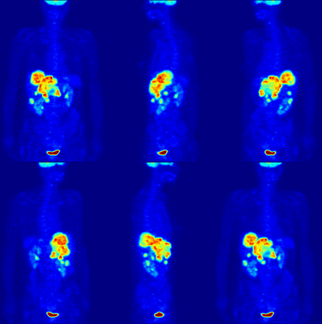
Positron emission tomography (PET) is a functional imaging technique that uses radioactive substances known as radiotracers to visualize and measure changes in metabolic processes, and in other physiological activities including blood flow, regional chemical composition, and absorption. Different tracers are used for various imaging purposes, depending on the target process within the body, such as: * Fluorodeoxyglucose ( 18F">sup>18FDG or FDG) is commonly used to detect cancer; * 18Fodium fluoride">sup>18Fodium fluoride (Na18F) is widely used for detecting bone formation; * Oxygen-15 (15O) is sometimes used to measure blood flow. PET is a common imaging technique, a medical scintillography technique used in nuclear medicine. A radiopharmaceutical—a radioisotope attached to a drug—is injected into the body as a tracer. When the radiopharmaceutical undergoes beta plus decay, a positron is emitted, and when the positron interacts with an ordinary electron, the tw ... [...More Info...] [...Related Items...] OR: [Wikipedia] [Google] [Baidu] |
Magnetic Resonance Imaging
Magnetic resonance imaging (MRI) is a medical imaging technique used in radiology to generate pictures of the anatomy and the physiological processes inside the body. MRI scanners use strong magnetic fields, magnetic field gradients, and radio waves to form images of the organs in the body. MRI does not involve X-rays or the use of ionizing radiation, which distinguishes it from computed tomography (CT) and positron emission tomography (PET) scans. MRI is a medical application of nuclear magnetic resonance (NMR) which can also be used for imaging in other NMR applications, such as NMR spectroscopy. MRI is widely used in hospitals and clinics for medical diagnosis, staging and follow-up of disease. Compared to CT, MRI provides better contrast in images of soft tissues, e.g. in the brain or abdomen. However, it may be perceived as less comfortable by patients, due to the usually longer and louder measurements with the subject in a long, confining tube, although ... [...More Info...] [...Related Items...] OR: [Wikipedia] [Google] [Baidu] |
Computed Axial Tomography
A computed tomography scan (CT scan), formerly called computed axial tomography scan (CAT scan), is a medical imaging technique used to obtain detailed internal images of the body. The personnel that perform CT scans are called radiographers or radiology technologists. CT scanners use a rotating X-ray tube and a row of detectors placed in a gantry to measure X-ray attenuations by different tissues inside the body. The multiple X-ray measurements taken from different angles are then processed on a computer using tomographic reconstruction algorithms to produce tomographic (cross-sectional) images (virtual "slices") of a body. CT scans can be used in patients with metallic implants or pacemakers, for whom magnetic resonance imaging (MRI) is contraindicated. Since its development in the 1970s, CT scanning has proven to be a versatile imaging technique. While CT is most prominently used in medical diagnosis, it can also be used to form images of non-living objects. The 1979 Nobel ... [...More Info...] [...Related Items...] OR: [Wikipedia] [Google] [Baidu] |
Medical Ultrasonography
Medical ultrasound includes Medical diagnosis, diagnostic techniques (mainly medical imaging, imaging) using ultrasound, as well as therapeutic ultrasound, therapeutic applications of ultrasound. In diagnosis, it is used to create an image of internal body structures such as tendons, muscles, joints, blood vessels, and internal organs, to measure some characteristics (e.g., distances and velocities) or to generate an informative audible sound. The usage of ultrasound to produce visual images for medicine is called medical ultrasonography or simply sonography, or echography. The practice of examining pregnant women using ultrasound is called obstetric ultrasonography, and was an early development of clinical ultrasonography. The machine used is called an ultrasound machine, a sonograph or an echograph. The visual image formed using this technique is called an ultrasonogram, a sonogram or an echogram. Ultrasound is composed of sound waves with frequency, frequencies greater than ... [...More Info...] [...Related Items...] OR: [Wikipedia] [Google] [Baidu] |
Medical Imaging
Medical imaging is the technique and process of imaging the interior of a body for clinical analysis and medical intervention, as well as visual representation of the function of some organs or tissues (physiology). Medical imaging seeks to reveal internal structures hidden by the skin and bones, as well as to diagnose and treat disease. Medical imaging also establishes a database of normal anatomy and physiology to make it possible to identify abnormalities. Although imaging of removed organ (anatomy), organs and Tissue (biology), tissues can be performed for medical reasons, such procedures are usually considered part of pathology instead of medical imaging. Measurement and recording techniques that are not primarily designed to produce images, such as electroencephalography (EEG), magnetoencephalography (MEG), electrocardiography (ECG), and others, represent other technologies that produce data susceptible to representation as a parameter graph versus time or maps that contain ... [...More Info...] [...Related Items...] OR: [Wikipedia] [Google] [Baidu] |


