|
Brodmann Area 12
Brodmann area 12 is a subdivision of the cerebral cortex of the guenon defined on the basis of cytoarchitecture. It occupies the most rostral portion of the frontal lobe. Brodmann-1909 did not regard it as homologous, either topographically or cytoarchitecturally, to rostral area 12 of the human. Distinctive features (Brodmann-1905): a quite distinct internal granular layer (IV) separates slender pyramidal cells of the external pyramidal layer (III) and the internal pyramidal layer (V); the multiform layer (VI) is expanded, contains widely dispersed spindle cells and merges gradually with the underlying cortical white matter; all cells, including the pyramidal cells of the external and internal pyramidal layers are inordinately small; the internal pyramidal layer (V) also contains spindle cells in groups of two to five located close to its border with the internal granular layer (IV). It is indirectly connected to the globus pallidus as well as the substantia nigra, due to effer ... [...More Info...] [...Related Items...] OR: [Wikipedia] [Google] [Baidu] |
Cerebral Cortex
The cerebral cortex, also known as the cerebral mantle, is the outer layer of neural tissue of the cerebrum of the brain in humans and other mammals. The cerebral cortex mostly consists of the six-layered neocortex, with just 10% consisting of allocortex. It is separated into two cortices, by the longitudinal fissure that divides the cerebrum into the left and right cerebral hemispheres. The two hemispheres are joined beneath the cortex by the corpus callosum. The cerebral cortex is the largest site of neural integration in the central nervous system. It plays a key role in attention, perception, awareness, thought, memory, language, and consciousness. The cerebral cortex is part of the brain responsible for cognition. In most mammals, apart from small mammals that have small brains, the cerebral cortex is folded, providing a greater surface area in the confined volume of the cranium. Apart from minimising brain and cranial volume, cortical folding is crucial for the brain ... [...More Info...] [...Related Items...] OR: [Wikipedia] [Google] [Baidu] |
Guenon
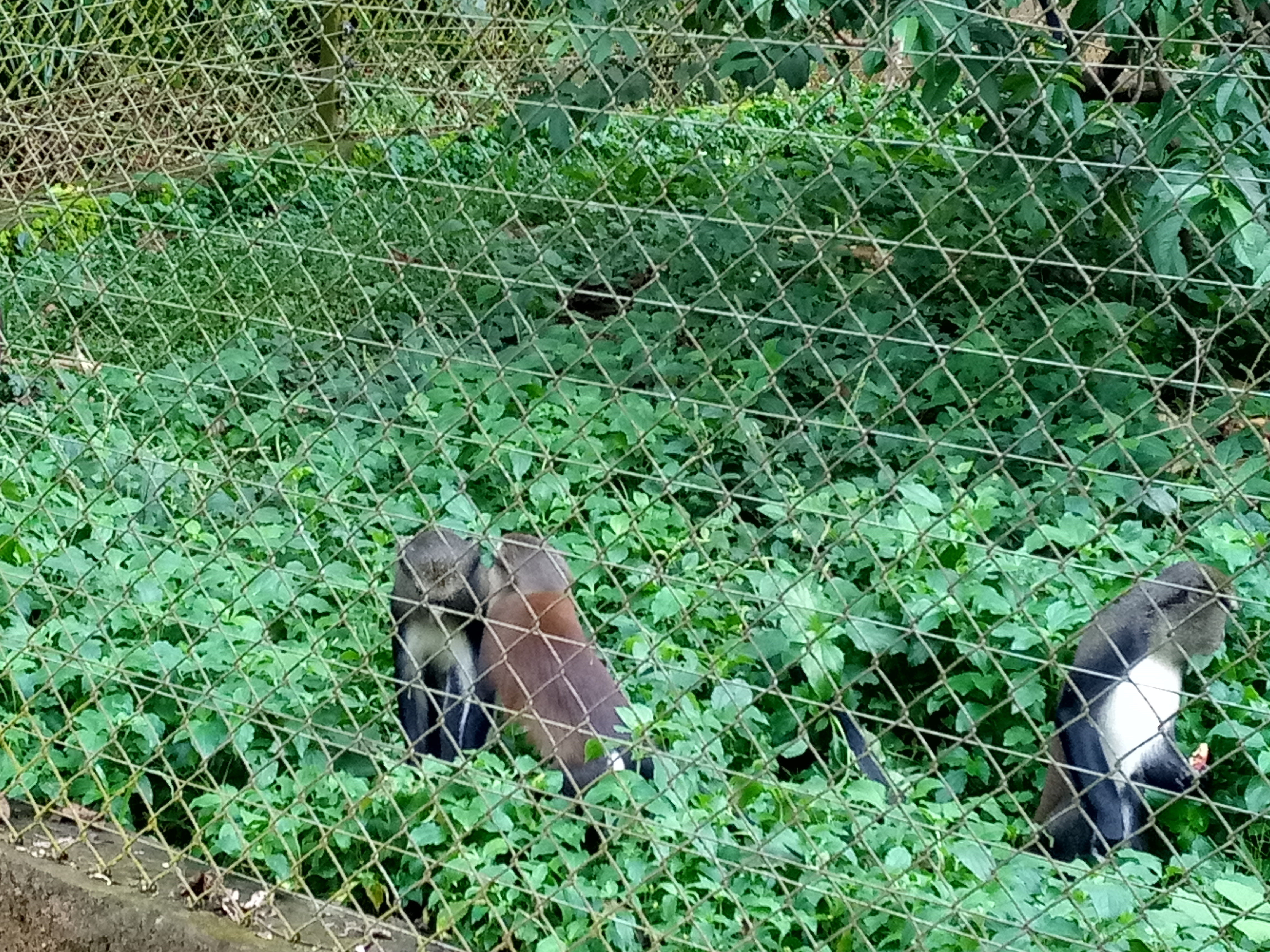
The guenons (, ) are Old World monkeys of the genus ''Cercopithecus'' (). Not all members of this genus have the word "guenon" in their common names; also, because of changes in scientific classification, some monkeys in other genera may have common names that include the word "guenon". Nonetheless, the use of the term guenon for monkeys of this genus is widely accepted. All members of the genus are endemic to sub-Saharan Africa, and most are forest monkeys. Many of the species are quite local in their ranges, and some have even more local subspecies. Many are threatened or endangered because of habitat loss. The species currently placed in the genus ''Chlorocebus'', such as vervet monkeys and green monkeys, were formerly considered as a single species in this genus, ''Cercopithecus aethiops''. In the English language, the word "guenon" is apparently of French origin. In French, ''guenon'' was the common name for all species and individuals, both males and females, from the gen ... [...More Info...] [...Related Items...] OR: [Wikipedia] [Google] [Baidu] |
Cytoarchitecture
Cytoarchitecture (Greek '' κύτος''= "cell" + '' ἀρχιτεκτονική''= "architecture"), also known as cytoarchitectonics, is the study of the cellular composition of the central nervous system's tissues under the microscope. Cytoarchitectonics is one of the ways to parse the brain, by obtaining sections of the brain using a microtome and staining them with chemical agents which reveal where different neurons are located. The study of the parcellation of ''nerve fibers'' (primarily axons) into layers forms the subject of myeloarchitectonics ( History of the cerebral cytoarchitecture Defining cerebral cytoarchitecture began with the advent of —the science of slicing a ...[...More Info...] [...Related Items...] OR: [Wikipedia] [Google] [Baidu] |
Frontal Lobe
The frontal lobe is the largest of the four major lobes of the brain in mammals, and is located at the front of each cerebral hemisphere (in front of the parietal lobe and the temporal lobe). It is parted from the parietal lobe by a groove between tissues called the central sulcus and from the temporal lobe by a deeper groove called the lateral sulcus (Sylvian fissure). The most anterior rounded part of the frontal lobe (though not well-defined) is known as the frontal pole, one of the three poles of the cerebrum. The frontal lobe is covered by the frontal cortex. The frontal cortex includes the premotor cortex, and the primary motor cortex – parts of the motor cortex. The front part of the frontal cortex is covered by the prefrontal cortex. There are four principal gyri in the frontal lobe. The precentral gyrus is directly anterior to the central sulcus, running parallel to it and contains the primary motor cortex, which controls voluntary movements of specific body parts ... [...More Info...] [...Related Items...] OR: [Wikipedia] [Google] [Baidu] |
Homology (biology)
In biology, homology is similarity due to shared ancestry between a pair of structures or genes in different taxa. A common example of homologous structures is the forelimbs of vertebrates, where the wings of bats and birds, the arms of primates, the front flippers of whales and the forelegs of four-legged vertebrates like dogs and crocodiles are all derived from the same ancestral tetrapod structure. Evolutionary biology explains homologous structures adapted to different purposes as the result of descent with modification from a common ancestor. The term was first applied to biology in a non-evolutionary context by the anatomist Richard Owen in 1843. Homology was later explained by Charles Darwin's theory of evolution in 1859, but had been observed before this, from Aristotle onwards, and it was explicitly analysed by Pierre Belon in 1555. In developmental biology, organs that developed in the embryo in the same manner and from similar origins, such as from matching p ... [...More Info...] [...Related Items...] OR: [Wikipedia] [Google] [Baidu] |
Topographical
Topography is the study of the forms and features of land surfaces. The topography of an area may refer to the land forms and features themselves, or a description or depiction in maps. Topography is a field of geoscience and planetary science and is concerned with local detail in general, including not only relief, but also natural, artificial, and cultural features such as roads, land boundaries, and buildings. In the United States, topography often means specifically ''relief'', even though the USGS topographic maps record not just elevation contours, but also roads, populated places, structures, land boundaries, and so on. Topography in a narrow sense involves the recording of relief or terrain, the three-dimensional quality of the surface, and the identification of specific landforms; this is also known as geomorphometry. In modern usage, this involves generation of elevation data in digital form (DEM). It is often considered to include the graphic representation of t ... [...More Info...] [...Related Items...] OR: [Wikipedia] [Google] [Baidu] |
Rostral Area 12
Brodmann area 12 is a subdivision of the cerebral cortex of the guenon defined on the basis of cytoarchitecture. It occupies the most rostral portion of the frontal lobe. Brodmann-1909 did not regard it as homologous, either topographically or cytoarchitecturally, to rostral area 12 of the human. Distinctive features (Brodmann-1905): a quite distinct internal granular layer (IV) separates slender pyramidal cells of the external pyramidal layer (III) and the internal pyramidal layer (V); the multiform layer (VI) is expanded, contains widely dispersed spindle cells and merges gradually with the underlying cortical white matter; all cells, including the pyramidal cells of the external and internal pyramidal layers are inordinately small; the internal pyramidal layer (V) also contains spindle cells in groups of two to five located close to its border with the internal granular layer (IV). It is indirectly connected to the globus pallidus as well as the substantia nigra, due to effer ... [...More Info...] [...Related Items...] OR: [Wikipedia] [Google] [Baidu] |
Granular Layer (cerebral Cortex)
The internal granular layer of the cortex, also commonly referred to as the granular layer of the cortex, is the layer IV in the subdivision of the mammalian cortex into 6 layers. The adjective internal is used in opposition to the external granular layer of the cortex, the term granular refers to the granule cells found here. This layer receives the afferent connections from the thalamus and from other cortical regions and sends connections to the other layers. The line of Gennari The line of Gennari (also called the "band" or "stria" of Gennari) is a band of myelinated axons that runs parallel to the surface of the cerebral cortex on the banks of the calcarine fissure in the occipital lobe. This formation is visible to the ... (occipital stripe) is also present in this layer. Cerebral cortex {{neuroanatomy-stub ... [...More Info...] [...Related Items...] OR: [Wikipedia] [Google] [Baidu] |
Pyramidal Cell
Pyramidal cells, or pyramidal neurons, are a type of multipolar neuron found in areas of the brain including the cerebral cortex, the hippocampus, and the amygdala. Pyramidal neurons are the primary excitation units of the mammalian prefrontal cortex and the corticospinal tract. Pyramidal neurons are also one of two cell types where the characteristic sign, Negri bodies, are found in post-mortem rabies infection. Pyramidal neurons were first discovered and studied by Santiago Ramón y Cajal. Since then, studies on pyramidal neurons have focused on topics ranging from neuroplasticity to cognition. Structure File:GFPneuron.png, Pyramidal neuron visualized by green fluorescent protein (gfp) File:Hippocampal-pyramidal-cell.png, A hippocampal pyramidal cell One of the main structural features of the pyramidal neuron is the conic shaped soma, or cell body, after which the neuron is named. Other key structural features of the pyramidal cell are a single axon, a large apical dendrite, ... [...More Info...] [...Related Items...] OR: [Wikipedia] [Google] [Baidu] |
Pyramidal Layer
A pyramid (from el, πυραμίς ') is a structure whose outer surfaces are triangular and converge to a single step at the top, making the shape roughly a pyramid in the geometric sense. The base of a pyramid can be trilateral, quadrilateral, or of any polygon shape. As such, a pyramid has at least three outer triangular surfaces (at least four faces including the base). The square pyramid, with a square base and four triangular outer surfaces, is a common version. A pyramid's design, with the majority of the weight closer to the ground and with the pyramidion at the apex, means that less material higher up on the pyramid will be pushing down from above. This distribution of weight allowed early civilizations to create stable monumental structures. Civilizations in many parts of the world have built pyramids. The largest pyramid by volume is the Great Pyramid of Cholula, in the Mexican state of Puebla. For thousands of years, the largest structures on Earth were pyrami ... [...More Info...] [...Related Items...] OR: [Wikipedia] [Google] [Baidu] |
Multiform Layer
The cerebral cortex, also known as the cerebral mantle, is the outer layer of neural tissue of the cerebrum of the brain in humans and other mammals. The cerebral cortex mostly consists of the six-layered neocortex, with just 10% consisting of allocortex. It is separated into two cortices, by the longitudinal fissure that divides the cerebrum into the left and right cerebral hemispheres. The two hemispheres are joined beneath the cortex by the corpus callosum. The cerebral cortex is the largest site of neural integration in the central nervous system. It plays a key role in attention, perception, awareness, thought, memory, language, and consciousness. The cerebral cortex is part of the brain responsible for cognition. In most mammals, apart from small mammals that have small brains, the cerebral cortex is folded, providing a greater surface area in the confined volume of the cranium. Apart from minimising brain and cranial volume, cortical folding is crucial for the brai ... [...More Info...] [...Related Items...] OR: [Wikipedia] [Google] [Baidu] |
Spindle Cell
Von Economo neurons (VENs), also called spindle neurons, are a specific class of mammalian cortical neurons characterized by a large spindle-shaped soma (or body) gradually tapering into a single apical axon (the ramification that ''transmits'' signals) in one direction, with only a single dendrite (the ramification that ''receives'' signals) facing opposite. Other cortical neurons tend to have many dendrites, and the bipolar-shaped morphology of von Economo neurons is unique here. Von Economo neurons are found in two very restricted regions in the brains of hominids (humans and other great apes): the anterior cingulate cortex (ACC) and the fronto-insular cortex (FI) (which each make up the salience network). In 2008, they were also found in the dorsolateral prefrontal cortex of humans. Von Economo neurons are also found in the brains of a number of cetaceans, African and Asian elephants, and to a lesser extent in macaque monkeys and raccoons. The appearance of von Econom ... [...More Info...] [...Related Items...] OR: [Wikipedia] [Google] [Baidu] |


