|
Brain Aging
Aging is a major risk factor for most common neurodegenerative diseases, including mild cognitive impairment, dementias including Alzheimer's disease, cerebrovascular disease, Parkinson's disease, and Lou Gehrig's disease. While much research has focused on diseases of aging, there are few informative studies on the molecular biology of the aging brain (usually spelled ageing brain in British English) in the absence of neurodegenerative disease or the neuropsychological profile of healthy older adults. However, research suggests that the aging process is associated with several structural, chemical, and functional changes in the brain as well as a host of neurocognitive changes. Recent reports in model organisms suggest that as organisms age, there are distinct changes in the expression of genes at the single neuron level. This page is devoted to reviewing the changes associated with healthy aging. Structural changes Aging entails many physical, biological, chemical, and psychologi ... [...More Info...] [...Related Items...] OR: [Wikipedia] [Google] [Baidu] |
Aging
Ageing ( BE) or aging ( AE) is the process of becoming older. The term refers mainly to humans, many other animals, and fungi, whereas for example, bacteria, perennial plants and some simple animals are potentially biologically immortal. In a broader sense, ageing can refer to single cells within an organism which have ceased dividing, or to the population of a species. In humans, ageing represents the accumulation of changes in a human being over time and can encompass physical, psychological, and social changes. Reaction time, for example, may slow with age, while memories and general knowledge typically increase. Ageing increases the risk of human diseases such as cancer, Alzheimer's disease, diabetes, cardiovascular disease, stroke and many more. Of the roughly 150,000 people who die each day across the globe, about two-thirds die from age-related causes. Current ageing theories are assigned to the damage concept, whereby the accumulation of damage (such as DNA ox ... [...More Info...] [...Related Items...] OR: [Wikipedia] [Google] [Baidu] |
Cerebral Cortex
The cerebral cortex, also known as the cerebral mantle, is the outer layer of neural tissue of the cerebrum of the brain in humans and other mammals. The cerebral cortex mostly consists of the six-layered neocortex, with just 10% consisting of allocortex. It is separated into two cortices, by the longitudinal fissure that divides the cerebrum into the left and right cerebral hemispheres. The two hemispheres are joined beneath the cortex by the corpus callosum. The cerebral cortex is the largest site of neural integration in the central nervous system. It plays a key role in attention, perception, awareness, thought, memory, language, and consciousness. The cerebral cortex is part of the brain responsible for cognition. In most mammals, apart from small mammals that have small brains, the cerebral cortex is folded, providing a greater surface area in the confined volume of the cranium. Apart from minimising brain and cranial volume, cortical folding is crucial for the brain ... [...More Info...] [...Related Items...] OR: [Wikipedia] [Google] [Baidu] |
Dendritic Spines
A dendritic spine (or spine) is a small membranous protrusion from a neuron's dendrite that typically receives input from a single axon at the synapse. Dendritic spines serve as a storage site for synaptic strength and help transmit electrical signals to the neuron's cell body. Most spines have a bulbous head (the spine head), and a thin neck that connects the head of the spine to the shaft of the dendrite. The dendrites of a single neuron can contain hundreds to thousands of spines. In addition to spines providing an anatomical substrate for memory storage and synaptic transmission, they may also serve to increase the number of possible contacts between neurons. It has also been suggested that changes in the activity of neurons have a positive effect on spine morphology. Structure Dendritic spines are small with spine head volumes ranging 0.01 μm3 to 0.8 μm3. Spines with strong synaptic contacts typically have a large spine head, which connects to the dendrite via a ... [...More Info...] [...Related Items...] OR: [Wikipedia] [Google] [Baidu] |
NeuroImage
''NeuroImage'' is a peer-reviewed scientific journal covering research on neuroimaging, including functional neuroimaging and functional human brain mapping. The current Editor in Chief is Michael Breakspear. Abstracts from the annual meeting of the Organization for Human Brain Mapping have been published as supplements to the journal. Members of the Organization for Human Brain Mapping are eligible for reduced subscription rates. In 2012, Elsevier launched an online-only, open access sister journal to ''NeuroImage'', entitled '' NeuroImage: Clinical''. Among prolific authors publishing numerous articles in the journal are Karl J. Friston, Arthur W. Toga, Paul M. Thompson and Karl Zilles. Related journals are Human Brain Mapping, Frontiers in Human Neuroscience, Cerebral Cortex, Magnetic Resonance in Medicine, Journal of Neuroscience and Journal of Cognitive Neuroscience. Abstracting and indexing The journal is abstracted and indexed in Scopus, Science Citation Index, Curre ... [...More Info...] [...Related Items...] OR: [Wikipedia] [Google] [Baidu] |
Sulcus (neuroanatomy)
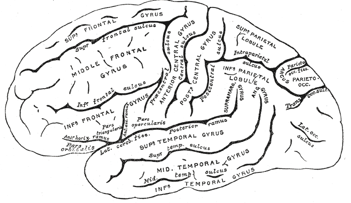
In neuroanatomy, a sulcus (Latin: "furrow", pl. ''sulci'') is a depression or groove in the cerebral cortex. It surrounds a gyrus (pl. gyri), creating the characteristic folded appearance of the brain in humans and other mammals. The larger sulci are usually called fissures. Structure Sulci, the grooves, and gyri, the folds or ridges, make up the folded surface of the cerebral cortex. Larger or deeper sulci are termed fissures, and in many cases the two terms are interchangeable. The folded cortex creates a larger surface area for the brain in humans and other mammals. When looking at the human brain, two-thirds of the surface are hidden in the grooves. The sulci and fissures are both grooves in the cortex, but they are differentiated by size. A sulcus is a shallower groove that surrounds a gyrus. A fissure is a large furrow that divides the brain into lobes and also into the two hemispheres as the longitudinal fissure. Importance of expanded surface area As the surfac ... [...More Info...] [...Related Items...] OR: [Wikipedia] [Google] [Baidu] |
Calcarine Sulcus
The calcarine sulcus (or calcarine fissure) is an anatomical landmark located at the caudal end of the medial surface of the brain of humans and other primates. Its name comes from the Latin "calcar" meaning "spur". It is very deep, and known as a complete sulcus. Structure The calcarine sulcus begins near the occipital pole in two converging rami. It runs forward to a point a little below the splenium of the corpus callosum. Here, it is joined at an acute angle by the medial part of the parieto-occipital sulcus. The anterior part of this sulcus gives rise to the prominence of the calcar avis in the posterior cornu of the lateral ventricle. The cuneus is above the calcarine sulcus, while the lingual gyrus is below it. Development In humans, the calcarine sulcus usually becomes visible between 20 weeks and 28 weeks of gestation. Function The calcarine sulcus is associated with visual cortex. It is where the primary visual cortex (V1) is concentrated. The central visua ... [...More Info...] [...Related Items...] OR: [Wikipedia] [Google] [Baidu] |
Occipital Cortex
The occipital lobe is one of the four major lobes of the cerebral cortex in the brain of mammals. The name derives from its position at the back of the head, from the Latin ''ob'', "behind", and ''caput'', "head". The occipital lobe is the visual processing center of the mammalian brain containing most of the anatomical region of the visual cortex. The primary visual cortex is Brodmann area 17, commonly called V1 (visual one). Human V1 is located on the medial side of the occipital lobe within the calcarine sulcus; the full extent of V1 often continues onto the occipital pole. V1 is often also called striate cortex because it can be identified by a large stripe of myelin, the Stria of Gennari. Visually driven regions outside V1 are called extrastriate cortex. There are many extrastriate regions, and these are specialized for different visual tasks, such as visuospatial processing, color differentiation, and motion perception. Bilateral lesions of the occipital lobe can lead ... [...More Info...] [...Related Items...] OR: [Wikipedia] [Google] [Baidu] |
Cingulate Gyrus
The cingulate cortex is a part of the brain situated in the medial aspect of the cerebral cortex. The cingulate cortex includes the entire cingulate gyrus, which lies immediately above the corpus callosum, and the continuation of this in the cingulate sulcus. The cingulate cortex is usually considered part of the limbic lobe. It receives inputs from the thalamus and the neocortex, and projects to the entorhinal cortex via the cingulum. It is an integral part of the limbic system, which is involved with emotion formation and processing, learning, and memory. The combination of these three functions makes the cingulate gyrus highly influential in linking motivational outcomes to behavior (e.g. a certain action induced a positive emotional response, which results in learning). This role makes the cingulate cortex highly important in disorders such as depression and schizophrenia. It also plays a role in executive function and respiratory control. Etymology The term ''cingul ... [...More Info...] [...Related Items...] OR: [Wikipedia] [Google] [Baidu] |
Parietal Lobes
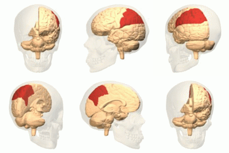
The parietal lobe is one of the four major lobes of the cerebral cortex in the brain of mammals. The parietal lobe is positioned above the temporal lobe and behind the frontal lobe and central sulcus. The parietal lobe integrates sensory information among various modalities, including spatial sense and navigation (proprioception), the main sensory receptive area for the sense of touch in the somatosensory cortex which is just posterior to the central sulcus in the postcentral gyrus, and the dorsal stream of the visual system. The major sensory inputs from the skin (touch, temperature, and pain receptors), relay through the thalamus to the parietal lobe. Several areas of the parietal lobe are important in language processing. The somatosensory cortex can be illustrated as a distorted figure – the cortical homunculus (Latin: "little man") in which the body parts are rendered according to how much of the somatosensory cortex is devoted to them. The superior parietal lobule and in ... [...More Info...] [...Related Items...] OR: [Wikipedia] [Google] [Baidu] |
Insular Cortex
The insular cortex (also insula and insular lobe) is a portion of the cerebral cortex folded deep within the lateral sulcus (the fissure separating the temporal lobe from the parietal and frontal lobes) within each hemisphere of the mammalian brain. The insulae are believed to be involved in consciousness and play a role in diverse functions usually linked to emotion or the regulation of the body's homeostasis. These functions include compassion, empathy, taste, perception, motor control, self-awareness, cognitive functioning, interpersonal experience, and awareness of homeostatic emotions such as hunger, pain and fatigue. In relation to these, it is involved in psychopathology. The insular cortex is divided into two parts: the anterior insula and the posterior insula in which more than a dozen field areas have been identified. The cortical area overlying the insula toward the lateral surface of the brain is the operculum (meaning ''lid''). The opercula are formed from parts o ... [...More Info...] [...Related Items...] OR: [Wikipedia] [Google] [Baidu] |
Voxel-based Morphometry
Voxel-based morphometry is a computational approach to neuroanatomy that measures differences in local concentrations of brain tissue, through a voxel-wise comparison of multiple brain images. In traditional morphometry, volume of the whole brain or its subparts is measured by drawing regions of interest (ROIs) on images from brain scanning and calculating the volume enclosed. However, this is time consuming and can only provide measures of rather large areas. Smaller differences in volume may be overlooked. The value of VBM is that it allows for comprehensive measurement of differences, not just in specific structures, but throughout the entire brain. VBM registers every brain to a template, which gets rid of most of the large differences in brain anatomy among people. Then the brain images are smoothed so that each voxel represents the average of itself and its neighbors. Finally, the image volume is compared across brains at every voxel. However, VBM can be sensitive to vari ... [...More Info...] [...Related Items...] OR: [Wikipedia] [Google] [Baidu] |
Biochemical
Biochemistry or biological chemistry is the study of chemical processes within and relating to living organisms. A sub-discipline of both chemistry and biology, biochemistry may be divided into three fields: structural biology, enzymology and metabolism. Over the last decades of the 20th century, biochemistry has become successful at explaining living processes through these three disciplines. Almost all areas of the life sciences are being uncovered and developed through biochemical methodology and research. Voet (2005), p. 3. Biochemistry focuses on understanding the chemical basis which allows biological molecules to give rise to the processes that occur within living cells and between cells,Karp (2009), p. 2. in turn relating greatly to the understanding of tissues and organs, as well as organism structure and function.Miller (2012). p. 62. Biochemistry is closely related to molecular biology, which is the study of the molecular mechanisms of biological phenomena.A ... [...More Info...] [...Related Items...] OR: [Wikipedia] [Google] [Baidu] |


