|
Brodmann's Area 9
Brodmann area 9, or BA9, refers to a cytoarchitecturally defined portion of the frontal cortex in the brain of humans and other primates. Its cytoarchitecture is referred to as granular due to the concentration of granule cells in layer IV. It contributes to the dorsolateral and medial prefrontal cortex. Functions The area is involved in short term memory, evaluating recency, overriding automatic responses, verbal fluency, error detection, auditory verbal attention, inferring the intention of others, inferring deduction from spatial imagery, inductive reasoning, attributing intention, sustained attention involved in counting a series of auditory stimuli, and displays lower levels of energy consumption in individuals suffering from bipolar disorder. The area found on the left hemisphere is at least partially responsible for working memory, empathy, idioms, processing pleasant and unpleasant emotional scenes, self criticisms and attention to negative emotions. On the right hemis ... [...More Info...] [...Related Items...] OR: [Wikipedia] [Google] [Baidu] |
Cytoarchitecture
Cytoarchitecture (from Greek κύτος 'cell' and ἀρχιτεκτονική 'architecture'), also known as cytoarchitectonics, is the study of the cellular composition of the central nervous system's tissues under the microscope. Cytoarchitectonics is one of the ways to parse the brain, by obtaining sections of the brain using a microtome and staining them with chemical agents which reveal where different neurons are located. The study of the parcellation of ''nerve fibers'' (primarily axons) into layers forms the subject of myeloarchitectonics (from Greek μυελός 'marrow' and ἀρχιτεκτονική 'architecture'), an approach complementary to cytoarchitectonics. History of the cerebral cytoarchitecture Defining cerebral cytoarchitecture began with the advent of histology—the science of slicing and staining brain slices for examination. It is credited to the Viennese psychiatrist Theodor Meynert (1833–1892), who in 1867 noticed regional variations in the ... [...More Info...] [...Related Items...] OR: [Wikipedia] [Google] [Baidu] |
Frontopolar Area 10
Brodmann area 10 (BA10, frontopolar prefrontal cortex, rostrolateral prefrontal cortex, or anterior prefrontal cortex) is the anterior-most portion of the prefrontal cortex in the human brain. BA10 was originally defined broadly in terms of its cytoarchitectonic traits as they were observed in the brains of cadavers, but because modern functional imaging cannot precisely identify these boundaries, the terms anterior prefrontal cortex, rostral prefrontal cortex and frontopolar prefrontal cortex are used to refer to the area in the most anterior part of the frontal cortex that approximately covers BA10—simply to emphasize the fact that BA10 does not include ''all'' parts of the prefrontal cortex. BA10 is the largest cytoarchitectonic area in the human brain. It has been described as "one of the least well understood regions of the human brain". Present research suggests that it is involved in strategic processes in memory recall and various executive functions. During human evolu ... [...More Info...] [...Related Items...] OR: [Wikipedia] [Google] [Baidu] |
List Of Regions In The Human Brain
The human brain anatomical regions are ordered following standard neuroanatomy hierarchies. Functional, connective, and developmental regions are listed in parentheses where appropriate. Hindbrain (rhombencephalon) Myelencephalon * Medulla oblongata ** Medullary pyramids ** Arcuate nucleus ** Olivary body *** Inferior olivary nucleus ** Rostral ventrolateral medulla ** Caudal ventrolateral medulla ** Solitary nucleus (Nucleus of the solitary tract) **Respiratory center- Respiratory groups *** Dorsal respiratory group *** Ventral respiratory group or Apneustic centre **** Pre-Bötzinger complex **** Botzinger complex **** Retrotrapezoid nucleus **** Nucleus retrofacialis **** Nucleus retroambiguus **** Nucleus para-ambiguus ** Paramedian reticular nucleus ** Gigantocellular reticular nucleus ** Parafacial zone ** Cuneate nucleus ** Gracile nucleus ** Perihypoglossal nuclei *** Intercalated nucleus *** Prepositus nucleus *** Sublingual nucleus ** Area postrema **Medul ... [...More Info...] [...Related Items...] OR: [Wikipedia] [Google] [Baidu] |
Dorsomedial Prefrontal Cortex
The dorsomedial prefrontal cortex (dmPFC or DMPFC is a section of the prefrontal cortex in some species' brain anatomy. It includes portions of Brodmann areas BA8, BA9, BA10, BA24 and BA32, although some authors identify it specifically with BA8 and BA9. Some notable sub-components include the dorsal anterior cingulate cortex (BA24 and BA32), the prelimbic cortex, and the infralimbic cortex. Functions Evidence shows that the dmPFC plays several roles in humans. The dmPFC is identified to play roles in processing a sense of self, integrating social impressions, theory of mind, morality judgments, empathy, decision making, altruism, fear and anxiety information processing, and top-down motor cortex inhibition. The dmPFC also modulates or regulates emotional responses and heart rate in situations of fear or stress and plays a role in long-term memory . Some argue that the dmPFC is made up of several smaller subregions that are more task-specific. The dmPFC is attribu ... [...More Info...] [...Related Items...] OR: [Wikipedia] [Google] [Baidu] |
Brodmann Area
A Brodmann area is a region of the cerebral cortex, in the human or other primate brain, defined by its cytoarchitecture, or histological structure and organization of cells. The concept was first introduced by the German anatomist Korbinian Brodmann in the early 20th century. Brodmann mapped the human brain based on the varied cellular structure across the cortex and identified 52 distinct regions, which he numbered 1 to 52. These regions, or Brodmann areas, correspond with diverse functions including sensation, motor control, and cognition. History Brodmann areas were originally defined and numbered by the German anatomist Korbinian Brodmann based on the cytoarchitectural organization of neurons he observed in the cerebral cortex using the Nissl method of cell staining. Brodmann published his maps of cortical areas in humans, monkeys, and other species in 1909, along with many other findings and observations regarding the general cell types and laminar organization of t ... [...More Info...] [...Related Items...] OR: [Wikipedia] [Google] [Baidu] |
Granule Cell
The name granule cell has been used for a number of different types of neurons whose only common feature is that they all have very small cell bodies. Granule cells are found within the granular layer of the cerebellum, the dentate gyrus of the hippocampus, the superficial layer of the dorsal cochlear nucleus, the olfactory bulb, and the cerebral cortex. Cerebellar granule cells account for the majority of neurons in the human brain. These granule cells receive excitatory input from mossy fibers originating from pontine nuclei. Cerebellar granule cells project up through the Purkinje layer into the molecular layer where they branch out into parallel fibers that spread through Purkinje cell dendritic arbors. These parallel fibers form thousands of excitatory granule-cell–Purkinje-cell synapses onto the intermediate and distal dendrites of Purkinje cells using glutamate as a neurotransmitter. Layer 4 granule cells of the cerebral cortex receive inputs from the thala ... [...More Info...] [...Related Items...] OR: [Wikipedia] [Google] [Baidu] |
Granular Layer (cerebral Cortex)
The internal granular layer of the cerebral cortex, also commonly referred to as the granular layer of the cortex, is the layer IV in the subdivision of the mammalian cerebral cortex into 6 layers. The adjective internal is used in opposition to the external granular layer of the cortex, the term granular refers to the granule cells found here. This layer receives the afferent connections from the thalamus The thalamus (: thalami; from Greek language, Greek Wikt:θάλαμος, θάλαμος, "chamber") is a large mass of gray matter on the lateral wall of the third ventricle forming the wikt:dorsal, dorsal part of the diencephalon (a division of ... and from other cortical regions and sends connections to the other layers. The line of Gennari (occipital stripe) is also present in this layer. See also * Granular layer * External granular layer (cerebral cortex) Cerebral cortex {{neuroanatomy-stub ... [...More Info...] [...Related Items...] OR: [Wikipedia] [Google] [Baidu] |
Pyramidal Layer
The cerebral cortex, also known as the cerebral mantle, is the outer layer of neural tissue of the cerebrum of the brain in humans and other mammals. It is the largest site of neural integration in the central nervous system, and plays a key role in attention, perception, awareness, thought, memory, language, and consciousness. The six-layered neocortex makes up approximately 90% of the cortex, with the allocortex making up the remainder. The cortex is divided into left and right parts by the longitudinal fissure, which separates the two cerebral hemispheres that are joined beneath the cortex by the corpus callosum and other commissural fibers. In most mammals, apart from small mammals that have small brains, the cerebral cortex is folded, providing a greater surface area in the confined volume of the cranium. Apart from minimising brain and cranial volume, cortical folding is crucial for the brain circuitry and its functional organisation. In mammals with small brains, ther ... [...More Info...] [...Related Items...] OR: [Wikipedia] [Google] [Baidu] |
Pyramidal Cell
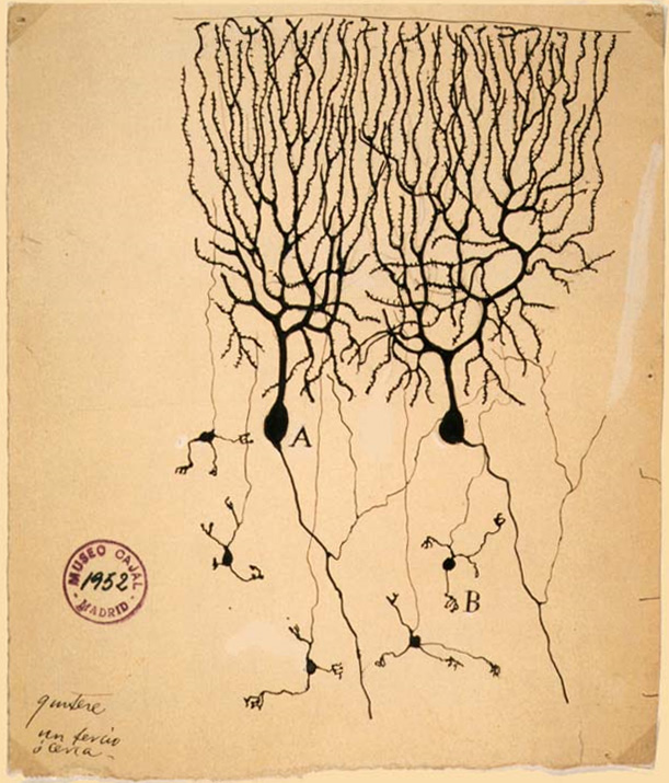
Pyramidal cells, or pyramidal neurons, are a type of multipolar neuron found in areas of the brain including the cerebral cortex, the hippocampus, and the amygdala. Pyramidal cells are the primary excitation units of the mammalian prefrontal cortex and the corticospinal tract. One of the main structural features of the pyramidal neuron is the conic shaped soma, or cell body, after which the neuron is named. Other key structural features of the pyramidal cell are a single axon, a large apical dendrite, multiple basal dendrites, and the presence of dendritic spines. Pyramidal neurons are also one of two cell types where the characteristic sign, Negri bodies, are found in post-mortem rabies infection. Pyramidal neurons were first discovered and studied by Santiago Ramón y Cajal. Since then, studies on pyramidal neurons have focused on topics ranging from neuroplasticity to cognition. Structure One of the main structural features of the pyramidal neuron is the conic sha ... [...More Info...] [...Related Items...] OR: [Wikipedia] [Google] [Baidu] |
Ganglion
A ganglion (: ganglia) is a group of neuron cell bodies in the peripheral nervous system. In the somatic nervous system, this includes dorsal root ganglia and trigeminal ganglia among a few others. In the autonomic nervous system, there are both sympathetic and parasympathetic ganglia which contain the cell bodies of postganglionic sympathetic and parasympathetic neurons respectively. A pseudoganglion looks like a ganglion, but only has nerve fibers and has no nerve cell bodies. Structure Ganglia are primarily made up of somata and dendritic structures, which are bundled or connected. Ganglia often interconnect with other ganglia to form a complex system of ganglia known as a plexus. Ganglia provide relay points and intermediary connections between different neurological structures in the body, such as the peripheral and central nervous systems. Among vertebrates there are three major groups of ganglia: * Dorsal root ganglia (also known as the spinal ganglia) contai ... [...More Info...] [...Related Items...] OR: [Wikipedia] [Google] [Baidu] |
Brodmann Area 8
Brodmann area 8 is one of Brodmann's cytologically defined regions of the brain. It is involved in planning complex movements. Human Brodmann area 8, or BA8, is part of the frontal cortex in the human brain. Situated just anterior to the premotor cortex ( BA6), it includes the frontal eye fields (so-named because they are believed to play an important role in the control of eye movements). Damage to this area, by stroke, trauma or infection, causes tonic deviation of the eyes towards the side of the injury. This finding occurs during the first few hours of an acute event such as cerebrovascular infarct (stroke) or hemorrhage (bleeding). Guenon The term Brodmann area 8 refers to a cytoarchitecturally defined portion of the frontal lobe of the guenon. Located rostral to the arcuate sulcus, it was not considered by Brodmann-1909 to be topographically homologous to the intermediate frontal area 8 of the human. Distinctive features (Brodmann-1905): compared to Brodmann area 6- ... [...More Info...] [...Related Items...] OR: [Wikipedia] [Google] [Baidu] |
Brodmann Area 6
Brodmann area 6 (BA6) is part of the frontal cortex in the human brain. Situated just anterior to the primary motor cortex ( BA4), it is composed of the premotor cortex and, medially, the supplementary motor area (SMA). This large area of the frontal cortex is believed to play a role in planning complex, coordinated movements. Brodmann area 6 is also called agranular frontal area 6 in humans because it lacks an internal granular cortical layer (layer IV). It is a subdivision of the cytoarchitecturally defined precentral region of cerebral cortex. In the human brain, it is located on the portions of the precentral gyrus that are not occupied by Brodmann area 4; furthermore, BA6 extends onto the caudal portions of the superior frontal and middle frontal gyri. It extends from the cingulate sulcus on the medial aspect of the hemisphere to the lateral sulcus on the lateral aspect. It is bounded rostrally by the granular frontal region and caudally by the gigantopyramidal area 4 (Br ... [...More Info...] [...Related Items...] OR: [Wikipedia] [Google] [Baidu] |


