|
Brain Activity And Meditation
Meditation and its effect on brain activity and the central nervous system became a focus of collaborative research in neuroscience, psychology and neurobiology during the latter half of the 20th century. Research on meditation sought to define and characterize various practices. The effects of meditation on the brain can be broken up into two categories: state changes and trait changes, respectively alterations in brain activities during the act of meditating and changes that are the outcome of long-term practice. Mindfulness meditation, a Buddhist meditation approach found in Zen and Vipassana, is frequently studied. Jon Kabat-Zinn describes mindfulness meditation as complete, unbiased attention to the current moment. Changes in brain state Electroencephalography Electroencephalography (EEG) has been used in many studies as a primary method for evaluating the meditating brain. Electroencephalography uses electrical leads placed all over the scalp to measure the collective e ... [...More Info...] [...Related Items...] OR: [Wikipedia] [Google] [Baidu] |
MRI Anterior Cingulate
Magnetic resonance imaging (MRI) is a medical imaging technique used in radiology to generate pictures of the anatomy and the physiological processes inside the body. MRI scanners use strong magnetic fields, magnetic field gradients, and radio waves to form images of the organs in the body. MRI does not involve X-rays or the use of ionizing radiation, which distinguishes it from computed tomography (CT) and positron emission tomography (PET) scans. MRI is a medical application of nuclear magnetic resonance (NMR) which can also be used for imaging in other NMR applications, such as NMR spectroscopy. MRI is widely used in hospitals and clinics for medical diagnosis, staging and follow-up of disease. Compared to CT, MRI provides better contrast in images of soft tissues, e.g. in the brain or abdomen. However, it may be perceived as less comfortable by patients, due to the usually longer and louder measurements with the subject in a long, confining tube, although "open" MR ... [...More Info...] [...Related Items...] OR: [Wikipedia] [Google] [Baidu] |
Functional Magnetic Resonance Imaging
Functional magnetic resonance imaging or functional MRI (fMRI) measures brain activity by detecting changes associated with blood flow. This technique relies on the fact that cerebral blood flow and neuronal activation are coupled. When an area of the brain is in use, blood flow to that region also increases. The primary form of fMRI uses the blood-oxygen-level dependent (BOLD) contrast, discovered by Seiji Ogawa in 1990. This is a type of specialized brain and body scan used to map neuron, neural activity in the brain or spinal cord of humans or other animals by imaging the change in blood flow (hemodynamic response) related to energy use by brain cells. Since the early 1990s, fMRI has come to dominate brain mapping research because it does not involve the use of injections, surgery, the ingestion of substances, or exposure to ionizing radiation. This measure is frequently corrupted by noise from various sources; hence, statistical procedures are used to extract the underlying si ... [...More Info...] [...Related Items...] OR: [Wikipedia] [Google] [Baidu] |
Hippocampus
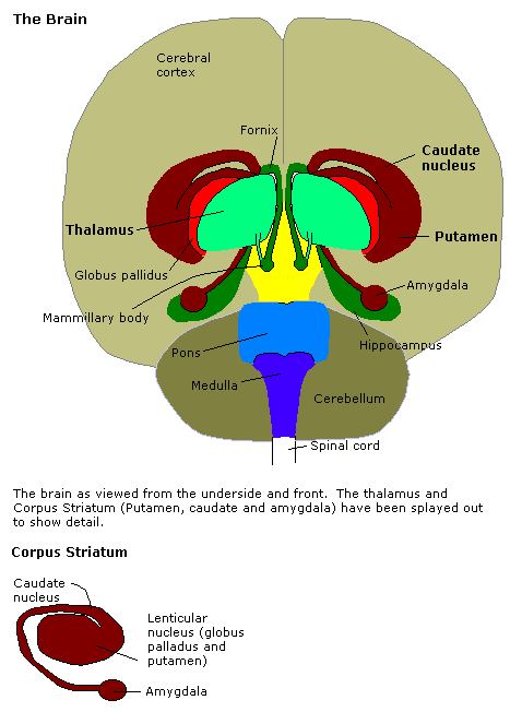
The hippocampus (: hippocampi; via Latin from Ancient Greek, Greek , 'seahorse'), also hippocampus proper, is a major component of the brain of humans and many other vertebrates. In the human brain the hippocampus, the dentate gyrus, and the subiculum are components of the hippocampal formation located in the limbic system. The hippocampus plays important roles in the Memory consolidation, consolidation of information from short-term memory to long-term memory, and in spatial memory that enables Navigation#Navigation in spatial cognition, navigation. In humans, and other primates the hippocampus is located in the archicortex, one of the three regions of allocortex, in each cerebral hemisphere, hemisphere with direct neural projections to, and reciprocal indirect projections from the neocortex. The hippocampus, as the medial pallium, is a structure found in all vertebrates. In Alzheimer's disease (and other forms of dementia), the hippocampus is one of the first regions of th ... [...More Info...] [...Related Items...] OR: [Wikipedia] [Google] [Baidu] |
Insular Cortex
The insular cortex (also insula and insular lobe) is a portion of the cerebral cortex folded deep within the lateral sulcus (the fissure separating the temporal lobe from the parietal lobe, parietal and frontal lobes) within each brain hemisphere, hemisphere of the mammalian brain. The insulae are believed to be involved in consciousness and play a role in diverse functions usually linked to emotion or the regulation of the body's homeostasis. These functions include compassion, empathy, taste, perception, motor control, self-awareness, cognitive functioning, interpersonal relationships, and awareness of homeostatic emotions such as Hunger (physiology), hunger, pain and fatigue. In relation to these, it is involved in psychopathology. The insular cortex is divided by the central sulcus of the insula, into two parts: the anterior insula and the posterior insula in which more than a dozen field areas have been identified. The cortical area overlying the insula toward the lateral su ... [...More Info...] [...Related Items...] OR: [Wikipedia] [Google] [Baidu] |
Sensory Cortex
The sensory cortex can refer sometimes to the primary somatosensory cortex, or it can be used as a term for the primary and secondary cortices of the different senses (two cortices each, on left and right hemisphere): the visual cortex on the occipital lobes, the auditory cortex on the temporal lobes, the primary olfactory cortex on the uncus of the piriform region of the temporal lobes, the gustatory cortex on the insular lobe (also referred to as the insular cortex), and the primary somatosensory cortex on the anterior parietal lobes. Just posterior to the primary somatosensory cortex lies the somatosensory association cortex or area, which integrates sensory information from the primary somatosensory cortex (temperature, pressure, etc.) to construct an understanding of the object being felt. Inferior to the frontal lobes are found the olfactory bulbs, which receive sensory input from the olfactory nerves and route those signals throughout the brain. Not all olfactory infor ... [...More Info...] [...Related Items...] OR: [Wikipedia] [Google] [Baidu] |
Brodmann Area 10
Brodmann area 10 (BA10, frontopolar prefrontal cortex, rostrolateral prefrontal cortex, or anterior prefrontal cortex) is the anterior-most portion of the prefrontal cortex in the human brain. BA10 was originally defined broadly in terms of its cytoarchitectonic traits as they were observed in the brains of cadavers, but because modern functional imaging cannot precisely identify these boundaries, the terms anterior prefrontal cortex, rostral prefrontal cortex and frontopolar prefrontal cortex are used to refer to the area in the most anterior part of the frontal cortex that approximately covers BA10—simply to emphasize the fact that BA10 does not include ''all'' parts of the prefrontal cortex. BA10 is the largest cytoarchitectonic area in the human brain. It has been described as "one of the least well understood regions of the human brain". Present research suggests that it is involved in strategic processes in memory recall and various executive functions. During human evol ... [...More Info...] [...Related Items...] OR: [Wikipedia] [Google] [Baidu] |
Meta-analysis
Meta-analysis is a method of synthesis of quantitative data from multiple independent studies addressing a common research question. An important part of this method involves computing a combined effect size across all of the studies. As such, this statistical approach involves extracting effect sizes and variance measures from various studies. By combining these effect sizes the statistical power is improved and can resolve uncertainties or discrepancies found in individual studies. Meta-analyses are integral in supporting research grant proposals, shaping treatment guidelines, and influencing health policies. They are also pivotal in summarizing existing research to guide future studies, thereby cementing their role as a fundamental methodology in metascience. Meta-analyses are often, but not always, important components of a systematic review. History The term "meta-analysis" was coined in 1976 by the statistician Gene V. Glass, Gene Glass, who stated ''"Meta-analysis refers t ... [...More Info...] [...Related Items...] OR: [Wikipedia] [Google] [Baidu] |
Hippocampus
The hippocampus (: hippocampi; via Latin from Ancient Greek, Greek , 'seahorse'), also hippocampus proper, is a major component of the brain of humans and many other vertebrates. In the human brain the hippocampus, the dentate gyrus, and the subiculum are components of the hippocampal formation located in the limbic system. The hippocampus plays important roles in the Memory consolidation, consolidation of information from short-term memory to long-term memory, and in spatial memory that enables Navigation#Navigation in spatial cognition, navigation. In humans, and other primates the hippocampus is located in the archicortex, one of the three regions of allocortex, in each cerebral hemisphere, hemisphere with direct neural projections to, and reciprocal indirect projections from the neocortex. The hippocampus, as the medial pallium, is a structure found in all vertebrates. In Alzheimer's disease (and other forms of dementia), the hippocampus is one of the first regions of th ... [...More Info...] [...Related Items...] OR: [Wikipedia] [Google] [Baidu] |
Superior Temporal Sulcus
The superior temporal sulcus (STS) is the sulcus separating the superior temporal gyrus from the middle temporal gyrus, in the temporal lobe of the mammalian brain. A sulcus (plural sulci) is a deep groove that curves into the largest part of the brain, the cerebrum, and a gyrus (plural gyri) is a ridge that curves outward of the cerebrum. The STS is located under the lateral fissure, which is the fissure that separates the temporal lobe, parietal lobe, and frontal lobe. The STS has an asymmetric structure between the left and right hemisphere, with the STS being longer in the left hemisphere, but deeper in the right hemisphere. This asymmetrical structural organization between hemispheres has only been found to occur in the STS of the human brain. The STS has been shown to produce strong responses when subjects perceive stimuli in research areas that include theory of mind, biological motion, faces, voices, and language. Language processing Spoken language processing ... [...More Info...] [...Related Items...] OR: [Wikipedia] [Google] [Baidu] |
Amygdala
The amygdala (; : amygdalae or amygdalas; also '; Latin from Greek language, Greek, , ', 'almond', 'tonsil') is a paired nucleus (neuroanatomy), nuclear complex present in the Cerebral hemisphere, cerebral hemispheres of vertebrates. It is considered part of the limbic system. In Primate, primates, it is located lateral and medial, medially within the temporal lobes. It consists of many nuclei, each made up of further subnuclei. The subdivision most commonly made is into the Basolateral amygdala, basolateral, Central nucleus of the amygdala, central, cortical, and medial nuclei together with the intercalated cells of the amygdala, intercalated cell clusters. The amygdala has a primary role in the processing of memory, decision making, decision-making, and emotions, emotional responses (including fear, anxiety, and aggression). The amygdala was first identified and named by Karl Friedrich Burdach in 1822. Structure Thirteen Nucleus (neuroanatomy), nuclei have been identif ... [...More Info...] [...Related Items...] OR: [Wikipedia] [Google] [Baidu] |
Frontal Cortex
The frontal lobe is the largest of the four major lobes of the brain in mammals, and is located at the front of each cerebral hemisphere (in front of the parietal lobe and the temporal lobe). It is parted from the parietal lobe by a groove between tissues called the central sulcus and from the temporal lobe by a deeper groove called the lateral sulcus (Sylvian fissure). The most anterior rounded part of the frontal lobe (though not well-defined) is known as the frontal pole, one of the three poles of the cerebrum. The frontal lobe is covered by the frontal cortex. The frontal cortex includes the premotor cortex and the primary motor cortex – parts of the motor cortex. The front part of the frontal cortex is covered by the prefrontal cortex. The nonprimary motor cortex is a functionally defined portion of the frontal lobe. There are four principal gyri in the frontal lobe. The precentral gyrus is directly anterior to the central sulcus, running parallel to it and contai ... [...More Info...] [...Related Items...] OR: [Wikipedia] [Google] [Baidu] |
Cingulate Cortex
The cingulate cortex is a part of the brain situated in the medial aspect of the cerebral cortex. The cingulate cortex includes the entire cingulate gyrus, which lies immediately above the corpus callosum, and the continuation of this in the cingulate sulcus. The cingulate cortex is usually considered part of the limbic lobe. It receives inputs from the thalamus and the neocortex, and projects to the entorhinal cortex via the cingulum. It is an integral part of the limbic system, which is involved with emotion formation and processing, learning, and memory. The combination of these three functions makes the cingulate gyrus highly influential in linking motivational outcomes to behavior (e.g. a certain action induced a positive emotional response, which results in learning). This role makes the cingulate cortex highly important in disorders such as depression and schizophrenia. It also plays a role in executive function and respiratory control. Structure Based on cerebral c ... [...More Info...] [...Related Items...] OR: [Wikipedia] [Google] [Baidu] |


