|
Anatomy Scan
The anomaly scan, also sometimes called the anatomy scan, 20-week ultrasound, or level 2 ultrasound, evaluates anatomic structures of the fetus, placenta, and maternal pelvic organs. This scan is an important and common component of routine prenatal care. The function of the ultrasound is to measure the fetus so that growth abnormalities can be recognized quickly later in pregnancy, to assess for congenital malformations and multiple pregnancies, and to plan method of delivery. Procedure This scan is conducted between 18 and 22 weeks' gestation, but most often performed at 19 weeks, as a component of routine prenatal care. Prior to 18 weeks' gestation, the fetal organs may be of insufficient size and development to allow for ultrasound evaluation. Scans performed beyond 22 weeks' gestation may limit the ability to seek pregnancy termination, depending on local legislation. Two-dimensional (2D) is used to evaluate fetal structures, placenta, and amniotic fluid volume. Maternal ... [...More Info...] [...Related Items...] OR: [Wikipedia] [Google] [Baidu] |
Fetus
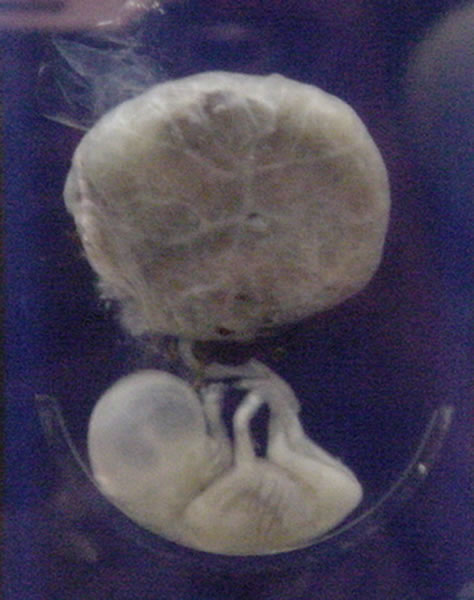
A fetus or foetus (; plural fetuses, feti, foetuses, or foeti) is the unborn offspring that develops from an animal embryo. Following embryonic development the fetal stage of development takes place. In human prenatal development, fetal development begins from the ninth week after fertilization (or eleventh week gestational age) and continues until birth. Prenatal development is a continuum, with no clear defining feature distinguishing an embryo from a fetus. However, a fetus is characterized by the presence of all the major body organs, though they will not yet be fully developed and functional and some not yet situated in their final anatomical location. Etymology The word '' fetus'' (plural '' fetuses'' or '' feti'') is related to the Latin '' fētus'' ("offspring", "bringing forth", "hatching of young") and the Greek "φυτώ" to plant. The word "fetus" was used by Ovid in Metamorphoses, book 1, line 104. The predominant British, Irish, and Commonwealth spelling ... [...More Info...] [...Related Items...] OR: [Wikipedia] [Google] [Baidu] |
Chorion
The chorion is the outermost fetal membrane around the embryo in mammals, birds and reptiles (amniotes). It develops from an outer fold on the surface of the yolk sac, which lies outside the zona pellucida (in mammals), known as the vitelline membrane in other animals. In insects it is developed by the follicle cells while the egg is in the ovary.Chapman, R.F. (1998) "The insects: structure and function", Section ''The egg and embryology''. Previewed in Google Bookon 26 Sep 2009. Structure In humans and other mammals (excluding monotremes), the chorion is one of the fetal membranes that exist during pregnancy between the developing fetus and mother. The chorion and the amnion together form the amniotic sac. In humans it is formed by extraembryonic mesoderm and the two layers of trophoblast that surround the embryo and other membranes; the chorionic villi emerge from the chorion, invade the endometrium, and allow the transfer of nutrients from maternal blood to feta ... [...More Info...] [...Related Items...] OR: [Wikipedia] [Google] [Baidu] |
Patau Syndrome
Patau syndrome is a syndrome caused by a chromosomal abnormality, in which some or all of the cells of the body contain extra genetic material from chromosome 13. The extra genetic material disrupts normal development, causing multiple and complex organ defects. This can occur either because each cell contains a full extra copy of chromosome 13 (a disorder known as trisomy 13 or trisomy D or T13), or because each cell contains an extra partial copy of the chromosome, or because there are two different lines of cells—one healthy with the correct number of chromosomes 13 and one that contains an extra copy of the chromosome—mosaic Patau syndrome. Full trisomy 13 is caused by nondisjunction of chromosomes during meiosis (the mosaic form is caused by nondisjunction during mitosis). Like all nondisjunction conditions (such as Down syndrome and Edwards syndrome), the risk of this syndrome in the offspring increases with maternal age at pregnancy, with about 31 years being th ... [...More Info...] [...Related Items...] OR: [Wikipedia] [Google] [Baidu] |
Edwards Syndrome
Edwards syndrome, also known as trisomy 18, is a genetic disorder caused by the presence of a third copy of all or part of chromosome 18. Many parts of the body are affected. Babies are often born small and have heart defects. Other features include a small head, small jaw, clenched fists with overlapping fingers, and severe intellectual disability. Most cases of Edwards syndrome occur due to problems during the formation of the reproductive cells or during early development. The rate of disease increases with the mother's age. Rarely, cases may be inherited from a person's parents. Occasionally, not all cells have the extra chromosome, known as mosaic trisomy, and symptoms in these cases may be less severe. An ultrasound during pregnancy can increase suspicion for the condition, which can be confirmed by amniocentesis. Treatment is supportive. After having one child with the condition, the risk of having a second is typically around one percent. It is the second-most com ... [...More Info...] [...Related Items...] OR: [Wikipedia] [Google] [Baidu] |
Fetal Kidney Perfusion And Fetal Breathing Activity
A fetus or foetus (; plural fetuses, feti, foetuses, or foeti) is the unborn offspring that develops from an animal embryo. Following embryonic development the fetal stage of development takes place. In human prenatal development, fetal development begins from the ninth week after fertilization (or eleventh week gestational age) and continues until birth. Prenatal development is a continuum, with no clear defining feature distinguishing an embryo from a fetus. However, a fetus is characterized by the presence of all the major body organs, though they will not yet be fully developed and functional and some not yet situated in their final anatomical location. Etymology The word ''fetus'' (plural ''fetuses'' or '' feti'') is related to the Latin '' fētus'' ("offspring", "bringing forth", "hatching of young") and the Greek "φυτώ" to plant. The word "fetus" was used by Ovid in Metamorphoses, book 1, line 104. The predominant British, Irish, and Commonwealth spelling is ''fo ... [...More Info...] [...Related Items...] OR: [Wikipedia] [Google] [Baidu] |
Lemon And Banana Sign For Neural Tube Defect Dr Wolfgang Moroder
The lemon (''Citrus limon'') is a species of small evergreen trees in the flowering plant family Rutaceae, native to Asia, primarily Northeast India (Assam), Northern Myanmar or China. The tree's ellipsoidal yellow fruit is used for culinary and non-culinary purposes throughout the world, primarily for its juice, which has both culinary and cleaning uses. The pulp and rind are also used in cooking and baking. The juice of the lemon is about 5% to 6% citric acid, with a pH of around 2.2, giving it a sour taste. The distinctive sour taste of lemon juice makes it a key ingredient in drinks and foods such as lemonade and lemon meringue pie. History The origin of the lemon is unknown, though lemons are thought to have first grown in Assam (a region in northeast India), northern Myanmar or China. A genomic study of the lemon indicated it was a hybrid between bitter orange (sour orange) and citron. Lemons are supposed to have entered Europe near southern Italy no later than ... [...More Info...] [...Related Items...] OR: [Wikipedia] [Google] [Baidu] |
Urinary Bladder
The urinary bladder, or simply bladder, is a hollow organ in humans and other vertebrates that stores urine from the kidneys before disposal by urination. In humans the bladder is a distensible organ that sits on the pelvic floor. Urine enters the bladder via the ureters and exits via the urethra. The typical adult human bladder will hold between 300 and (10.14 and ) before the urge to empty occurs, but can hold considerably more. The Latin phrase for "urinary bladder" is ''vesica urinaria'', and the term ''vesical'' or prefix ''vesico -'' appear in connection with associated structures such as vesical veins. The modern Latin word for "bladder" – ''cystis'' – appears in associated terms such as cystitis (inflammation of the bladder). Structure In humans, the bladder is a hollow muscular organ situated at the base of the pelvis. In gross anatomy, the bladder can be divided into a broad , a body, an apex, and a neck. The apex (also called the vertex) is directed fo ... [...More Info...] [...Related Items...] OR: [Wikipedia] [Google] [Baidu] |
Ovaries
The ovary is an organ in the female reproductive system that produces an ovum. When released, this travels down the fallopian tube into the uterus, where it may become fertilized by a sperm. There is an ovary () found on each side of the body. The ovaries also secrete hormones that play a role in the menstrual cycle and fertility. The ovary progresses through many stages beginning in the prenatal period through menopause. It is also an endocrine gland because of the various hormones that it secretes. Structure The ovaries are considered the female gonads. Each ovary is whitish in color and located alongside the lateral wall of the uterus in a region called the ovarian fossa. The ovarian fossa is the region that is bounded by the external iliac artery and in front of the ureter and the internal iliac artery. This area is about 4 cm x 3 cm x 2 cm in size.Daftary, Shirish; Chakravarti, Sudip (2011). Manual of Obstetrics, 3rd Edition. Elsevier. pp. 1-16. . The ovari ... [...More Info...] [...Related Items...] OR: [Wikipedia] [Google] [Baidu] |
Fallopian Tube
The fallopian tubes, also known as uterine tubes, oviducts or salpinges (singular salpinx), are paired tubes in the human female that stretch from the uterus to the ovaries. The fallopian tubes are part of the female reproductive system. In other mammals they are only called oviducts. Each tube is a muscular hollow organ that is on average between 10 and 14 cm in length, with an external diameter of 1 cm. It has four described parts: the intramural part, isthmus, ampulla, and infundibulum with associated fimbriae. Each tube has two openings a proximal opening nearest and opening to the uterus, and a distal opening furthest and opening to the abdomen. The fallopian tubes are held in place by the mesosalpinx, a part of the broad ligament mesentery that wraps around the tubes. Another part of the broad ligament, the mesovarium suspends the ovaries in place. An egg cell is transported from an ovary to a fallopian tube where it may be fertilized in the ampulla of th ... [...More Info...] [...Related Items...] OR: [Wikipedia] [Google] [Baidu] |
Amniotic Fluid
The amniotic fluid is the protective liquid contained by the amniotic sac of a gravid amniote. This fluid serves as a cushion for the growing fetus, but also serves to facilitate the exchange of nutrients, water, and biochemical products between mother and fetus. For humans, the amniotic fluid is commonly called water or waters (Latin liquor amnii). Development Amniotic fluid is present from the formation of the gestational sac. Amniotic fluid is in the amniotic sac. It is generated from maternal plasma, and passes through the fetal membranes by osmotic and hydrostatic forces. When fetal kidneys begin to function around week 16, fetal urine also contributes to the fluid. In earlier times, it was believed that the amniotic fluid was composed entirely of fetal urine. The fluid is absorbed through the fetal tissue and skin. After 22 to 25 week of pregnancy, keratinization of an embryo's skin occurs. When this process completes around the 25th week, the fluid is primarily absor ... [...More Info...] [...Related Items...] OR: [Wikipedia] [Google] [Baidu] |
Umbilical Cord
In placental mammals, the umbilical cord (also called the navel string, birth cord or ''funiculus umbilicalis'') is a conduit between the developing embryo or fetus and the placenta. During prenatal development, the umbilical cord is physiologically and genetically part of the fetus and (in humans) normally contains two arteries (the umbilical arteries) and one vein (the umbilical vein), buried within Wharton's jelly. The umbilical vein supplies the fetus with oxygenated, nutrient-rich blood from the placenta. Conversely, the fetal heart pumps low-oxygen, nutrient-depleted blood through the umbilical arteries back to the placenta. Structure and development The umbilical cord develops from and contains remnants of the yolk sac and allantois. It forms by the fifth week of development, replacing the yolk sac as the source of nutrients for the embryo. The cord is not directly connected to the mother's circulatory system, but instead joins the placenta, which transfers materi ... [...More Info...] [...Related Items...] OR: [Wikipedia] [Google] [Baidu] |
Cervix
The cervix or cervix uteri (Latin, 'neck of the uterus') is the lower part of the uterus (womb) in the human female reproductive system. The cervix is usually 2 to 3 cm long (~1 inch) and roughly cylindrical in shape, which changes during pregnancy. The narrow, central cervical canal runs along its entire length, connecting the uterine cavity and the lumen of the vagina. The opening into the uterus is called the internal os, and the opening into the vagina is called the external os. The lower part of the cervix, known as the vaginal portion of the cervix (or ectocervix), bulges into the top of the vagina. The cervix has been documented anatomically since at least the time of Hippocrates, over 2,000 years ago. The cervical canal is a passage through which sperm must travel to fertilize an egg cell after sexual intercourse. Several methods of contraception, including cervical caps and cervical diaphragms, aim to block or prevent the passage of sperm through the cervic ... [...More Info...] [...Related Items...] OR: [Wikipedia] [Google] [Baidu] |


